OPERATIVE TREATMENT OF
NON-CAVITATED APPROXIMAL CARIOUS LESIONS OF
PERMANENT CHILDREN’S TEETH
Rossiza I. Kabakchieva1, Natalia H. Gateva1, Hristina D. Mihaylova2
1) Department of Paediatric Dentistry, Faculty of Dental Medicine, Medical Uni-versity - Sofia, Bulgaria
2) Department of Imaging and Oral Diagnostic, Faculty of Dental Medicine, Medical University - Sofia, Bulgaria
Journal of IMAB - Annual Proceeding (Scientific Papers)2014, vol. 20, issue 5 Journal of IMAB
ISSN: 1312-773X
http://www.journal-imab-bg.org
SUMMARY
Aim: To apply and follow up in clinical conditions the success rate of microinvasive technique of infiltration with low viscosity resin ICON® (DMG) of non cavitated approximal caries lesions of permanent children’s teeth for a period of one year.
Material and methods: The study included 18 children aged 716 years. They were divided into two groups -children with medium and high caries risk. The survey in-clude 20 teeth with approximal non-cavitated enamel lesions up to the outer third of dentin (E1, E2, D1 – according to the manufacturer’s instructions of ICON®). The size of the lesions was determined using bitewing radiographs and the activity - by Papilla Bleeding Index. The clinical application of the infiltr+ant (ICON® Caries Infiltrant Proximal, DMG) was conducted according to the manufacturer’s instructions. Bitewing radiographs were made at 6 and 12 months after infiltration in order to evaluate the success of the method. A test of the difference between two relative pro-portions and alternative test for analysis of the results were used.
Results: Our study confirm the hypothesis that this method of infiltration is equally successful for permanent teeth in patients with moderate caries risk as well as those at high caries risk.
Conclusion: This study is the first survey regarding the success of the application of ICON® for treatment of non-cavitated approximal carious lesions in permanent dentition of children in the country. Research in this direction should continue in order to increase the conviction that caries can be controlled and arrested in its earliest stages.
Key words: caries infiltration, permanent teeth, non-cavitated approximal carious lesions,
Philosophy of minimally invasive dentistry integrates prevention, remineralization, and minimal preparation during operative treatment, which actually guarantees the preserva-tion of healthy tooth structures [1]. At its core minimally in-vasive treatment includes early caries detection and non-op-erative treatment of non-cavitated carious lesions [2].
It is known from epidemiology of caries that during childhood caries in fissures and pits dominate according to the indicator DMFT/dft. During adolescence, the proportion
of proximal caries increases and dominates the calculation of the if DMFS-index is observed [3, 4]. Studies have found that about 50% more of approximal carious lesions were identified using bitewing radiographs compared with clini-cally diagnosed by visual examination [5, 6]. Authors rec-ommend radiographic studies should be carried out selec-tively, based on the individual caries risk [7, 8, 9]. Pitts and Rimmer [10] found in a study that 60% of approximal car-ies which is radiographically proved as carcar-ies in the outer third of dentin have not yet cavitated enamel surface. The ability of clinicians to interpret radiographs has the greatest influence in the decision for the type of treatment [11]. Diagnosting caries as “active” or “inactive” has also a basic role for the treatment. Results of “Papilla Bleeding Index” (PBI) are also important indicators of proximal caries activ-ity [12, 13].
Strategy for non-surgical treatments using “procedures for remineralization” “cover with sealants” or “infiltration with low viscosity resin” for initial carious lesions are an ef-fective alternative for operative restorative approaches [14, 15, 16, 17], but they are not real alternatives in the attempt to reduce caries activity.
A relatively new technique for microinvasive treatment of non-cavitated carious lesions is the infiltration technique with low-viscosity resin. This microinvasive treatmet‘s method has great potential in Paediatric Dentistry [16, 18, 19]. The initial carious lesions can be arrested using resin in-filtration [17, 20, 21, 22, 23]. Inin-filtration of caries lesions with this material is not intended to cover the lesion, but to fill the lesion‘s pores, the more deeper, the better [24, 25, 26, 27]. The main aim of the caries infiltration technique or “car-ies infiltration” is to use the possibility of this low viscos-ity light-curing resin material called “infiltrant” [28, 29] to penetrate into the pores of “the body of the lesion” and to close them. The method can be used for arresting the non-cavitated carious lesions developing on smooth approximal surfaces of the deciduous and permanent teeth. Current clini-cal results show good effectiveness of the method [15, 24, 29, 30, 31, 32, 33].
The aim of this study is to apply and follow-up clini-cally the success rate of the microinvasive technique of in-filtration with low viscosity resin ICON® (DMG) of noncavitated approximal carious lesions in permanent chil-dren’s teeth for a period of one year.
http://www.journal-imab-bg.org 627 To achieve the aim, the following tasks were set:
- to apply in clinical setting the microinvasive infil-tration technique for non-cavitated approximal carious lesions on 20 permanent premolars and molars using professional kit ICON® (DMG, Hamburg, Germany)
- determine the success rate of this infiltration tech-nique in two groups of children - children with moderate and children at high caries risk.
Working hypothesis that we tested during the study was that the method of infiltration with ICON is equally suc-cessful for children with moderate and children at high car-ies risk in the permanent dentition.
MATERIAL AND METHODS:
The study included 18 children aged 7-16 years with permanent teeth which have non-cavitated approximal le-sions. Informed consent for participation was signed by par-ents.
Criteria for selection of children:
- Children at moderate and high caries risk - indi-vidual assessment (assessment was made applying the tool for caries risk assessment used in the Department of Paedi-atric Dentistry, FDM, MU-Sofia).
- Children with active participation – children, who after the procedure should maintain oral hygiene by brush-ing, fluoride toothpaste and dental floss.
Criteria for selecting teeth
- permanent teeth with non-cavitated approximal cari-ous lesions in the enamel up to the outer third of dentin ac-cording to bitewing X-rays (E1, E2, D1 – acac-cording to the manufacturer’s instructions of ICON®).
Bitewing radiographs for diagnosis of non-cavitated approximal lesion and evaluation of its depth were made with X-ray apparatus for dental X-rays Siemens, model 8458747 x 1744 with the following parameters: 70 kV/7 mA. Films Primax RDX-58E soft with size 2/3 and 3/4 cm were used with X-ray holders Kerr/Kwik - bite with ring. The radio-graphic criteria for determining the depth of approximal cari-ous lesions (by Kidd et al. [7] were compared with those of the manufacturer of Icon (E1, E2, D1):
D1: radiolucency in the outer half of the enamel = E1 D2: radiolucency in the inner half of enamel, includ-ing lesions that extend to, but not beyond the enamel-dentin border = E2
D3: radiolucency in dentine – radiolucency beyond enamel-dentine border, but no visible involvement of den-tin = D1
Interpretation of the X-ray image was made by a spe-cialist radiologist, using light box and magnification of 2.5 x.
- “Active lesion” – according to the Papilla Bleeding Index (PBI) score of Saxer and Muhleman (score1, 2, 3, 4). Bleeding after probing the sulcus is a sign of inflammation. We accept the hypothesis that when there is inflamed gingiva and papilla, the approximal carious lesions is active.
Clinical application of infiltrant (ICON® Caries Infiltrant Proximal, DMG) is done according to the manu-facturer’s instructions:
- teeth are professionally cleaned using dental
prophy-laxis paste and floss;
- teeth are separated with special separators in order to create access to the proximal carious lesions;
- etch gel is applied for 120 s and after that it is re-moved by washing with air - water spray. The lesion is dried rigorously for a further 15 seconds with an air spray. To im-prove the drying of the lesion, an ethanol (Icon-dry) is placed over it for 15 seconds and evaporated using a pressurized air (for15 seconds);
- application of infiltrant (ICON® -Infiltrant) with a new applicator is made, The applicator is removed after 3 minutes and the contact area is cleaned from the residue of the material with interdental floss. Infiltrant is light cured for 40 seconds in the buccal, oral, and the occlusal surfaces. In order to improve the impermeability of the coating the infiltrant should be additionally applied for a second time for 1 minute. After removing the excess of the resin and after light curing the separating wedge is removed.
Filling in the patient’s card
ICON material is not radiopaque. To record the infil-trated surfaces and the depth of the lesions during treatment, a card is completed and this card accompanies the patient at review appointments. This card is a component of the pro-fessional kit and contains:
- header - the name of the patient; - dental chart - the treated area is charted;
- the treated tooth, the treated surface, the depth of the lesion and the date of the operation are recorded;
- the results of the bitewing radiographies are re-corded too .
Instruction for patients. After the procedure is com-pleted, the children and their parents were instructed to main-tain personal oral hygiene using a toothbrush and fluoride toothpaste and interdental floss according to the routine meth-ods.
Clinical examination was done by two doctors, experts in paediatric dentistry. “Calibration” of the research team was done in advance.
Criteria for assessing the success of infiltration held on 6 and 12 months follow ups:
- there is no difference between the size of carious le-sions on the follow up radiographs taken on the 6 and 12 months compared to those from the diagnostic radiographs The diagnostic and follow up radiographs at 6 and 12 months are made using the same equipment and parameters, which are entered in an individual card that accompanies the pa-tient in order all the radiographies to be comparable.
- no inflammation (bleeding) of the papilla adjacent to the treated carious lesions is noticed - PBI of Saxer and Muhleman - code 0.
Statistical methods:
In order to analyse the hypothesis that the method of infiltration with ICON® is equally successful in children at medium and high caries risk two tests for assessment of the proportions of success were used:
Test for difference between two relative shares -based on approximation by a normal distribution;
distri-bution (Fisher exact p estimation). The test was used on the basis of simulation constructed from aggregate data.
RESULTS:
Table 1 shows the distribution of groups of children and teeth with approximal lesions, according to the risk of caries - a group of children and teeth with a moderate and high risk of caries development. Bitewing radiographs deter-mined the depth of carious lesions - E1, E2, D1.
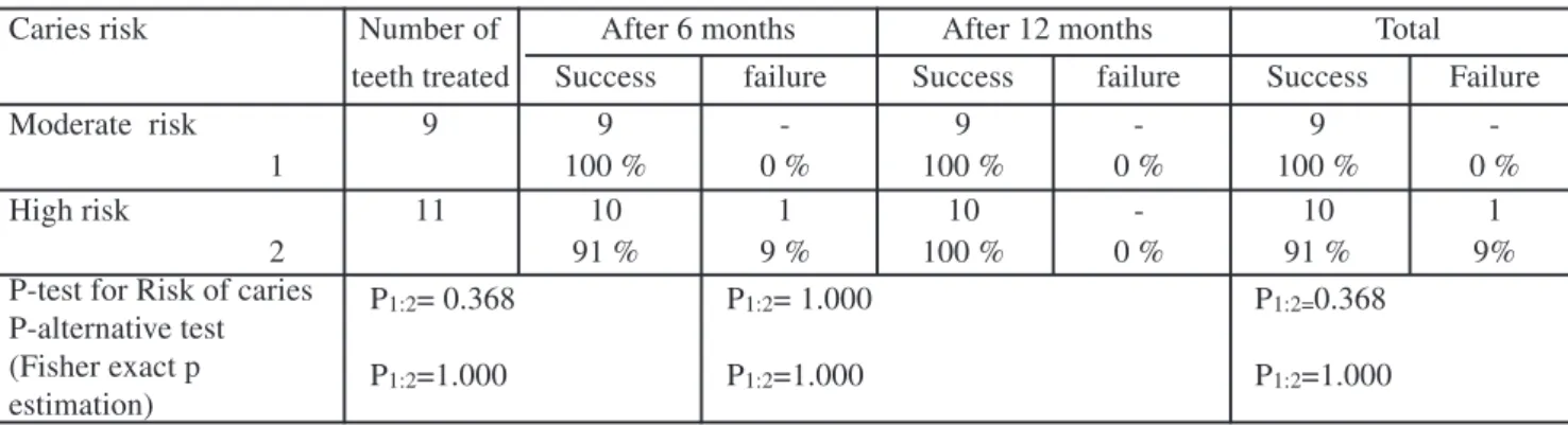
The results of “success” and “failure” treatment after an application of non-cavitated approximal carious lesions with infiltrant ICON® and one year of follow-up are pre-sented in Table 2.
Table 1. Distribution of children and teeth with approximal non-cavitated carious lesions
Table 2. Level of success rate and failure after treatment of non-cavitated approximal lesions in permanent teeth with ICON®
Caries risk Number of After 6 months After 12 months Total
teeth treated Success failure Success failure Success Failure
Moderate risk 9 9 - 9 - 9
1 100 % 0 % 100 % 0 % 100 % 0 %
High risk 11 10 1 10 - 10 1
2 91 % 9 % 100 % 0 % 91 % 9%
P-test for Risk of caries P1:2= 0.368 P1:2= 1.000 P1:2=0.368 P-alternative test
(Fisher exact p P1:2=1.000 P1:2=1.000 P1:2=1.000
estimation)
The result from the assessment of the criteria “dif-ference between the amount of caries lesions” is actually a result made from the include analysis of 60 bitewing graphs; 20 of them were diagnostic, and 40 - control radio-graphs (20 radioradio-graphs were made 6 months after, and 20 were made 12 months after the application). No radio-graphic criteria were found to confirm the increase of size of the caries lesions in the permanent teeth with a moder-ate caries risk 6 months and 12 months after the applica-tion. In the group of permanent teeth with a high caries risk 6 months after the application only one tooth showed ex-pansion of carious lesion. The other successful infiltrations successfully done, remained unchanged after the 12 months. All teeth with infiltration were assessed with code 0 ac-cording to PBI. At the end of the one-year follow-up the results showed that the method of infiltration in teeth with moderate caries risk was 100% successful and 91% in teeth with high caries risk.
Tests applied to assess the differences between the occurrence of success of treatment 6 months and 12 months
after the application and respectively the total score for both groups (of moderate and high caries risk) in permanent teeth showed no evidence of occurrence of a statistically signifi-cant difference between the two groups for all three meas-urements - 6 months, 12 months and total (P> 0.05). Our study confirmed the hypotheses that the tested method of infiltration of permanent teeth is equally successful for pa-tients at moderate and those at high risk of caries develop-ment.
DISCUSSION:
Treatment of caries using infiltration with low vis-cosity resin is a relatively new microinvasive treatment method with enormous potential in paediatric dentistry [14, 34, 35]. Short-term clinical results show good effectiveness of the method in caries lesions of approximal surfaces in permanent teeth. Stationing of development of the lesion within 1 to 2 years is reported [3, 30, 36, 37].
The high scores of “success” after administration of caries infiltration technique with ICON® in permanent den-Caries risk Noncavitated approximal
Study Groups Moderate High lesions
Children teeth Children teeth E1 E2 D1
Children with permanent teeth
8 9 10 11 10 7 3
http://www.journal-imab-bg.org 629 tition according to our trial we can explain with the
follow-ing: children with permanent dentition have higher effi-ciency of caries prevention – consciousness for food and oral prevention and generally they are more responsible. And this leads to lower risk of deepening of the approximal lesions. The possibility of failure of the infiltrant requires strict professional control in order to prevent complications of dental caries. Another prerequisite for a possible failure is the inaccuracy of diagnostic X-ray method; sometimes the clinician could be misled to use the technique of infil-tration. The survey results show that ICON® is suitable for use in the treatment of permanent teeth with an initial approximal caries. Our hypothesis that infiltration is equally successful nevertheless the degree of caries risk makes the caries infiltration a method of choice in the ambition for using minimally invasive methods of treatment of caries.
We understand that our experience in the application of the technique of infiltration is currently too short. We need to continue the follow of the cases in time and to in-crease their number in order to become more confident and
convinced of the long-term best clinical outcome of infil-tration technique for approximal lesions in the permanent dentition.
CONCLUSION
Progress in the evidence-based clinical dentistry pro-vides sufficient facts for reducing the invasive techniques for dental caries treatment especially in childhood. Due to this, there is a great interest in the development and intro-duction into clinical practice the alternative non-invasive or minimally invasive methods, materials and procedures that are equivalent or even more effective than those based on recovery after the drilling. According to the knowledge of the staff, the present study is the first survey concerning the success of the ICON® to in treatment of non cavitated approximal carious lesions in permanent dentition of chil-dren in the country. Research in this direction should tinue in order to increase the belief that caries can be con-trolled and arrested in its earliest stages.
1. Dawson AS, Makinson OF. Den-tal treatment and denDen-tal health. Part 2. An alternative philosophy and some new treatment modalities in operative dentistry. Aust Dent J 1992 Jun;37(3): 205-10. [PubMed]
2. White J, Eakle WS. Rationale and Treatment Approach in Minimally Invasive Dentistry, J Am Dent Assoc. 2000 Jun;131 Suppl:13S-19S [PubMed] [CrossRef]
3. Fejerskov O, Nyvad B, Kidd E. Clinical and histological manifestations of dental caries. In: Fejerskov O, Kidd E. Dental caries: The disease and its clinical management. Copenhagen: Blackwell Munksgaard 2003: 71-97.
4. Marsh PD, Nyvad B. The oral microflora and biofilms on teeth. In: Feierskov O, Kidd E. Dental caries: The disease and its clinical manage-ment. Copenhagen: Blackwell Munks-gaard 2003: 29-49
5. Varlinkova-Nikolova K, R. Kabaktchieva, H. Mihailova. Clinically undetected approximal and occlusal caries in Bulgarian preschool children, 12th congress of the BaSS, Istanbul,
Turkey, 12-14 April, 2007, abstr. p.19. 6. Kidd EA, Pitts NB. A reappraisal of the value of the bitewing radiograph in the diagnosis of posterior
approximal caries. Br Dent J. 1990 Oct 6; 169(7):195-200
7. Kidd EA, Mejáre I, Nyvad B. Clinical and radiographic diagnosis. In: Feierskov O, Kidd E.: Dental caries: The disease and its clinical manage-ment. Copenhagen: Blackwell Munks-gaard 2003: 111-128
8. Splieth CH. Revolutions in pediatric dentistry, Quintessence. 2011; 89-101
9. Wenzel A, Pitts N, Verdonschot EH, Karlsbeek H. Developments in ra-diographic caries diagnosis. J Dent. 1993; 21:131-140
10. Pitts NB, Rimmer PA. An in vivo comparison of radiographic and directly assessed clinical caries status of posterior approximal surfaces in pri-mary and permanent teeth. Caries Res. 1992; 26(2):146–152. [PubMed]
11. Mileman PA, Mulder E, van der Weele L. Factors influencing the like-lihood of successful decisions to treat dentin caries from bitewing radoigraphs. Community Dent Oral Epidemiol. 1992 Aug;20(4):175-180. [PubMed]
12. Ekstrand KR, Bruun G, Bruun M. Plaque and gingival status as indicators for caries progression on approximal surfaces. Caries Res. 1998;
32: 41-45. [PubMed]
13. Thylstrup A., Birkeland JM.: Prognosis of caries. In: Textbook of Clinical Cariology, ed 2, Copenhagen: Munksgaard, 1994: 383-392
14. Paris S, Dorfer CE, Meyer-Lueckel H. Surface conditioning of natural enamel caries lesions in decidu-ous teeth in preparation for resin infil-tration. J Dent. 2010 Jan;38(1):65-71. [PubMed] [CrossRef]
15. Paris S, Hopfenmuller W, Meyer-Lueckel H. Resin Infiltration of Caries Lesions: an Efficacy Rando-mized Trial. J Dent Res. 2010 Aug; 89(8):823-826. [PubMed] [CrossRef]
16. Phark JH, Duarte S Jr, Meyer-Lueckel H, Paris S. Caries Infiltration With Resins: A Novel Treatment Op-tion for Interproximal Caries. Compend Contin Educ Dent. 2009 Oct;30 Spec No 3:13-17. [Pubmed]
17. Splieth CH, Ekstrand KR, Alkilzy M, Clarkson J, Meyer-Lueckel H, Martignon S, et al. Sealants in Den-tistry: Outcomes of the ORCA Satur-day Afternoon Symposium 2007. Car-ies Res. 2010 Mar;44(1):3-13. [PubMed] [CrossRef]
18. Kugel G., Arsenault P., Papas A.: Treatment Modalities for Caries Management, Including a New Resin REFERENCES:
Acknowledgements:
Address for correspondence: Assoc. Prof. Dr Natalia Gateva PhD. Faculty of Dentistry, Medical University 1, Sv. G. Sofiiski boul., 1431 Sofia, Bulgaria Infiltration System. Compend Contin
Educ Dent 2009 Oct;30 Spec No. 3:1-10. [PubMed]
19. Shivanna V, Shivakumar B. Novel Treatment of white spot lesions: A report of two cases. J Conserv Dent 2011 Oct-Dec;14(4):423-426. [PMC] [CrossRef]
20. Academy of Operative Den-tistry. Recommendations for clinical practice: fissure caries. Oper Dent 2001; 26: 324-327
21. Gomez SS, Basili CP, Emilson CG. A 2-year clinical evaluation of sealed noncavitated approximal poste-rior carious lesions in adolescents. Clin Oral Investig. 2005 Dec;9(4):239-243. [PubMed] [CrossRef]
22. Griffin SO, Oong E, Kohn W, Vidakovic B, Gooch BF, Bader J, et al. The effectiveness of sealants in man-aging caries lesions. J Dent Res. 2008 Feb; 87(2):169-174. [PubMed] [CrossRef]
23. Martigton S, Ekstrand KR, Ellwood R. Efficacy of sealing proxi-mal early active lesions: an 18-month clinical study evaluated by conven-tional and subtraction radiography. Caries Res. 2006; 40(5):382-388. [PubMed] [CrossRef]
24. Ekstrand KR, Bakhshandeh A, Martignon S. Treatment of proximal superficial caries lesions on primary molar teeth with resin infiltration and fluoride varnish versus fluoride varnish only: efficacy after 1 year. Caries Res 2010; 44(1):41-46. [PubMed] [CrossRef]
25. Meyer-Lueckel H, Paris S. Pro-gression of artificial enamel caries le-sions after infiltration with experimen-tal light curing resins. Caries Res. 2008; 42(2):117–124. [Pubmed] [CrossRef]
26. Paris S, Meyer-Lueckel H, Kielbassa AM. Resin infiltration of natural caries lesions. J Dent Res. 2007 Jul;86(7):662-666. [PubMed]
27. Robinson C, Brookes SJ, Kirkham J, Wood SR, Shore RC. In vitro studies of the penetration of ad-hesive resins into artificial caries-like lesions. Caries Res 2001 Mar-Apr;35(2): 136-141. [PubMed] [CrossRef]
28. Borges BC, de Souza Borges J, de Araujo LS, Machado CT, Dos Santos AJ, de Assunçao Pinheiro IV. Update on Nonsurgical, Ultraconserva-tive Approaches to Treat EffecUltraconserva-tively Non-Cavitated Caries Lesions in Per-manent Teeth. Eur J Dent. 2011 Apr;5(2): 229-236. [PubMed]
29. Kim S, Kim EY, Jeong TS, Kim JW. The evaluation of resin infiltration for masking labial enamel white spot lesions. Int J Paediatr Dent. 2011 Jul; 21(4):241-248. [PubMed] [CrossRef]
30. Kantovitz KR, Pascon FM, Nobre-dos-Santos M, Puppin-Rontani RM. Review of the effects of infiltrants and sealers on non-cavitated enamel le-sions. Oral Health Prev Dent. 2010; 8(3):295-305. [PubMed] [CrossRef]
31. Kielbassa AM, Müller J, Gernhardt CR. Closing the gap be-tween oral hygiene and minimally
in-vasive dentistry: A review on the resin infiltration technique of incipient (proximal) enamel lesions. Quintes-sence Int. 2009 Sep;40(8):663-681. [PubMed]
32. Paris S, Bitter K, Naumann M, Dörfer CE, Meyer-Lueckel H. Resin infiltration of proximal caries lesions differing in ICDAS codes. Eur J Oral Sci. 2011 Apr;119(2):182–186. [PubMed] [CrossRef]
33. Paris S, Meyer-Lueckel H. Resin infiltration of caries lesions: an efficacy randomized trial. J Dent Res. 2010 Aug; 89(8):823-826. [PubMed] [CrossRef]
34. Kidd EA, Fejerkov OM. What constitutes dental caries. Histopathol-ogy of carious enamel and dentin re-lated to the action of cariogenic biofilms? J Dent Res. 2004 Jul;83 Spec No C:C35-C38. [PubMed] [CrossRef]
35. Paris S, Meyer-Lueckel H, Colfen H, Kielbassa M. Resin infiltra-tion of artificial enamel caries lesions with experimental light curing resins. Dent Mater J. 2007 Jul;26(4):582-588. [PubMed] [CrossRef]
36. Davila JM, Buonocore MG, Greeley CB, Provenza DV. Adhesive penetration in human artificial and natural white spots. J Dent Res. 1975 Sep-Oct;54(5):999-1008. [PubMed] [CrossRef]
37. Meyer-Lueckel H, Paris S. Im-proved resin infiltration of natural car-ies lesion. J Dent Res. 2008 Dec; 87(12):1112-6. [PubMed] [CrossRef]
Please cite this article as: Kabakchieva RI, Gateva NH, Mihaylova HD. Non-operative treatment of non-cavitated approximal carious lesions of permanent children’s teeth. J of IMAB. 2014 Oct-Dec;20(5):626-630.
doi: http://dx.doi.org/10.5272/jimab.2014205.626