O R I G I N A L A R T I C L E UDC: 504.5::549.25]::616.314-02 DOI: 10.2298/VSP1308751C
Environmental lead pollution and its possible influence on tooth loss
and hard dental tissue lesions
Zaga enje okoline olovom i njegov uticaj na gubitak zuba i
lezije tvrdih zubnih tkiva
Desanka Ceniü-Miloševiü, Ivan Mileusniü, Veljko Kolak, Djordje Pejanoviü, Tamara Ristiü, Ankica Jakovljeviü, Milica Popoviü, Dragana Pešiü, Irena Melih
Institute of Stomatology, Faculty of Stomatology, Panþevo, Serbia
Abstract
Bacground/Aim. Environmental lead (Pb) pollution is a global problem. Hard dental tissue is capable of accumulat-ing lead and other hard metals from the environment. The aim of this study was to investigate any correlation between the concentration of lead in teeth extracted from inhabitants of Panÿevo and Belgrade, Serbia, belonging to different age groups and occurrence of tooth loss, caries and non-carious lesions. Methods. A total of 160 volunteers were chosen consecutively from Panÿevo (the experimental group) and Belgrade (the control group) and divided into 5 age sub-groups of 32 subjects each. Clinical examination consisted of caries and hard dental tissue diagnostics. The Decayed Missing Filled Teeth (DMFT) Index and Significant Caries Index were calculated. Extracted teeth were freed of any organic residue by UV digestion and subjected to voltam-metric analysis for the content of lead. Results. The aver-age DMFT scores in Panÿevo (20.41) were higher than in Belgrade (16.52); in the patients aged 31–40 and 41–50 years the difference was significant (p < 0.05) and highly signifi-cant in the patients aged 51–60 (23.69 vs 18.5, p < 0.01). Non-carious lesions were diagnosed in 71 (44%) patients from Panÿevo and 39 (24%) patients from Belgrade. The concentrations of Pb in extracted teeth in all the groups from Panÿevo were statistically significantly (p < 0.05) higher than in all the groups from Belgrade. In the patients from Panÿevo correlations between Pb concentration in extracted teeth and the number of extracted teeth, the num-ber of carious lesions and the numnum-ber of non-carious le-sions showed a statistical significance (p < 0.001, p < 0.01 and p < 0.001, respectively). Conclusion. According to cor-relations between lead concentration and the number of extracted teeth, number of carious lesions and non-carious lesions found in the patients living in Panÿevo, one possible cause of tooth loss and hard dental tissue damage could be a long-term environmental exposure to lead.
Key words:
environmental pollution; lead; dmf index; serbia.
Apstrakt
Uvod/Cilj. ZagaĀenost životne sredine olovom predstavlja globalni problem. þvrsto zubno tkivo ima sposobnost da akumulira olovo i ostale teške metale iz okruženja. Cilj ovog rada bio je da se ispitaju korelacije izmeĀu koncentracije olova u izvaĀenim zubima stanovnika Panÿeva i Beograda, pripadnika razliÿitih starosnih grupa, i uÿestalosti gubitka zuba, karijesa i nekarijesnih ošteýenja tvrdog zubnog tkiva.
Metode. Za ovo istraživanje konsekutivno je odabrano 160 volontera iz Panÿeva (eksperimentalna grupa) i Beograda (kontrolna grupa) podeljenih u pet starosnih podgrupa od po 32 ispitanika. Kliniÿkim pregledom dijagnostikovani su karijes i nekarijesna ošteýenja. Izraÿunati su karijes ekstrahi-rani plombiekstrahi-rani (KEP) indeks i znaÿajni karijes indeks. Or-ganski sadržaj ekstrahovanih zuba uklonjen je UV digesti-jom pre odreĀivanja koncentracije olova voltametrijskom analizom. Rezultati. Proseÿni KEP indeks bio je veýi kod ispitanika iz Panÿeva (20,41) nego kod ispitanika iz Beogra-da (16,52); kod ispitanika starih 31–40 i 41–50 godina razli-ka je bila znaÿajna (p < 0,05), a kod ispitanika starih 51–60 godina veoma znaÿajna (23,69 i 18,5, p < 0,01). Nekarijesna ošteýenja dijagnostikovana su kod 71 (44%) ispitanika iz Panÿeva i 39 (24%) iz Beograda. Koncentracija olova kod svih ispitanika iz Panÿeva bila je znaÿajno veýa (p < 0,05) nego kod svih ispitanika iz Beograda. U eksperimentalnoj grupi (Panÿevo) korelacije izmeĀu koncentracije olova u ek-strahovanim zubima i broja ekstrahovanih zuba, broja kari-jesnih zuba i broja zuba sa nekarijesnim ošteýenjima bile su statistiÿki znaÿajne (p < 0,001, p < 0,01 i p < 0,001, respek-tivno). Zakljuÿak. Imajuýi u vidu pronaĀene korelacije iz-meĀu koncentracije olova u ekstrahovanim zubima i broja ekstrahovanih zuba, broja karijesnih zuba i broja zuba sa nekarijesnim ošteýenjima kod ispitanika iz Panÿeva, može se zakljuÿiti da je jedan od moguýih uzroka gubitka zuba i ne-karijesnih ošteýenja dugotrajna izloženost životnoj sredini zagaĀenoj olovom.
Kljuÿne reÿi:
Introduction
The main causes of environmental pollution are pro-duction and use of energy, industrial chemicals and increased agricultural activity. As a result, all biological organisms, in-cluding humans, live in a chemically polluted environment. The city of Pan evo is a modern, small-sized city and one of the most powerful industrial centers in Serbia, and this in-evitably brings with it the frequent occurrence of air pollu-tion and contaminapollu-tion of water and soil. The existence of these risk factors in the environment has a negative impact on public health. An analysis of morbidity in the adult population of Pan evo showed that this population most of-ten suffers from respiratory diseases. The Institute of Public Health in Pan evo has monitored concentrations of SO2, NO2, NH3 and soot in the air in a 10-year period (1991– 2001). The results showed that concentrations of soot and NH3 were high above the legal limit during this period.
Environmental lead (Pb) pollution is a global problem. Pb is one of the most important and widely distributed pol-lutants in the environment 1–3 and a great part of this pollu-tion comes from vehicle exhaust fumes through the combus-tion of leaded petrol. This, and other human activities such as the extensive use of Pb in industry, has resulted in its redis-tribution in the environment and, hence, the contamination of air, water and food. Consequently, the levels of Pb content in blood, saliva and other human organs 4 are significantly in-creased. The levels of Pb in blood and saliva reflect recent exposure. Long-term deposition of Pb is much greater in skeletal tissues than in soft tissues 5. Heavy metals, ie Pb and cadmium (Cd), have no known physiological functions and are toxic even in low concentrations 6.
It has been demonstrated in a number of studies 7–15, that hard dental tissues have the capacity to accumulate Pb and other heavy metals from the environment.
Hard dental tissue lesions include caries and non-carious lesions (abrasion, erosion, attrition). The mechanism of caries development has been the subject of many studies during pre-vious years and consequently general and local predisposing factors are now well-known 16. In recent years, the subject of many studies has been the determination of the effects of heavy metals on the occurrence and incidence of caries. In addition, it has been speculated for some time that environmental pollu-tion, especially by acid fumes, could also be one of the factors involved in the occurrence and incidence of non-carious le-sions. A high incidence of tooth-structure damage significantly affects the functional ability of chewing, mental and work ca-pacity of individuals, causes diseases of the digestive tract and other systems and organs and also represents a serious medical, social and economic problem of the global society.
The principal hypothesis of this study was that pollution of the environment by lead cause dlong-lasting changes in teeth through its deposition in dental hard tissues. Therefore, the aim of this study was to find a correlation between the concentration of lead in teeth extracted from inhabitants of Pan evo and Belgrade belonging to different age groups and occurrence of tooth loss, caries and non-carious lesions in the same groups.
Methods
This study was undertaken on 160 patients of both sexes from Pan evo (the experimental group) and 160 pa-tients of both sexes from Belgrade (the control group). The volunteers were selected from patients who visited the Insti-tute of Stomatology at the Faculty of Stomatology, Pan evo, Serbia. The primary criterion for inclusion and subsequent sample collection was that these patients had been living in either Pan evo or Belgrade for a period of at least 15 years prior to the study beginning. They had to be in good general health with no signs of disease or medication use. The vol-unteers were then divided into five separate age subgroups for each city, with 32 volunteers in each group.
The study proposal was submitted to the Research Eth-ics Committee (Approval Protocol No. 1323/1-2008, ac-cording to Resolution sections 3, 7, and 8 of the National Commission of Ethics in Research). Patients had to sign an informed consent form prior to the inclusion in the study. Additionally, signed permission for collection of samples of extracted teeth (only if extraction was necessary as a thera-peutic procedure) had to be obtained from each participant in the study.
Clinical examination
The patients were clinically examined by the stan-dardized procedure for dental examination, using a dental mirror, a straight or proximal dental probe. Dental caries lesions were diagnosed by standard criteria and marked in universal templates for dental status. The Klein-Palmer system Decayed Missing Filled Teeth (DMFT) was applied in assessing the prevalence of dental caries. Also, for each of the age groups of patients a Significant Caries Index (SiC) was calculated, which represents the mean values of the DMFT index for one third of respondents with the highest DMFT values, using tables recommended by the World Health Organisation (WHO) 17. Information related to clinically diagnosed loss of enamel and dentin of a non-carious etiology, the so-called non-non-carious lesions, was re-corded for each patients.
Collection of samples – extracted teeth
Following a detailed examination, all teeth to be ex-tracted were carefully evaluated for the presence of fillings or caries. The final decision for tooth extraction was reached following careful consideration of periodontal status and re-storative possibilities. In many cases the cause for extraction was either subsequent orthodontic therapy or progressive periodontal disease. The weight of each sample was at least 0.5 g which is a cut-off value for valid chemical analysis.
Chemical analysis
All chemical analyses were done in an independent laboratory, Department of Ecotoxicology at the Institute of Public Health, Pan evo.
sam-ples to a constant mass was done under laboratory conditions for 48 h at 80oC. Dried samples were then finely ground to grains under 1 mm in diameter. For further voltammetric analyses all organic substances had to be removed from the sample. This was done by UV digestion (MILESTONE SK-10, Milestone, Sorisole, Italy). Batches of 0.5 g of the sam-ples were diluted for 30 minutes using 7 mL of 65% HNO3 and 1 mL of 30% H2O2 at 200oC. After cooling to room tem-perature, the digested samples were transferred directly to the appropriate vessel for further analysis. The concentra-tions of Pb in the final digested solution of samples were determined by the PS control system for voltammetry 797 VA Computrace (Metrohm, Herisau, Switzerland). This method was chosen because it can distinguish between dif-ferent oxidation states of metal ions as well as between free and bound metal ions, which provides important information regarding the bioavailability and toxicity of Pb. Validation of the voltammeter was done using the GLP Wizard of the ma-chine. Chemical analysis was done using a Pb-ion standard. The decisive parameters for the validation of the measuring instrument are the accuracy and the scatter of the result. Both values are calculated automatically by the internal software of the 797 VA Computrace. Electronic validation included: current at – or + 200 mV: measured values – or + 2 μA: tol-erances from -1.6 μA to -2.4 μA. Peak voltage: measured values -497 mV; tolerances: from -450 mV to +450 mV. Chemical validation included: measuring: 20 mL H2O + 0.5 mL KCl + 100 μL Pb standard. Electrolyte was c (KCl) = 3 mol/L, and standard: (Pb) = 1 g/L. Accuracy was 0.95 – 1.05 g/L and scatter was ± 3%. Sensitivity of the method was achieved to the level of Pb of 0.0005–2.5 μg/m3.
Reagents used for the voltammetric determination of lead were: suprapure NaOH, suprapure acetic acid, suprapure KCl and Pb stock solution- (Pb) = 1g/L (commercially available). Solutions used were: KCl-acetate: c(KCl) = 1.5
mol/L, c(CH3COONa) = 0.5 mol/L, 55.9 g KCl + 25 mL NaOH + 14.2 mL CH3COOH filled up to 500 mL with high purity water, standard solution (Pb) = 0.5 mg/L; diluted solution for Pb was prepared using c (HNO3) = 0.014 mol/L.
The detection limit for Pb used in this study, as stated by the manufacturer, was 0.02 μg/g. The detection limit for atomic spectroscopy for the same metal was 0.2 μg/g.
The statistical significance was calculated by the Stu-dent’s t-test and its modification by Bonferroni 18.The proc-essor CORR from the SAS package, version 6.4 19, was used to estimate correlations between trait pairs (the number of extracted teeth and concentration of lead; the number of carious lesions and concentration of lead; the number of non carious lesions and concentration of lead) within locality. Correlations were computed as Pearson product-moment cor-relations.
Results
All the results were statistically processed and shown in tables. Each investigated parameter is represented by mean value and statistical significance and separately marked for both sexes since no sex-related differences were found.
The mean values of the number of extracted teeth in all the five age groups, from Pan evo and from Belgrade are given in Table 1. It was clear that the number of extracted teeth was statistically significantly (p < 0.001) higher in groups III, IV and V from Pan evo than in groups III, IV and V from Belgrade. The number of extracted teeth in the group I from Pan evo was higher (no statistical significance) than in the group I from Belgrade, whereas the number of ex-tracted teeth was higher (no statistical significance) in the group II from Belgrade than in the group II from Pan evo.
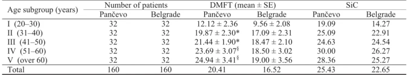
Table 2 shows the mean values of the DMFT index for each of the five groups of patients from the experimental and
Table 1 Total number and mean values of extracted teeth from Panþevo and Belgrade
Extracted teeth
Pan evo Belgrade
Age subgroups (years)
total mean ± SE total mean ± SE
I (20–30) 72 2.25 ± 0.75 44 1.37 ± 0.63
II (31–40) 166 5.18 ± 1.20 233 7.25 ± 1.42
III (41–50) 332* 10.37 ± 2.12 169 5.28 ± 1.80
IV (51–60) 428* 13.37 ± 3.45 68 2.12 ± 0.61
V (over 60) 452* 14.12 ± 3.85 158 4.93 ± 1.48
*Statistically significant;SE = standard error.
Table 2 Mean values of the Decayed Missing Filled Teeth (DMFT) index and significant caries (SiC) index in the patients from
Panþevo and Belgrade
Number of patients DMFT (mean ± SE) SiC
Age subgroup (years)
Pan evo Belgrade Pan evo Belgrade Pan evo Belgrade
I (20–30) 32 32 12.12 ± 2.36 9.56 ± 2.08 19.09 14.27
II (31–40) 32 32 19.87 ± 2.30* 17.09 ± 2.31 25.09 22.91
III (41–50) 32 32 21.44 ± 1.90* 18.47 ± 2.10 24.63 24.54
IV (51–60) 32 32 23.69 ± 3.07§ 18.50 ± 3.02 30.00 26.27
V (over 60) 32 32 24.94 ± 3.41§ 19.00 ± 3.56 28.36 25.27
Total 160 160 20.41 16.52 25.43 22.65
SE = standard error; *statistically significant; §
control groups, as well as the SiC index values. The average value of the DMFT index in the experimental group and in the control group was 20.41 and 16.52, respectively. It is notable that the index value increased with the age of pa-tients and that it was higher in each subgroup of papa-tients from the experimental group, compared to the control group. In the group I the difference was not statistically significant (p> 0.05), while in the groups II and III the difference be-tween the coefficient of DMFT patients from the experi-mental group and the control group was statistically signifi-cant (p< 0.05). The most striking differences were in the group IV, where the recorded value of the DMFT index in the experimental group was 23.69 vs 18.50 in the control group, which was a highly statistically significant difference (p< 0.01). Also in the group V the recorded value of the DMFT index in the experimental group was 24.94 vs 19.00 in the control group, which was an extremely statistically significant difference (p< 0.001). The SiC index values were also higher in all age subgroups from the experimental group than in the control group (Table 2).
Non-carious lesions were diagnosed in 71 patients (44%) from the experimental group and 39 patients (24%) from the control group (Table 3).
The mean concentrations of Pb (presented in μg/g) in extracted teeth in all age subgroups from Pan evo and from Belgrade are given in Table 4. The concentrations of Pb in extracted teeth in all the groups from Pan evo were statisti-cally significantly (p< 0.05) higher than in all the groups from Belgrade. When the measured levels of Pb did not reach the threshold values for the method used, they were marked as ‘undetectable’ in the corresponding tables.
Table 4 Mean values of lead concentrations in extracted teeth
Pb concentrations (μg/g), mean ± SE
Age subgruops Pan evo Belgrade
I (20–30) 1.57 ± 0.35 0.61 ± 0.14
II (31–40) 4.48 ± 1.12 0.39 ± 0.09 III (41–50) 4.60 ± 0.78 0.80 ± 0.22 IV (51–60) 11.10 ± 2.43 4.15 ± 1.76 V (over 60) 26.61 ± 3.89 4.86 ± 2.00
SE = standard error.
Correlation determination was done three times, sepa-rately for the patients from Pan evo and from Belgrade, with two variables. The first correlation was done between the number of extracted teeth from 32 patients in each of the 5
subgroups, and the concentration of Pb in 8 teeth from each of the five subgroups. The second correlation was done be-tween the number of carious lesions from 32 patients in each of the 5 subgroups, and the concentration of Pb in 8 teeth from each of the 5 subgroups. The third correlation was done between the number of non-carious lesions from 32 patients in each of the 5 subgroups, and the concentration of Pb in 8 teeth from each of the 5 subgroups. For the patients from Belgrade all correlations were negative and without statisti-cal significance. However, all the correlations for patients from Pan evo showed a statistical significance (the number of extracted teeth and the concentration of Pb in extracted teeth – p< 0.001, the number of carious lesions and the con-centration of Pb in extracted teeth – p< 0.01 and the number of non-carious lesions and the concentration of Pb in ex-tracted teeth – p< 0.001).
Discussion
In this study it was noticed that the patients from Pan evo had fewer teeth than those from Belgrade although the patients from both cities had similar oral hygiene habits and visited dentists at approximately the same intervals.
Therefore, one possible cause of tooth loss in the patients from Pan evo aged over 40 years could be the long-term ex-posure to a polluted environment.
The value of the DMFT index and the SiC index values, both in Pan evo and Belgrade, must be considered as ex-tremely high, given some of the values that WHO has de-fined as acceptable 20. Namely, as expressed by DMFT in-dex, 6 is the acceptable value of oral health for members of the group aged 35–44 years, while in the experimental group this value was as much as 20, and in the control group 17. These results are worse than in many well-developed coun-tries (Turkey 12.62, Austria 14.7, Germany 16.1, UK 16.6, Denmark 16.7), however, they are similar to, for example, Norway (20.5) and Canada (20.0) 21, 22. For people over 65 years of age, WHO considers acceptable DMFT to be 12, while in the experimental group this value was as much as 25 and in the control group it was 19. This low level of oral health in patients aged over 60 years resulted from the high DMFT index values in all age groups and also from a large number of extracted teeth.
An alarming fact is that the most frequent component of the DMFT index in the experimental group was extracted teeth, with the proportion of 44% vs 25% in the control
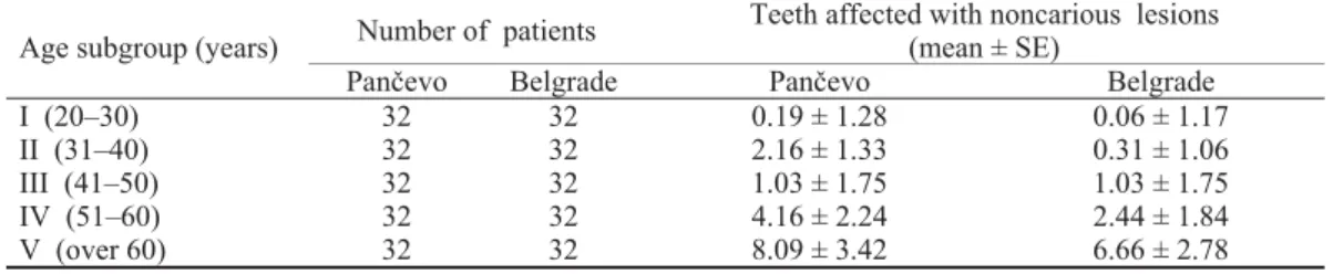
Table 3 Mean number of teeth affected with non-carious lesions
Number of patients Teeth affected with noncarious lesions (mean ± SE)
Age subgroup (years)
Pan evo Belgrade Pan evo Belgrade
I (20–30) 32 32 0.19 ± 1.28 0.06 ± 1.17
II (31–40) 32 32 2.16 ± 1.33 0.31 ± 1.06
III (41–50) 32 32 1.03 ± 1.75 1.03 ± 1.75
IV (51–60) 32 32 4.16 ± 2.24 2.44 ± 1.84
V (over 60) 32 32 8.09 ± 3.42 6.66 ± 2.78
group (which is also a high percentage). A large number of people prefer to have teeth extracted rather than undergo some kind of conservative treatment, partly due to fear and partly because of low income. Besides caries, the reasons for the large number of extracted teeth can also be found in a low level of health education.
Abrasions of anterior teeth and premolars and wedge-shaped erosions where the loss of tooth substance extends to both the enamel and dentin, were the most frequently diag-nosed non-carious lesions in the patients from the experimen-tal group, while abrasions were the most frequently diagnosed non-carious lesions in the patients from the control group. In our sample of younger patients (up to 50 years of age) pre-sented with less non-carious lesions, therefore, the standard er-ror was higher. The explanation for the representation of abra-sions, could be related to early loss of molar teeth and, there-fore, over-loading of the remaining teeth during mastication 23. The most frequently cited etiological factor in forming cervical erosions is chemical etching, more precisely, acid dissolution of hard dental tissues 24. Studies have confirmed that cervical erosions are more frequent in people who are exposed to acids in the workplace or living environment, in competitive swim-mers, in people who frequently consume acidic drinks, or use chemicals for oral hygiene which chelate calcium. Due to fre-quent vomiting, a significant frequency of dental erosive le-sions was noticed in patients with gastrointestinal problems, bulimia and anorexia neurosa, in pregnant women and alco-holics 25–28. Many different studies in recent years, have con-firmed that ''bending'' or ''flexing'' of teeth caused by eccentric occlusal forces is one of the factors which could explain the occurrence of cervical lesions 29–35 while numerous epidemiol-ogical studies consider inappropriate teeth brushing technique as one of the reasons for cervical erosions etiology 31, 36. One of the possible reasons for higher number of non-carious le-sions in the older patients from this investigation could be heavy metals accumulation during the years.
This study showed that the concentration of Pb in ex-tracted teeth in all the groups of patients from Pan evo was higher compared to the patients from Belgrade. Additionally, the concentration of Pb increased rapidly for the older pa-tients from both cities, indicating that the concentration of Pb is age-dependent. Other studies also showed that concentra-tions of Pb are age-dependent. Baranowska et al. 37 and Nowak and Chmielnicka 38 found a positive correlation be-tween age and Pb in human teeth.
Teeth are not a uniform mass of calcified tissues and it has been well-established that Pb is not homogeneously dis-tributed within a fully developed tooth, with Pb levels in dentine being significantly higher than in enamel 39. Fur-thermore, Arora et al. 40 presented the spatial distribution of
Pb in the roots of human primary teeth while other authors measured the Pb content in whole teeth 41.
In this study a consistent relationship was demonstrated between a long-term environmental Pb exposure and its in-corporation into hard dental tissues. This is similar to the re-sults of de Almeida et al. 42, although they measured the content of Pb in the surface enamel of deciduous teeth sam-pled in vivo from children living in uncontaminated and in lead-contaminated areas of Brazil using different methods. It had been shown previously that heavy metals can be incor-porated into dental tissues if there is exposure during the process of dentinogenesis 43. Therefore, unlike bone, in which the mineral phase is subject to turnover, once formed, teeth provide a permanent, cumulative and relatively stable record of environmental exposure 44.
It should be noted that in the patients living in Pan evo statistically significant correlations between the concentra-tion of Pb and the number of extracted teeth (p < 0.001), the number of carious lesions (p < 0.01) and the number of non-carious lesions (p < 0.001) were found, whereas the same correlations in the patients from Belgrade were of no statisti-cal significance. This evidence suggests that the possible cause of tooth loss and hard dental tissue lesions (carious and non-carious) in the patients from Pan evo could be a long-term environmental exposure to lead.
Conclusion
Significantly higher values of the DMFT index and higher frequency of non-carious lesions were recorded in the patients from Pan evo. The concentration of lead in ex-tracted teeth increased rapidly in the older patients from both Pan evo and Belgrade, indicating that the deposition of lead is age-dependent. According to the correlations between the concentration of lead in extracted teeth and the number of extracted teeth, the number of carious lesions and non-carious lesions found in the patients living in Pan evo, one possible cause of tooth loss and hard dental tissue damage could be a long-term environmental exposure to lead. That means that polluted environment is one of the factors that cannot be ignored, but also requires confirmation by further comprehensive basic research.
Acknowledgments
The study was supported by the Ministry of Education, Science and Technological Development, the Republic of Serbia (Project Number 21045), conducted between April 2008 and December 2010 as “The Effects of a Chemically Polluted Environment on Oral Tissues and Teeth of Patients from the town of Pan evo, Serbia”.
R E F E R E N C E S
1. Frank RM, Sargentini-Maier ML, Turlot JC, Leroy MJ.
Compari-son of lead levels in human permanent teeth from Strasbourg, Mexico City and rural zones of Alsace. J Dent Res 1990; 69(1): 90î3.
2. Cleymaet R, Bottenberg P, Slop D, Clara R, Coomans D. Study of
3. Cleymaet R, Bottenberg P, Retief DH, Slop D, Michotte Y, Coomans D. In vivouse of a dual acid etch biopsy for the evaluation of lead profiles in human surface enamel. Caries Res 1991; 25(4): 256î63.
4. Karahalil B, Aykanat B, Ertas N. Dental lead levels in children
from two different urban and suburban areas of Turkey. Int J Hyg Environ Health 2007; 210(2): 107î12.
5. Hu H, Rabinowitz M, Smith D. Bone lead as a biological marker in
epidemiologic studies of chronic toxicity; conceptual paradigms. Environ Health Perspect 1998; 106(1): 1î8.
6. Alomary A, Al-Momani IF, Massadeh AM. Lead and cadmium in
human teeth from Jordan by atomic absorption spectrometry: Some factors influencing their concentrations. Sci Total Environ 2006; 369(1î3): 69î75.
7. Anttila A, Anttila AT. Trace-element content in the enamel
sur-face and in whole enamel of deciduous incisors by proton-induced X-ray emission of children from rural and urban Fin-nish areas. Arch Oral Biol 1987; 32(10): 713î7.
8. Frank RM, Sargentini-Maier ML, Leroy MJ, Turlot JC. Age-related
lead increase in human permanent teeth demonstrated by energy dispersive X-ray fluorescence. J Trace Elem Electrolytes Health Dis 1988; 2(3): 175î9.
9. Heilmann HH, Kuhl K, Steckhan F, Steckhan F.The gradients of
the inorganic components in the enamel and dentin of the de-ciduous teeth. II. A comparison of the concentrations of zinc,magnesium, calcium and lead. Zahn Mund Kieferheilkd Zentralbl 1990; 78(7): 587î92. (German)
10. Cleymaet R, Bottenberg P, Retief DH, Slop D, Michotte Y, Coomans D.
In vivo use of a dual acid etch biopsy for the evaluation of lead profiles in human surface enamel. Caries Res 1991; 25(4): 256î63.
11. Cleymaet R, Collys K, Retief DH, Michotte Y, Slop D, Taghon E, et al.
Relationship between lead in surface tooth enamel, blood and saliva from children residing in the vicinity of a non-ferrous metal plant in Belgium. Br J Ind Med 1991; 48(10): 702î9.
12. Cleymaet R, Quartier E, Slop D, Retief DH, Smeyers-Verbeke J,
Coomans D. Model for assessment of lead content in human
sur-face enamel. J Toxicol Environ Health 1991; 32(2): 111î27.
13. Cleymaet R, Retief DH, Quartier E, Slop D, Coomans D, Michotte Y.
A comparative study of the lead and cadmium content of sur-face enamel of Belgian and Kenyan children. Sci Total Environ 1991; 104(3): 175î89.
14. Hernandez-Guerrero JC, Jimenez-Farfan MD, Belmont R,
Ledesma-Montes C, Baez A. Lead levels in primary teeth of children living
in Mexico City. Int J Paediatr Dent 2004; 4(3): 175î81.
15. Gomes VE, Rosario de Sousa M da L, Barbosa FJr, Krug FJ, Pereira
Saraiva M da C, Cury JA, et al. In vivo studies on lead content of
deciduous teeth superficial enamel of preschool children. Sci Total Environ 2004; 320(1): 25î35.
16. Jakovljevic A, Ristic N. Dental pathology . 1st ed. Pancevo: Faculty
of Stomatology; 2008. (Serbian).
17. WHO Oral Health Country/Area Profile Programme [online database]. Available from:
http://www.whocollab.od.mah.se/expl/siccalculation.xls. [ac-cessed 2011 January 13].
18. Wallenstein S, Zucker CL, Fleiss JL. Some statistical methods
use-ful in circulation research. Circ Res 1980; 47(1): 1î9.
19. SAS/STAT user s guide [computer programme]. Version 6.4. Cary NC: SAS Institute Inc; 1989.
20. WHO Oral Health Country/Area Profile Programme [online database]. Available from:
http://www.whocollab.od.mah.se/countriesalphab.html [ac-cessed 2011 February 9].
21. Namal N, Can G,Vehid S, Koksal S, Kaypmaz A. Dental health
status and risk factors for dental caries in adults in Istanbul, Tur-key. East Mediterr Health J 2008; 14(1): 110î8.
22. Nishi M, Stjernsward J, Carlsson P, Bratthall D. Caries experience of
some countries and areas expressed by the Significant Caries In-dex. Community Dent Oral Epidemiol 2002; 30(4): 296î301.
23.Jakovljevic A, Cenic-Milosevic D, Kolak V. Frequency of
noncari-ous lesions at residents of Pancevo. Proceedings of The FirstSymposium Dentists of Vojvodina; Novi Sad; Serbia; 2009 June 4î6; Novi Sad: 2009. p. 49.
24.Leviteh LC, Bader JD, Shugars DA, Heymann HO. Non-carious
cervical lesions. J Dent1994; 22(4): 195î207.
25.Chikte UM, Josie-Perez AM, Cohen TL. A rapid epidemiological
assessment of dental erosion to assist in settling an industrial dispute. J Dent Assoc SAfr1998; 53(1): 7î1.
26.Dülgergil CT, Erdemir EO, Ercan E, Erdemir A. An industrial
dental-erosion by chromic acid: A case report. Eur J Dent 2007; 1(2): 119î22.
27.Jarvinen VK, Rytomaa II, Heinonen OP. Risk faktors in dental
erosion. J Dent Res1991; 70(6): 942î7.
28.Mayhew RB, Jessee SA, Martin RE. Association of occlusal,
periodontal and dietary factors with the presence of non-carious cervical dental lesions. Am J Dent 1998; 11(1): 29î32.
29.Estafan A, Furnari PC, Goldstein G, Hittelman EL. In vivo
cor-relation of non-carious cervical lesions and occlusal wear. J Prosthet Dent 2005; 93(3): 221î6.
30.Grippo JO, Simring M. Dental ‘erosion’ revisited. . J Am Dent
Assoc 1995; 126(5): 619î30.
31.Hattab FN, Yassin OM. Etiology and diagnosis of tooth wear:
A literature review and presentation of selected cases. Int J Prosthodont2000; 13(2): 101î7.
32.Litonjua LA, Andreana S, Bush PJ, Tobias TS, Cohen RE.
Non-carious cervical lesions and abfractions: a re-evaluation. J Am Dent Assoc 2003; 134(7): 845î50.
33.Mayhew RB, Jessee SA, Martin RE. Association of occlusal,
periodontal and dietary factors with the presence of non-carious cervical dental lesions. Am J Dent 1998; 11(1): 29î32.
34.Pegoraro LF, Milczewsky Scolaro J, Cesar Conti P, Telles D, Pegoraro
TA. Non-carious cervical lesions in adults. Prevalence and oc-clusal aspects. J Am Dent Assoc 2005; 136(12): 1694î700
.
35.Telles D, Pegoraro LF, Pereira JC. Prevalence of non-carions
cer-vical lesions and their relation to occlusal aspects: a clinical study. J Esthet Dent 2000; 12(1): 10î5.
36.Litonjua LA, Andreana S, Bush PJ, Tobias TS, Cohen RE. Wedged
cervical lesions produced by toothbrushing. Am J Dent 2004; 17: 237î40.
37.Baranowska I, Barchanski L, Bak M, Smolec B, Mzyk Z. X-Ray
fluorescence spectrometry in multielemental analysis of hair and teeth. Pol J Environ 2004; 13(6): 639î46.
38.Nowak B, Chmielnicka J. Relationship of lead and cadmium to
essential elements in hair, teeth and nails of environmentally exposed people. Ecotoxicol Environ Saf 2000; 46(3): 265î74.
39.Grobler SR, Theunissen FS, Kotze TJ. The relation between lead
concentration in human dental tissues and in blood. Arch Oral Biol 2000; 45(7): 607î9.
40.Arora M, Chan SW, Kennedy BJ, Sharma A, Crisante D, Walker
DM. Spatial distribution of lead in the roots of human primary teeth. J Trace Elem Med Biol 2004; 18(2): 135î9.
41.Rabinowitz MB. Relating tooth and blood lead levels in
chil-dren. Bull Environ Contam Toxicol 1995; 55(6): 853î7.
42.De Almeida CGR, Pereira SMdaC, Barbosa FJr, Krug FJ, Cury JA,
de Sousa RMdaL, et al. Lead contents in the surface enamel of
deciduous teeth sampled in vivo from from children in un-contaminated and in lead-un-contaminated areas. Environ Res 2007; 104(3): 337î45.
43.Gdula-Argasinska J, Appleton J, Sawicka-Kapusta K, Spence B.
Further investigation of the heavy metal content of the teeth of the bank as an exposure indicator of environmental pollu-tion in Poland. Environ Pollut 2004; 131(1): 71î9.
44.Tvinnereim HM, Eide R, Riise T, Wesenberg GR, Fosse G, Steinnes
E. Lead in primary teeth from Norway: changes in lead level from the 1970s to the 1990s. Sci Total Environ 1997; 207(2î3): 165î77.