Extremity reconstruction using a free ibula lap
after oncological resection
Reconstrução de extremidades com retalho livre de fíbula após ressecções oncológicas
ABSTRACT
Background: Primary tumors of the long bones are rare, accounting for 0.2–1% of malignant tumors. In the past, amputation was the standard treatment and had a large impact on patient morbidity and mortality. With advances in surgical techniques and mul
-tidisciplinary involvement, conservative surgery of the limbs has become the treatment of choice, and reconstruction using a microsurgical fibula flap is the most commonly used technique. In this study, we aimed to present the experience of the National Cancer Institute (INCA) with limb reconstruction using a microsurgical fibula flap following tumor resection from the long bones. Methods: We retrospectively analyzed 7 cases of free fibular flap surgery at the INCA from 1997 to 2009 for the reconstruction of defects of the extremities after bone tumor resection. We evaluated the following parameters: gender, age, diagnosis, tumor location, resection size and type, reconstruction size and type, vessels used for the anastomosis, postoperative complications, disease status at the last visit, follow-up, and time to ambulation. Results: Seven patients with a mean age of 11.8 years (range, 5–14 years) underwent extremity reconstruction with a free fibula flap with 100% bone viability. The lesions were located within the femur, tibia, or humerus. Osteosarcoma was the most common tumor type. The average return to ambulation was 14.7 months. Conclusions: The use of a free fibula flap is an excellent alternative for limb reconstruction and features a high bone healing rate, early ambulation, good functionality, and a low complication rate.
Keywords: Fibula. Extremities. Transplantation, homologous. Reconstructive surgical pro -cedures.
RESUMO
Introdução: O tumor primário de ossos longos é raro, correspondendo de 0,2% a 1% dos tumores malignos. No passado, a amputação era o tratamento padrão, ocasionando grande impacto na morbidade e na mortalidade desses pacientes. Com o avanço das técnicas cirúr
-gicas e o envolvimento multidisciplinar, a cirurgia conservadora dos membros tornou-se o tratamento de escolha, sendo a reconstrução com retalho microcirúrgico de fíbula a mais This study was performed at the
Instituto Nacional de Câncer, Rio de Janeiro, RJ, Brazil. Submitted to SGP (Sistema de Gestão de Publicações/Manager Publications System) of RBCP (Revista Brasileira de Cirurgia Plástica/Brazilian Journal of Plastic Surgery). Article received: May 12, 2012 Article accepted: August 7, 2012
1. Resident Physician, Plastic Surgery and Microsurgery Department of the Instituto Nacional de Câncer, Rio de Janeiro, RJ, Brazil.
2. Plastic Surgeon, full member of the Associação Brasileira de Cirurgia Craniomaxilofacial/Brazilian Association of Craniomaxillofacial Surgery, associate member of the Sociedade Brasileira de Cirurgia Plástica/Brazilian Society of Plastic Surgery (SBCP), Specialist in Reconstructive Microsurgery in Onco-logy at the Instituto Nacional de Câncer, Master from the Faculdade de Medicina (FAMED), Preceptor of the Plastic Surgery Residency and Postgraduate Course in Craniomaxillofacial Surgery at the Hospital de Clínicas de Porto Alegre, Porto Alegre, RS, Brazil.
3. Orthopedist, Connective Bone Tissue Service of the Instituto Nacional de Câncer and Instituto Nacional de Traumatologia e Ortopedia, Rio de Janeiro, RJ, Brazil.
4. Orthopedist, Connective Bone Tissue Service of the Instituto Nacional de Câncer and Chief of Orthopedic Oncology Center of the Instituto Nacional de Traumatologia e Ortopedia, Rio de Janeiro, RJ, Brazil.
5. Full member of the SBCP, Master of Medicine, Universidade Estadual de Campinas, Plastic Surgeon at the University Hospital of the Universidade Federal do Rio de Janeiro, Program Coordinator, Residency in Plastic Surgery at the Instituto Nacional de Câncer, Rio de Janeiro, RJ, Brazil.
6. PhD, Full Member of the SBCP, Plastic Surgeon, Plastic Surgery and Microsurgery Service of the Instituto Nacional de Câncer, President of the SBCP - Regional Rio de Janeiro, Rio de Janeiro, RJ, Brazil.
Eduardo ravasio
Machado1
ciro Paz Portinho2
robErto andré torrEs dE
vasconcElos3
WaltEr MEohas4
Juliano carlos sbalchiEro5
Paulo robErto dE
utilizada. Este trabalho tem como objetivo apresentar a experiência do Instituto Nacional de Câncer (INCA) nas reconstruções de membros com retalho microcirúrgico de fíbula após ressecções de tumores de ossos longos. Método: Foi realizada análise retrospectiva de 7 casos de retalho livre de fíbula operados no INCA, no período de 1997 a 2009, para recons
-trução de defeitos de extremidades após ressecções de tumores ósseos. Foram avaliados os seguintes parâmetros: sexo, idade, diagnóstico, localização do tumor, tipo e tamanho da ressecção, tipo e tamanho da reconstrução, vasos utilizados para anastomose, complicações pós-operatórias, estado da doença na última consulta, seguimento e tempo até deambulação. Resultados: No total, 7 pacientes com média de idade de 11,8 anos (variando de 5 anos a 14 anos) foram submetidos a reconstrução de extremidades com retalho livre de fíbula, com 100% de viabilidade e consolidação óssea. As lesões eram localizadas em fêmur, tíbia ou úmero. O tumor mais comum foi o osteossarcoma. O tempo médio de retorno à deambulação foi de 14,7 meses. Conclusões: O uso do retalho livre de fíbula é uma excelente alternativa para reconstrução de membros, apresentando alta taxa de consolidação óssea, deambulação precoce, boa funcionalidade e baixa taxa de complicações.
Descritores: Fíbula. Extremidades. Transplante homólogo. Procedimentos cirúrgicos re -construtivos.
INTRODUCTION
Primary malignant tumors of the long bones are rare, accoun ting for 0.2–1% of malignant tumors; osteosarcoma and Ewing’s sarcoma are the most frequent of these types of
tumors1-3.
Amputation, the classical treatment of such tumors, pro -vided excellent local disease control but had a great impact on patient morbidity and mortality.
Conservative limb surgery has emerged as an alternative, and recent studies have shown a similar eficacy of this pro -cedure to amputation after neoadjuvant chemotherapy1,2. Conservative limb surgery is appropriate for approximately 90% of patients1. Contraindications include the involvement of key neurovascular trunks, large muscle involvement, infection, and lack of patient motivation for rehabilitation and other surgical procedures4.
With advances in surgical techniques and multidiscipli
-nary involvement of plastic surgery, orthopedics, pathology, medical oncology, and physiotherapy, conservative limb sur -gery became the treatment of choice for tumors of the long bones. Surgeons performing this procedure should always respect 3 principles: 1. the chance of recurrence should not be greater than amputation; 2. it should not affect overall patient survival; and 3. the rehabilitation period should not postpone further treatment1,3,4.
There are many techniques for conservative limb surge ry. Microsurgical ibular and iliac crest laps are more com -monly used than autologous bone grafts and allografts. These latter techniques do not require microsurgery and present with high infection rates, pseudoarthrosis, and fracture, which leads to graft failure in up to 50% of cases1-5. Thus,
based on the experience gained with the use of free laps in the reconstruction of limbs in trauma and jaw reconstruc
-tions, free fibula transplantation is becoming increasingly common in orthopedic reconstructions after oncological re sections since they are preferable to iliac crest free flaps for creating straight and long cortical bones and because a skin island can be included when necessary5-7.
To date, few studies have evaluated the efficacy of ex -tremity reconstruction using a free fibula flap after onco
-logical resection1,8,9. Therefore, in this study, we aim to pre sent the experience of the Instituto Nacional de Câncer (INCA) in the reconstruction of limbs using this flap after the resection of long bone tumors.
METHODS
We retrospectively analyzed 7 patients who received a free ibular lap at the INCA between 1997 and 2009 for the reconstruction of defects of the extremities after bone tumor resection.
Hospital records were evaluated for patient gender, age, diagnosis, tumor location, resection size, reconstruction size, blood vessels used for the anastomosis, postoperative com -plications, disease status at the last visit, follow-up, and time to ambulation.
RESULTS
and humerus (n = 2), and 5 patients were male. The mean age was 11.8 years (range, 5–14 years). The most common tumor was osteosarcoma (n = 6).
All reconstructions were performed using a bone lap without a skin island that had an average size of 15.8 cm (range, 10–20 cm) (Table 2). All patients underwent neoad
-juvant and ad-juvant chemotherapy.
Two patients developed a lap fracture in the late postoperative period due to trauma and both experienced
consolidation after treatment. One patient had chronic pain and another presented with exposure of the synthesis material after 7 months, which was removed without lap damage. Two patients died – one as a result of metastatic disease and the other due to local recurrence.
The average return to ambulation was 14.2 months (range, 6–30 months). Four patients were free of disease at the end of this study.
Figures 1 and 2 illustrate some cases in this series.
Table 1 – Demographic data.
Patient Sex Age (years) Localization Diagnosis Flap Disease Status
1 M 13 Left femur Osteosarcoma Free Fibula Death/lung metastasis
2 M 13 Left femur Osteosarcoma Free Fibula + allograft Free
3 F 5 Left femur Osteosarcoma Free Fibula Free
4 F 13 Right tibia Osteosarcoma Free Fibula Death/local recurrence
5 M 13 Right humerus Osteosarcoma Free Fibula Free
6 M 14 Left tibia Osteosarcoma Free Fibula Free
7 M 12 Right humerus Ewing Sarcoma Free Fibula Free
F = female; M = male.
Table 2 – Study results.
Patient Resection type
Resection size (cm)
Flap size
(cm) Flap type
Vessels
used Complications Follow-up
Time to deambulation
1 the left femurDiaphysis of 19.5 × 5.3 × 5 20 Bone Fibular + femoral Chronic pain 4 years 6 months2 years,
2 the left femurDiaphysis of 18 × 9 × 6 19 AllograftBone + Fibular + femoral the synthesis Exposure of
material 8 years 10 months
3
Distal femur and tibial
plateau 13 × 2 × 1.5 14 Bone
Fibular +
femoral Flap fracture by trauma 9 years 13 months
4 Right tibia and ibula 9 × 4 and 9 × 1.5 10 Bone Fibular + anterior
tibial None
1 year and 6
months 6 months
5 Proximal third of the right humerus
13.5 × 8 × 6 15 Bone Fibular + brachial Flap fracture by trauma 8 years Not applicable
6 Distal third of
the left tibia 13.5 × 5 × 3 15 Bone
Fibular +
anterior
tibial None 10 months 10 months
7
Proximal and middle third
of the right humerus
DISCUSSION
The essential goal of treating virtually all long bone tumors is appropriate surgical resection combined with chemotherapy along with lower morbidity and improved survival. With the progress of such treatment modalities, there has been a signiicant reduction in mortality over re cent decades. Currently, up to 75% of patients without metas ta ses at the time of admission and 50% of those with isolated pulmonary metastases are cured2. Therefore, oncological re section-related reconstruction is mandatory and requires appropriate integration between the orthopedics and plastic surgery department4,10-12. In this series, all patients received neoadjuvant and adjuvant chemotherapy.
Several long bone reconstruction techniques, such as can cellous bone graft, allografts, the use of prostheses, and external ixation techniques, have been used with this purpo se in mind13-15. Cancellous bone graft reconstruction may be suitable for small defects surrounded by good amounts of soft tissue. However, it has the disadvantage of a long period required for revascularization between the graft and the recipient site and may cause pathological fractures, pseu
-doarthrosis, and nonconsolidation, similar to that observed when using allografts15,16.
The external ixation technique takes a long time to treat extensive bone defects and produces long-term complica
-tions in patients undergoing bone resec-tions for osteosarcoma due to post-chemotherapy immunosuppression17.
Vascularized bone transplantation is the best reconstruc
-tion method. This technique differs from the others because revascularization of the lap occurs immediately on comple
-tion of the anastomosis, thereby retaining osteoblastic and osteoclastic activity with the same potential consolidation of a simple fracture, and is thus associated with a remodeling process that leads to rapid lap incorporation18. Use of the free ibula flap is more appropriate for this type of reconstruc -tion, with the advantages being the involvement of a long tubular bone (25 cm), easy dissection, and the low associated morbidity of the donor area.
Chang and Weber19 demonstrated that the use of a free lap associated with an allograft improves this integration, preventing the incidence of nonconsolidation and shortening the functional limb rehabilitation. In this series, we used the allograft in one case, in which ambulation occurred 10 months after surgery.
Figure 1 – Case 2. In A, osteosarcoma of the left femur. In B, the receiving area. In C, the ibula lap. In D, allograft.
In E, free ibula lap + allograft ixed by the receiving area.
In F, perfusion scintigraphy performed on postoperative day 20
showing good perfusion of the lap and the allograft.
A
C
E F
B
D
E
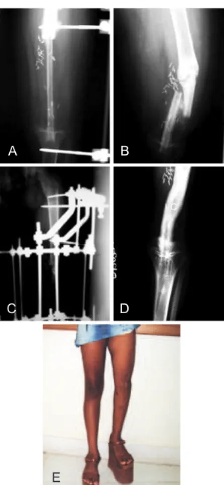
Figure 2 – Case 3. In A, radiography performed on immediate
postoperative reconstruction of defect of the left femur and tibial plateau with a ibular free lap. In B, radiograph showing lap
fracture in the late postoperative period. In C, radiograph after fracture reduction and external ixation. In D, radiograph
showing lap consolidation and hypertrophy. In E, late postoperative appearance.
C
A
C
B
Clemens et al.20 recently demonstrated a higher rate of con solidation and therefore earlier rehabilitation when only the free lap was used compared to the allograft/free lap (100% vs. 82.4%) and recommended the use of this kind of reconstruction in cancer patients. In this series, 100% of the flaps were viable, there was excellent integration of the flap to the receptor site, and there were no cases of pseudoar thro sis or nonconsolidation. There were 2 cases of delayed lap fracture caused by external trauma, both of which presented with normal consolidation after orthopedic ixation. The mean time to ambulation was 14.2 months, similar to those of global reports2,19,20.
The mean flap size in this study was 15.8 cm (range, 10–20 cm). No evidence of functional impairment was pre -sent in the donor area. The mean age of patients was lower than those in more recent studies, which may be most likely due to the small sample size.
Despite the small number of cases in this study, our re -sults are in agreement with reported data showing that the free lap is the method of choice for limb reconstruction in patients with cancer since it provides better consolidation and greater resistance to infection compared to allograft and endo prosthesis, thus promoting earlier rehabilitation and facilitating further treatment of the underlying disease.
CONCLUSIONS
The use of a free ibula lap is an excellent alternative in the therapeutic armamentarium for limb reconstruction after orthopedic resection of oncological long bones since it displays a high bone healing rate, early ambulation, good functionality, and a low complication rate while favoring adjuvant treatment follow-up. The patients in this series had excellent functional and oncological results.
REFERENCES
1. Bach AD, Kopp J, Stark GB, Horch RE. The versatility of the free osteo-cutaneous ibula lap in the reconstruction of extremities after sarcoma resection. World J Surg Oncol. 2004;2:22.
2. Chen CM, Disa JJ, Lee HY, Mehrara BJ, Hu QY, Nathan S, et al. Re -construction of extremity long bone defects after sarcoma resection with vascularized ibula laps: a 10-year review. Plast Reconstr Surg. 2007;119(3):915-24.
3. Weiland AJ, Daniel RK, Riley LH Jr. Application of the free vasculari
-zed bone graft in the treatment of malignant or aggressive bone tumors. Johns Hopkins Med J. 1977;140(3):85-96.
4. Evans GRD, Goldberg DP. The management and reconstructive con
-cerns of extremity malignances. Principles of extremity microvascular reconstruction. In: Schusterman MA, ed. Microsurgical reconstruction of the cancer patient. Philadelphia: Lippincott-Raven Publishers; 1997. p.233-47.
5. Donati D, Di Liddo M, Zavatta M, Manfrini M, Bacci G, Picci P, et al. Massive bone allograft reconstruction in high-grade osteosarcoma. Clin Orthop Relat Res. 2000;(377):186-94.
6. Helmstedter CS, Goebel M, Zlotecki R, Scarborough MT. Pathologic fractures after surgery and radiation for soft tissue tumors. Clin Orthop Relat Res. 2001;(389):165-72.
7. Lee KS, Han SB, Baek JR. Free vascularized osteocutaneous ibular graft to the tibia in 51 consecutive cases. J Reconstr Microsurg. 2004; 20(4):277-84.
8. Minami A, Usui M, Ogino T, Minami M. Simultaneous reconstruction of bone and skin defects by free ibular graft with a skin lap. Microsurgery. 1986;7(1):38-45.
9. Hsu RW, Wood MB, Sim FH, Chao EY. Free vascularised ibular graf
-ting for reconstruction after tumour resection. J Bone Joint Surg Br. 1997;79(1):36-42.
10. Steinau HU, Buttemeyer R, Vogt P, Hussmann J, Hebebrand D. Limb salvage and reconstructive procedures in soft tissue sarcomas of the extremities. Recent Results Cancer Res. 1995;138:31-9.
11. Arndt CA, Crist WM. Common musculoskeletal tumors of childhood and adolescence. N Engl J Med. 1999;341(5):342-52.
12. Drake DB. Reconstruction for limb-sparing procedures in soft tissue sarcomas of the extremities. Clin Plast Surg. 1995;22(1):123-8. 13. Choong PF, Sim FH. Limb-sparing surgery for bone tumors: new deve
-lopments. Semin Surg Oncol. 1997;13(1):64-9.
14. Aberg M, Rydholm A, Holmberg J, Wieslander JB. Reconstruction with a free vascularized ibular graft for malignant bone tumor. Acta Orthop Scand. 1988;59(4):430-7.
15. Enneking WF, Morris JL. Human autologous cortical bone transplants. Clin Orthop Relat Res. 1972;87:28-35.
16. Mankin HJ, Gebhardt MC, Tomford WW. The use of frozen cadaveric allografts in the management of patients with bone tumors of the ex
-tremities. Orthop Clin North Am. 1987;18(2):275-89.
17. Ozaki T, Nakatsuka Y, Kunisada T, Kawai A, Dan’ura T, Naito N, et al. High complication rate of reconstruction using Ilizarov bone transport method in patients with bone sarcomas. Arch Orthop Trauma Surg. 1998;118(3):136-9.
18. Ozaki T, Hillmann A, Wuisman P, Winkelmann W. Reconstruction of tibia by ipsilateral vascularized ibula and allograft. 12 cases with ma -lignant bone tumors. Acta Orthop Scand. 1997;68(3):298-301. 19. Chang DW, Weber KL. Use of a vascularized ibula bone lap and
intercalary allograft for diaphyseal reconstruction after resection of primary extremity bone sarcomas. Plast Reconstr Surg. 2005;116(7): 1918-25.
20. Clemens MW, Chang EI, Selber JC, Lewis VO, Oates SD, Chang DW. Composite extremity and trunk reconstruction with vascularized ibula lap in postoncologic bone defects: a 10-year experience. Plast Reconstr Surg. 2012;129(1):170-8.
Correspondence to: Eduardo Ravasio Machado