Artigo
*e-mail: [email protected]
#Artigo em homenagem ao Prof. Otto R. Gottlieb (31/8/1920-19/6/2011)
VOLATILE AND NON-VOLATILE COMPOUNDS AND ANTIMICROBIAL ACTIVITY OF Mansoa difficilis (Cham.) Bureau & K. Schum. (Bignoniaceae)#
Giselle Maria Skelding Pinheiro Guilhon*, Elisângela Sarmento da Silva e Lourivaldo da Silva Santos Faculdade de Química, Universidade Federal do Pará, Av. Augusto Corrêa, 1, 66075-900 Belém – PA, Brasil Maria das Graças Bichara Zoghbi
Coordenação de Botânica, Museu Paraense Emílio Goeldi, CP 399, 66077-901 Belém – PA, Brasil Isabella Santos Araújo
Universidade Estadual de Feira de Santana, Av. Transnordestina, s/n, 44036-900 Feira de Santana – BA, Brasil Ana Paula Trovatti Uetanabaro
Universidade Estadual de Santa Cruz, Rodovia Ilhéus-Itabuna, s/n, 45662-900 Ilhéus – BA, Brasil
Recebido em 30/4/12; aceito em 1/10/12; publicado na web em 9/11/12
Essential oil from the leaves of Mansoa difficilis was analyzed by GC/MS. Oct-1-en-3-ol (49.65%) was the major compound, but diallyl di- and trisulfide were also present (0.85 and 0.37%, respectively), justifying the garlic-like odor of the crushed leaves. The hexane and methanol extracts of the leaves and stems afforded as main constituents a mixture of linear hydrocarbons, spinasterol, stigmasterol, ursolic and oleanolic acids, two apigenin derivatives and verbascoside. The hexane and methanol extracts of leaves were tested for antimicrobial activity against ten microorganisms. The hexane extract was active against both Psedomonas aeruginosa and Staphylococcus aureus.
Keywords: Mansoa difficilis; chemical composition; antimicrobial inhibition.
INTRODUCTION
Bignoniaceae is predominantly a neotropical family and compri-ses almost 800 species and 104 genera.1 Around 316 species, grouped into 55 genera, are known in Brazil.2 Among the genus Mansoa, M. alliacea (Lam.) A. H. Gentry, M. angustidens (DC.) Bureau & K. Schum., M. difficilis (Cham.) Bureau & K. Schum. and M. standleyi (Steyerm.) A. H. Gentry are common in the North of Brazil.
Mansoa species are known for their pungent garlic-like smell when their vegetative and reproductive organs are crushed, parti-cularly M. alliacea. Mansoa difficilis, locally known as cipó-sino, cipó-de-alho-do-mato and cipó-una, also has a garlic-like odor, but not as strong as that of M. alliacea and M. standleyi, whose essential oils are rich in organosulfur compounds.3,4
Antimicrobial activities have been observed in many species of Bignoniaceae: extracts, and essential oils of Mansoaalliacea were found to be active against Alterniaria brassicae,5Drechslera oryzae,6 Colletotrichum capsici, Curvularia lunata, Alternaria alternata, A. brassicae, A. brassicola, A.carthami, Fusarium oxysporum and F. udum.7 Crude extracts of M. hirsuta DC. inhibited the growth of standardized cultures of Aspergillus niger and Fusarium oxyspo-rum.8 Extracts of M. hymenaea (DC.) A. H. Gentry have shown high antifungal activity, especially against Trichophyton mentagrophytes, Microsporum gypseum,9,10Trichophyton rubrum, and dermatophyte fungi.8
A survey of the literature revealed that no studies on the volatile and non-volatile compounds or antimicrobial activity of M. difficilis have been published to date. This species has been used in handicraft as a raw material for baskets, fishing and planting equipment.11 The aim of this study was to characterize the chemical composition of volatile and non-volatile compounds of M. difficilis and to test the hexane and methanol extracts of the leaves against a diverse range
of organisms comprising Gram-positive and Gram-negative bacteria and yeasts.
EXPERIMENTAL
Botanical material
The samples of M. difficilis were collected in the municipality of Santa Luzia do Pará, in the Northeast of the State of Pará (Brazil), at their full flowering stage, in June 2008. A voucher specimen was deposited in the Herbarium of the Museu Paraense Emílio Goeldi (MG190,032).
Extractions of volatile and non-volatile compounds
Samples of fresh leaves (300.7 g) were hydrodistilled for 3 h using a Clevenger-type apparatus with maintenance of the cooling water at 12 oC. The oils obtained were centrifuged for 5 min (3,000 rpm), dried over Na2SO4, centrifuged again, and immediately submitted to GC/MS analysis. A solution containing 2 µL of the oil in 1 mL of hexane was immediately prepared for gas chromatography analysis. The total oil yield was expressed in percentage (volume/mass) on the basis of free water material. The amount of water was measured using infrared light on a Mater 50 device.
and MdLMB:6.0 g) and stems (MdSMD:5.8 g, MdSMEt:5.3 g and MdSMB:3.3 g).
Analysis of volatile compounds
The oil was analyzed using a Shimadzu GC/MS Model QP 2010 Plus, equipped with a Rtx-5MS (30 m x 0.25 mm; 0.25 µm film thickness) fused silica capillary column. Helium was used as the carrier gas adjusted to 1.2 mL min-1; with splitless injection of 1 µL of a hexane solution; injector and interface temperature were 250 °C; oven temperature programmed was 60-240 °C at 3 °C min-1. EIMS: electron energy, 70 eV; ion source temperature was 200 oC. Identification of the compounds were made by comparing their GC mass and retention data with those held in the NIST-05 library and cited in the literature data.12,13 Retention indices were calculated us-ing n-alkane standard solutions (C8-C26) available from Fluka S. A., under the same chromatographic conditions. Quantitative data were obtained from the electronic integration of the total ion chromatogram (TIC) peak areas.
Classic chromatographic procedures
The MdLH (15.0 g), MdSH (1.7 g), MdLMD (3.0 g), MdSMD (3.5 g), MdLMEt (12.0 g) and MdSMEt (3.5 g) were fractionated by column chromatography (CC) on silica (70-230 mesh) using mixtures of hexane, EtOAc and methanol with increasing order of polarity as mobile phase.
Column chromatography of MdLH (40 g) on silica afforded eight fractions (A-H) that were eluted with hexane (A) and hexane-EtOAc 2.5% (B-D), hexane-EtOAc 4% (E), hexane-EtOAc 10.0% (F), hexa-ne-EtOAc 15.0% (G) and hexahexa-ne-EtOAc 50% (H). Compounds 1 (400 mg) and 2 (440 mg) were the main constituents of fractions A and B, respectively. Fractions C-G were further fractionated by CC on silica using mixtures of hexane and EtOAc as eluents: fraction C (hexane--EtOAc 1.5%) afforded compounds 3 (13 mg) and 4 (30 mg); fraction D (hexane-EtOAc 1.25%) yielded compound 5 (8 mg); fraction E (hexane-EtOAc 10%) yielded 6 (45 mg); fraction F (hexane-EtOAc 3%) led to a mixture of 7 and 8 (357 mg) purified by crystallization with hexane-EtOAc; fraction G (hexane-EtOAc 12-14%) afforded a mixture of 9 and 10 (95 mg). CC on silica of MdLMD (3 g) led to isolation of 11 (8 mg) when using hexane-EtOAc 9% as the eluent and to additional quantities of the mixture of 9 and 10 (1,100 mg). CC fractioning of MdLMEt (12 g) on silica with further purification on Sephadex LH-20 led to compounds 12 (198 mg) and a mixture of 13 and 14 (45 mg).
Similar CC procedures of MdSH (1.7 g) led to the isolation of 1 (30 mg), 6 (145 mg), 7-8 and 15 (175 mg) and 9-10. CC of MdSMD (3.5 g) on silica using EtOAc-MeOH 5% as the eluent afforded the mixture of 16-17 (13 mg). CC of MdSMEt (3.5 g) led to additional quantities of 13-14 (423 mg).
Structural determination of non-volatile constituents
Structures of the isolated compounds were proposed based on the analysis of 1H- and 13C-NMR spectral data and GC-MS and by comparison with literature data. NMR spectra were recorded on a Mercury 300 – Varian instrument using CDCl3, CD3OD, C5D5N or DMSO-d6 as solvents.
Microbial strains
The following bacteria and yeasts were used for the experiments: Escherichia coli CCMB 261 (sensitive to trimethoprim and resistant
to sulphonamide), Pseudomonas aeruginosa CCMB 268, Salmonella sp. CCMB 281, Staphylococcus aureus CCMB 262 (resistant to strep-tomycin and dihydrostrepstrep-tomycin), Staphylococcus aureus CCMB 263, Staphylococcus aureus CCMB 285, Bacillus cereus CCMB 282, Candida albicans CCMB 286, Candida albicans CCMB 266 and Candida parapsilosis CCMB 288 (resistant to amphotericin-B). All microorganisms were cultured on Müeller-Hinton agar (MHA). The bacterial strains were cultured at 37 oC for 24 h and yeasts at 28 oC for 48 h. All the microbial tests were performed in triplicate.
Minimum inhibitory concentration (MIC)
The minimum inhibitory concentration (MIC) of the hexane and methanol extracts from the leaves of M. dificillis was determined based on a microdilution method in 96 multi-well microtiter plates.14 All microbial tests were performed in MHA. The extracts were dis-solved in a DMSO-water solution (1:1) and sterilized by filtration through a cellulose acetate membrane (0.22 mm). Serial dilutions from 10 to 0.078 mg mL-1 of the extracts were prepared. Each well received 10 µL of suspension of each micro-test. The purity of the suspension of the inoculums was verified in a simultaneous incuba-tion. After the period of incubation, 50 µL of triphenyl tetrazolium chloride 2-3-5 (TTC) was added to a final concentration of 0.40 mg mL-1 (final concentration; assays with yeasts) and 30 µL of rezasu-rine (RZ, assays with bacteria) to a final concentration of 0.01% for qualitative analysis of microbial growth in the wells in order to determine the antimicrobial activity of each dilution of the samples. Nystatin (20 mg mL-1) and chloramphenicol (10 mg mL-1) were used as positive controls. Controls were performed to test the viability of microorganisms and the sterility of the culture medium. The MIC was considered the lowest extract concentration where there was no visible microbial growth after the color indicator (TTC and RZ) step.
Minimal microbicidal concentration (MMC)
Petri dishes containing MHA were used for this assay. Volumes of 5 µL from each MIC well were transferred to MHA and cultured at 28 °C for 48 h (yeasts) and at 37 °C for 24 h (bacteria). The MMC was considered the lowest extract concentration where there was no cellular growth.
RESULTS AND DISCUSSION
Volatile compounds
were lower than those of the oils of M. alliacea3 and M. standleyi.4 According to Campbell and coworkers,17 cyclic polysulfides, such as 5-methyl-1,2,3,4-tetrathiane, are formed during gas chromatographic procedures when the temperature injection is over 170 oC; therefore, this constituent was probably formed during the analysis and is not actually present in the oil of M. difficilis.
Non-volatile compounds
Classic chromatographic procedures of the hexane and metha-nol extracts of leaves and stems led to the identification of known compounds. Linear hydrocarbons that varied from C9 to C22 (1)12 and squalene (2)18 were identified, together with mixtures of fatty alcohols (3), fatty acid methyl esters (4), phytol fatty esters (5) and fatty acids (6)19 that were not analyzed further. The steroids spinas-terol (7), stigmasspinas-terol (8), sitosspinas-terol (15), 3-O-β -D-glucopiranosyl-stigmasterol (16) and 3-O-β-D-glucopiranosyl-sitosterol (17) were identified,20 as well as the two triterpenoids ursolic (9) and oleanolic acids (10)21 and the diterpenoid phytol (11).22 A mix-ture of two flavones, 7-O-β-glucopiranosyl-apigenin (13) and
7-O-β-glucopiranosyl-6-hydroxyapigenin (14),23 and the glycoside verbascoside (12)24 were identified.
The chemical composition of the non-volatile compounds from the stems of M. difficilis closely resembled that found for the leaves, except for verbascoside (12) which was only identified in the leaves. Ursolic acid was also previously isolated from some Bignoniaceae species, such as from Clytostoma ramentaceum (Mart. ex DC.) Bureau & K. Schum., Arrabidaea triplinervia (Mart. ex DC.) Baill. ex Bureau and Arrabidaea samydoides Sandw.8,25 The occurrence of verbascoside was previously reported in some Bignoniaceae genera, such as Arrabidaea,26Deplanchea, Jacaranda, Mussatia, Tecoma, Newboldia,27 and also the species Barnettia kerri (Barnett & Sandwith) Santisuk and Markhamia stipulata (Wall.) Seem.,28 but this is the first report of the compound in Mansoa. The structures of some of the identified compounds from M. difficilis are shown in Figure 1.
Antimicrobial activity
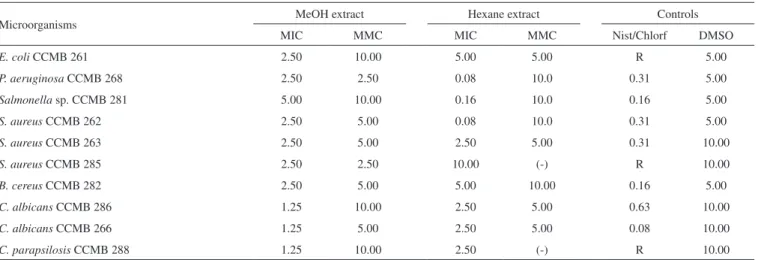
The MIC and MCC data on the hexane and methanol extracts obtained from the leaves of M. difficilis areshown in Table 2. Both extracts showed inhibition of the tested microorganisms. The hexane extract showed higher activity than the methanol extract, inhibiting Salmonella sp. CCMB 281 (MIC = 0.16 mg mL-1), P. aeruginosa CCMB 268 (MIC = 0.08 mg mL-1), S. aureus CCMB 262 (MIC = 0.08 mg mL-1). The best results were observed against P. aeruginosa 268 CCMB and S. aureus CCMB 262, where the values of MIC were lower than the control (chloramphenicol) (Table 2). The methanol extract showed higher inhibition against yeasts than the bacteria tested and the best result was observed against C. albicans CCMB 266 (MIC = 1.25 mg mL-1 and MMC 5.00 mg mL-1).
According to Fontanay and coworkers,29 MIC values below 10 µg mL-1 are considered good and those around 50 µg mL-1 moderate, for antibacterial activity. MIC values that equal hundreds of µg mL-1 indicate that the compound has no activity. Thus, it can be considered that the hexane extract of M. dificillis has moderate activity against P. aeruginosa CCMB268 and S. aureus CCMB 262.
Linear hydrocarbons (C9 to C22) (1), sterols (7, 8, 15), ursolic (9)
Table 1. Constituents (%) identified in the essential oil of the leaves of
Mansoa difficilis
Constituents RI* %
(3Z)-hexenol 867 3.49
oct-1-en-3-ol 979 49.65
octan-3-ol 997 1.65
diallyl disulfide 1082 0.85
linalool 1102 14.93
3-hexenyl butanoate 1188 0.88
α-terpineol 1194 1.19
methyl salicylate 1198 0.29
nerol 1230 0.43
(3Z)-hexenyl 2-methyl butanoate 1234 0.65
geraniol 1256 1.25
safrole 1292 0.99
diallyl trisulfide 1303 0.37
1,2-dihydro-1,1,6-trimethylnaphthalene 1357 0.20
5-methyl-1,2,3,4-tetrathiane 1367 0.26
(3Z)-hexenyl hexanoate 1383 1.16
(Z)-β-damascenone 1388 1.82
(2E)-hexenyl caproate 1391 0.27
(E)-β-damascenone 1419 0.43
β-caryophyllene 1424 0.56
(E)-α-ionone 1432 0.20
(E)-β-ionone 1491 0.42
(E)-nerolidol 1563 0.11
(3Z)-hexenyl benzoate 1571 0.42
pentadecanal 1717 0.59
6,10,14-trimethyl-2-pentadecanone 1848 0.30
cis-phytol 2117 12.83
butyl palmitate 2190 0.29
Total 96.48
*RI on Rtx-5MS
and oleanolic (10) acids isolated from the hexane extract of M. diffici-lis, as well as verbascoside (12), found in the methanol extract of M. difficilis probably contribute to the antimicrobial activity of the extracts. According to Sharma30, homologous series of n-alkanes exhibited rela-tively strong antibacterial action against S. aureus and Klebsiella and moderate action against Staphylococcus albus, Steptococcus viridans, E. coli and Pseudomonas pyocyanea; it also was postulated that the antibacterial activity did not vary with the molecular weights of the tested mixtures of n-alkanes. In the same study, Sharma showed that the common phytosterols stigmasterol and sitosterol showed strong action against Gram-positive bacteria and a moderate response against Gram-negative bacteria. Spinasterol showed activity against multiple antibiotic-resistant Helicobacter pylory31 and broad activity against opportunistic Candida species, Cryptococcus gattii and Sporothrix schenckii.32 Antimicrobial action of oleanolic acid against 21 micro-organisms (6 Gram-positive and 12 Gram-negative bacteria and 3 Candida species) has been reported.33 Ursolic acid has been shown to have antimicrobial activity against S. aureus, Gram-negative bacteria and Microsporium lenosum.34 Ursolic acid was identified as one of the active compounds in rosemary that inhibits the growth of S. aureus, E. coli, Lactobacillus brevis, Psedomonas fluorescens, Rhodotorula glutinis and Kluyveromyces bulgaricus at a concentration of 150 µg mL-1.35 In a recent review, Krystyna and coworkers36 revealed the appreciable antibacterial activity of ursolic and oleanolic acids. Verbascoside is known for its antibacterial activity against Proteus mirabilis and S. aureus including one methicillin-resistant strain.37 A mixture of the isomeric compounds verbascoside and isoverbascoside was active against 5 Gram-positive bacteria (S. aureus, Micrococcus luteus, Bacillus subtilis, Bacillus mycoides, Enterococcus faecalis), 2 Gram-negative bacteria (E. coli and Serratia marcensis), and one yeast (C. albicans), whereas P. aeruginosa and Mycobacterium smegmatis were found to be resistant.26
CONCLUSION
This is the first report on the volatile and non-volatile compounds of M. difficillis including antimicrobial activity evaluation. This study highlighted another source of organosulfur and other compounds from the Mansoa genus. The results of the present investigation demonstrate that M. difficilis shows growth inhibition against seve-ral bacteria and yeasts. The hexane extract proved especially active against P. aeruginosa and S. aureus, including streptomycin- and dihydrostreptomycin-resistant strains.
ACKNOWLEDGMENTS
The authors are grateful to MCT/PPG-7/USAID, Conselho Nacional de Desenvolvimento Científico e Tecnológico (CNPq, Brazil) and Fundação de Amparo à Pesquisa no Estado do Pará (FAPESPA/PA) for the financial support, to O. C. do Nascimento from the Museu Paraense Emílio Goeldi for the vegetal sample collection, and to the Coleção de Culturas de Microrganismos da Bahia (CCMB), Universidade Estadual de Feira de Santana, (Bahia, Brazil) for the microbial strains. E. S. da Silva is grateful to CNPq for the fellowship.
REFERENCES
1. Fischer, E.; Theisen, I.; Lohmann, L. G. In The families and genera of vascular plants; Kadereit, J. W., ed.; Springer-Verlag: Heidelberg, 2004, vol. 7.
2. Gentry, A. H.; Flora Neotropica1980, 25, 1.
3. Zoghbi, M. G. B.; Ramos, L. S.; Maia, J. G. S.; Silva, M. L.; Luz, A. I. R.; J. Agric. Food Chem.1984, 32, 1009; Zoghbi, M. G. B.; Andrade, E. H. A.; Maia, J. G. S.; Flav. Fragr. J. 2002, 17, 133.
4. Zoghbi, M. G. B.; Pereira, R. A.; Lima, G. S. L.; Guilhon, G. M. S. P.; J. Essent. Oil Res. 2010, 22, 247.
5. Bhupendra, K. R.; Taneja, V.; Singh, U. P.; Pharm. Biol. 1999, 37, 13. 6. Chaturvedi, R.; Dikshit, A.; Dixit, S. N.; Tropical Agriculture 1987, 64,
318.
7. Rana, B. K.; Taneja, V.; Singh, U. P.; Pharm. Biol. 1999, 37, 13. 8. Rocha, A. D.; Oliveira, A. B.; Souza Filho, J. D.; Lombardi, J. A.; Braga,
F. C.; Phytother. Res. 2004, 18, 463.
9. Freixa, B.; Vila, R.; Vargas, L.; Lozano, N.; Adzet, T.; Canigueral, S.; Phytother. Res. 1998, 12, 427.
10. Chirunthorn, R.; Supavita, T.; Intaraksa, N.; Kummee, S.; Junkong, N.; Chisorn, B.; Itharat, A.; J. Sci. Technol. 2005, 27 (Suppl. 2), 489. 11. Oliveira, J.; Zoghbi, M. G. B. In Cipó-de-alho: aspectos botânicos,
químicos e moléculas bioativas; Souza Filho, A. P.; Nascimento, J. L. M., eds.; Embrapa: Brasília, 2012, cap. 4.
12. Adams, R. P.; Identification of Essential Oil Components by Gas Chromatography/Mass Spectrometry, 4th ed., Allured Pub. Corp.: Carol Stream, 2007.
13. Yayli, N.; Güleç, C.; Üçüncü, O.; Yasar, A.; Ülker, S.; Coşkunçelebi, K.; Terzioğlu, S.; Turk. J. Chem. 2006, 30, 71.
14. Sarker, S. D.; Nahar, L.; Kumarasamy, Y.; Methods2007, 42, 321. 15. Aboutab, E. A.; Hashem, F. A.; Sleem, A. A.; Maamoun, A. A.; Plant
Product Res. J.2010, 14, 19.
Table 2. Minimum inhibitory concentration (MIC) and minimum microbicide concentration (CMM), in mg mL-1, of the methanol and hexane extracts of
Mansoa difficilis
Microorganisms MeOH extract Hexane extract Controls
MIC MMC MIC MMC Nist/Chlorf DMSO
E. coli CCMB 261 2.50 10.00 5.00 5.00 R 5.00
P. aeruginosa CCMB 268 2.50 2.50 0.08 10.0 0.31 5.00
Salmonella sp. CCMB 281 5.00 10.00 0.16 10.0 0.16 5.00
S. aureus CCMB 262 2.50 5.00 0.08 10.0 0.31 5.00
S. aureus CCMB 263 2.50 5.00 2.50 5.00 0.31 10.00
S. aureus CCMB 285 2.50 2.50 10.00 (-) R 10.00
B. cereus CCMB 282 2.50 5.00 5.00 10.00 0.16 5.00
C. albicans CCMB 286 1.25 10.00 2.50 5.00 0.63 10.00
C. albicans CCMB 266 1.25 5.00 2.50 5.00 0.08 10.00
C. parapsilosis CCMB 288 1.25 10.00 2.50 (-) R 10.00
16. Guilhon, G. M. S. P.; Bittencourt, R. M.; Lima, G. S. L.; Zoghbi, M. G. B. In Cipó-de-alho: aspectos botânicos, químicos e moléculas bioativas; Souza Filho, A. P.; Nascimento, J. L. M., eds.; Embrapa: Brasília, 2012, cap. 4; Zoghbi, M. G. B.; Pereira, R. A.; Oliveira, J. In ref. 11, cap. 5. 17. Campbell, C.; Gries, R.; Khaskin, G.; Gries, G.; J. Appl. Entomol. 2011,
135, 374.
18. Breitmaier, E.; Voelter, W.; Carbon-13 NMR Spectroscopy, 3rd ed., VCH: Weinheim, 1987.
19. Ragasa, C. Y.; Hofilena, J.; Rideout, J. A.; Philippine J. Sci. 2004, 133, 1; Knothe, G.; Kenar, J. A.; Eur. J. Sci. Technol. 2004, 106, 88; Ragasa, C. Y.; Lim, K.; Philippine J. Sci.2005, 134, 63; Asahi Research Center; Handbook of proton-NMR spectra and data, Academic Press: Tokyo, 1985.
20. Forgo, P.; Kover, K. E.; Steroids 2004, 69, 43; Zhang, X.; Cambrai, A.; Miesh, M.; Roussi, S.; Raul, F.; Aoude-Werner, D.; Marchioni, E.; J. Agric. Food Chem. 2006, 54, 1196; Sultanova, N. A.; Abilov, Z. A.; Shul’ts, E. E.; Omurkamzinova, V. B.; Chem. Nat. Compd. 2004, 40, 192; Kojima, H.; Sato, N.; Hatano, A.; Ogura, H.; Phytochemistry 1990, 29, 2351; Zhang, L.; Yang, X-D.; Xu, L-Z.; Zou, Z-M.; Yang, S-L.; J. Asian Natural Products Res. 2005, 7, 649.
21. Ahmad, V. U.; Rahman, A.; Handbook of natural products data: penta-cyclic triterpenoids, Elsevier: Amsterdam, 1994,vol. 2.
22. Rahman, A-U.; Ahmad, V. U.; 13C-NMR of Natural Products, Plenum
Press: New York, 1994, vol. 2.
23. Oyama, K.-I.; Kondo, T.; Tetrahedron 2004,60, 2025; Peng, Z. F.; Strack, D.; Baumert, A.; Subramaniam, R.; Goh, N. K.; Chia, T. F.; Tan, S. N.; Chia, L.S.; Phytochemistry 2003, 62, 219.
24. Krebs, H. C.; Habermehl, G. G.; Magn. Reson. Chem. 1992, 30, 56. 25. Leite, J. P. V.; Lombardi, J. A.; Chiari, E.; Oliveira, A. B.; Rev. Bras.
Farmacogn. 2001, 11, 77; Pauletti, P. M.; Bolzani, V. S.; Young, M. C. M.; Quim. Nova 2003, 26, 641.
26. Lima, C. S. A.; Amorim, E. L. C.; Sena, K. X. F. R.; Chiappeta, A. A.; Nunes, X. P.; Agra, M. F.; Cunha, E. V. L.; Silva, M. S.; Barbosa Filho, J. M.; Rev. Bras. Cien. Farm. 2003, 39, 77.
27. Davioud, E.; Bailleul, F.; Delaveau, P.; Debray, M. M.; Planta Med.
1989, 55, 87; Jimenez, C.; Villaverde, M. C.; Riguera, R.; Castedo, L.; Stermitz, F. R.; Phytochemistry 1987, 26, 1805; Jimenez, C.; Villaverde, M. C.; Riguera, R.; Castedo, L.; Stermitz, F. R.; Phytochemistry 1988, 27, 2947; Jimenez, C.; Villaverde, M. C.; Riguera, R.; Castedo, L.; Stermitz, F. R.; J. Nat. Prod. 1989, 52, 408; Guiso, M.; Marra, C.; Piccioni, F.; Nicoletti, M.; Phytochemistry 1997, 45, 193; Gafner, S.; Wolfender, J. L.; Nianga, M.; Hostettmann, K.; Phytochemistry 1997, 44, 687.
28. Garcez, F. R.; Garcez, W. S.; Mahmoud T. S.; Figueiredo, P. O.; Resende, U. M.; Quim. Nova 2007, 30, 1887.
29. Fontanay, S.; Grare, M.; Mayer, J.; Finance, C.; Duval, R. M.; J. Ethnopharmacol.2008, 120, 272.
30. Sharma, R. K.; Bioorg. Chem. 1993, 21, 49.
31. Wang, Y-C.; Li, W-Y.; Deng-Chyang Wu, D-C.; Wang, J-J.; Wu, C-H.; Liao, J-J.; Lin, C-K.; Evidence-Based Complementary and Alternative Medicine (2011), doi:10.1093/ecam/nep147.
32. Johann, S.; Mendes, B. G.; Missau, F. C.; Resende, M. A.; Pizzolatti, M. G.; Braz. J. Microbiol. 2011, 42, 1065.
33. Kuete, V.; Eyong, K. O.; Folefoc, G. N.; Beng, V. P.; Hussain, H.; Krohn, K.; Nkengfack, A. E.; Pharmazie 2007, 62, 552.
34. Liu, J.; J. Ethnopharmacol. 1995, 49, 57.
35. Collins, M. A.; Charles, H. P.; Food Microbiol.1987, 4, 311.
36. Krystyna, I. W.; Grudniak, A. M.; Fiecek, B.; Kraczkiewicz-Dowjat, A.; Kurek, A.; Cent. Eur. J. Biol. 2010, 5, 543.