1 Rehabilitation Sciences Graduate Program, Universidade Federal de Minas Gerais (UFMG), Belo Horizonte, MG, Brazil 2 Physical Therapy Department, Universidade Federal de Minas Gerais (UFMG), Belo Horizonte (MG), Brazil
Received: 07/01/2013 Revised: 09/20/2013 Accepted: 10/23/2013
a r t i c l e
Assessment of muscular strength with the modified
sphygmomanometer test: what is the best method and
source of outcome values?
Lucas A. C. Souza1, Júlia C. Martins1, Juliana B. Moura2, Luci F. Teixeira-Salmela1,2, Fátima V. R. De Paula1,2, Christina D. C. M. Faria1,2
ABSTRACT | Background: Tests that are usually employed for the clinical assessment of muscular strength have notable disadvantages. The Modiied Sphygmomanometer Test (MST) is a promising method because it is low-cost and provides objective measures. Objectives: To investigate the most adequate method and sources of outcome values for the assessment of strength with the MST. Method: Methodological study with 40 healthy adults (22.98±2.26 years), who did not practice physical activity regularly. The strength of the lexors and extensors of the elbow and knee, the handgrip of the dominant side and anterior trunk lexors were randomly assessed with portable dynamometers and the MST (bag and cuff adaptations, and sphygmomanometer without adaptation) by a single examiner. An independent examiner read and recorded the values. The sources of the investigated outcome values were the irst trial and the means of two and three trials. One-way ANOVAs and Pearson Correlation Coeficients were used for the analyses (α=0.05).
Results: For the MST methods applied to assess all muscular groups, similar values were found for all sources of outcome
values (0.01<F≤0.26; 0.77≤p≤1.00) with signiicant and positive correlations between the measures obtained with the
dynamometers (0.51≤r≤0.94; p≤0.003). Conclusions: All MST methods showed adequate results for the assessment
of strength in healthy individuals, and after familiarization, only one trial was suficient to provide reliable measures. The sphygmomanometer without adaptation is not time consuming, compared to the other adaptations, and showed the capability of measuring higher values of strength. The bag method was easily trained to be used and stabilized.
Keywords: rehabilitation; muscular strength; assessment; upper extremity; lower extremity; trunk.
HOW TO CITE THIS ARTICLE
Souza LAC, Martins JC, Moura JB, Teixeira-Salmela LF, De Paula FVR, Faria CDCM. Assessment of muscular strength with the modiied sphygmomanometer test: what is the best method and source of outcome values?. Braz J Phys Ther. 2014 Mar-Apr; 18(2):191-200. http://dx.doi.org/10.1590/S1413-35552012005000149
Introduction
Given the need for an objective assessment of
muscular strength within clinical settings and the
limitations of commonly used methods to measure
this outcome, such as the manual muscle testing (MMT) and the portable dynamometry, the Modiied Sphygmomanometer Test (MST) is an interesting alternative. Although the MMT is widely used within clinical contexts, it is a descriptive, subjective and
less sensitive method1. The portable dynamometer
is capable of providing accurate, valid, reliable,
and sensitive measurements of strength2-4; however, its relatively high cost has hindered its use in most
clinical contexts, including those in which there are insuficient inancial resources. The MST provides
objective, reliable, and valid measurements of
strength5-7, and it has a low cost relative to the dynamometer.
The MST involves an aneroid sphygmomanometer, a device that is portable, easily obtained, and
commonly acquired by healthcare professionals for
the measurement of blood pressure. This is a quick and easy test, which follows procedures similar to those used with the MMT and portable dynamometers6.
However, some type of adaptation to the conventional
sphygmomanometer is usually required8.
A systematic review showed that the MST has been used with the sphygmomanometer adapted
reliability of the measurements obtained with these
adaptations have been investigated for the upper
limb (UL) muscles in healthy adults7,9, the elderly6,
and individuals with rheumatoid arthritis10 as well
as in the lower limb (LL) and trunk muscles in individuals with rheumatoid arthritis5 and low back pain11. The results were signiicant and of moderate to high magnitude12. However, it is unknown
whether this method is better than the others. In addition, the use of the MST without adaptation of the sphygmomanometer could expand its use, but
has not yet been investigated.
Most studies using the MST8 used the mean of
three trials to obtain a inal result. However, no study has investigated the best way to operationalize the test, that is, to determine how many repetitions are required to obtain reliable measurements. Should lower number of repetitions be sufficient, the feasibility of using the MST within clinical settings will be greater13.
The MST has the potential to be used within
clinical contexts because it uses portable and low-cost equipment that is easily accessible to healthcare professionals and provides objective measurements
of strength. Therefore, the establishment of the best method for its use is required. Thus, the present study aimed to investigate the best method (bag, cuff, and non-adapted sphygmomanometer) and the most appropriate sources of outcome values (irst trial and means of two and three trials) for the assessment of strength with the MST.
Method
Participants
Healthy young adults of both genders were recruited from the general community, according to the following criteria: Had ages between 20 to 30 years, physical activity levels rated as insuficient
or inactive14, and were able to perform the proposed tests. Health conditions that could affect the measurement of strength and the presence of pain
during the evaluations were used as exclusion criteria. All participants provided consent, based upon previous approval from the University Ethical Research Committee of the Universidade Federal de Minas Gerais (UFMG), Belo Horizonte, MG, Brazil (# 04 92.0.203.000-10).
Procedures
After veriication of eligibility, demographic and physical data were collected from all participants. Their dominant UL was determined as the limb most frequently used to write15, while the dominant LL as
the one they would more easily kick a ball16. In total,
40 subjects were included, whose demographic and physical characteristics are given in Table 1.
The strength of the elbow and knee lexor and extensor muscles on the dominant side was evaluated with a Microfet2® digital manual dynamometer (Hoggan Health Industries, UT, USA), and grip
strength with the SAEHAN® hydraulic hand dynamometer (SAEHAN Corporation, Korea, Model SH5001). A DuraShockTM Tycos® aneroid sphygmomanometer (Welch Allyn Inc., NY, USA, Model DS-44) was used for the evaluation of all muscular groups by three methods (bag and cuff adaptations and non-adapted sphygmomanometer).
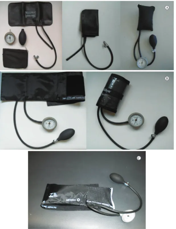
For the bag method adaptation, the inlatable part
of the outer velcro constituting the cuff of the
equipment was removed, and this structure was
folded into three equal parts and placed in a cotton
bag with a zipper5. After being adaptated, the
modiied sphygmomanometer was 3.5 cm long, 10 cm wide, and 7 cm thick (Figure 1A). For the cuff method adaptation, the inlatable part, inserted into the velcro cuff, was folded into four equal parts, and the remainder of the cuff was wrapped around the inlatable part5 and ixed with adhesive tape. Once
adapted, the modiied sphygmomanometer had the following dimensions: 14 cm long, 6 cm wide, and 4.5 cm thick (Figure 1B). The dimensions of the non-adapted sphygmomanometer were 27 cm long, 14 cm wide, and 9 cm thick (Figure 1C).
All employed equipments (portable dynamometers and sphygmomanometer) were acquired for the present study and arrived factory-calibrated, according to the manufacturers’ instructions. Before the strength assessments, the principal investigator randomized the equipment order, by drawing. After
the adaptation of the sphygmomanometer and before
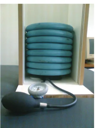
its use for data collection, calibration procedures were performed with 5 kg weights to verify whether
the equipment provided consistent measurements
throughout the study, for possible adjustments of systematic errors, if necessary. Due to the methodological nature of the study, the identiication
of systematic error could be the loss of calibration of
the sphygmomanometer, which was used for all the MST measurements. This calibration procedure was
performed according to previous recommendations6,
as follows: The sphygmomanometer was inlated to 100 mm Hg and its valve was kept closed to remove the folds from the inflatable portion. Then, the pressure was reduced to 20 mm Hg, and the valve was closed again to prevent leakage6, providing a
measurement range between 20-304 mm Hg. To stack the weight plates on the equipment, a wood apparatus was built to keep them aligned (Figure 2). All weights were numbered and consistently stacked on top of
each other in the same sequence. The correlation
between the weights (plates) and the values in mm Hg was high (0.97≤r≤1.00; p≤0.001), with a 2-13%
coeficient of variation. No systematic errors were
observed.
After calibration, the irst examiner (examiner 1)
performed the strength assessments and the second
examiner (examiner 2) independently read and recorded all the measurement values. The use of two
independent observers is recommended to ensure
the internal validity of methodological studies, and for this reason, this procedure was adopted17. Both
examiners were previously trained in their respective functions: examiner 1 to perform all procedures established for the strength measurements (detailed later), and examiner 2 to correctly read and record the strength values obtained with the dynamometers
and sphygmomanometer. The manometer of the
sphygmomanometer was analogic with measurement scale intervals of 2 mmHg.
The strength assessments followed the order of the equipment determined by randomization. The entire procedure for data collection, including the positioning of the participant, the body segment, and the used equipment, as well as the verbal feedback provided during the tests, was standardized, following
recommendations in the literature18-23 and detailed
below. For all equipment/assessment methods, the same procedures were adopted to ensure the
internal validity of the study17. Thus, the only
carried out change was related to the device/method: portable dynamometers or sphygmomanometer with three different methods (bag and cuff adaptations and without adaptation).
The following positions were adopted: for the evaluation of elbow lexors and extensors, the subjects remained in supine position with their forearms in the neutral position and elbows lexed at 90°18; for
the knee lexors and extensors, they were seated on a table, with their legs hanging naturally over the edge of the table at approximately 90 degrees and
hands on the thighs20; the handgrip measurements
were obtained with the subject seated on a chair without armrests, feet supported, shoulders adducted, forearms in the neutral position and elbows lexed 90°19; the assessment of the anterior trunk lexors was
conducted with individuals sitting on a chair without armrests, feet supported, knees bent at 90° and hands relaxed and resting on the thighs23. The participants
were instructed not to perform compensatory
movements during the strength tests.
The equipment placement was as follows: elbow lexors, distal and anterior to the forearm24; elbow
extensors, distal and posterior to the forearm24; trunk
lexors, on the sternum below the jugular notch23;
knee lexors, distal and posterior to the leg24, and
knee extensors, distal and anterior to the leg24. The
sphygmomanometer was positioned parallel to the segment in a way to resist the movement of the tested
muscle group.
Immediately prior to the strength assessments, a demonstration and familiarization with the equipment and procedures were performed. During testing, the subjects were instructed to perform a maximal isometric contraction for 5 s, and the peak force value was recorded. The volunteers received verbal
encouragement to initiate the movement and to hold
the contraction: “one, two, three, and now! Force!... Table 1. Subjects’ demographic and physical characteristics
(n=40).
Variables Results
Age (years): mean ± SD; range [min–max] 22.98±2.2620-28 Body mass index (kg/m2): mean ± SD 21.52±3.00
Gender
Men: number (%) 18 (45%)
Women: number (%) 22 (55%) Dominant Upper Limb
Right: number (%) 33 (82.5%) Left: number (%) 7 (17.5%) Dominant Lower Limb
Right: number (%) 36 (90%)
Left: number (%) 4 (10%)
Figure 1. Sphygmomanometer adaptation methods for the assessment of muscular strength: (A) bag method; (B) cuff method; (C) without adaptation.
force!... force!... relax!”21. Three trials were obtained
for each assessed muscle group, and a 15 s rest-interval was provided between the repetitions22. The
muscle groups were always evaluated in the same
Statistical analysis
Descriptive analysis and normality tests were
performed for the categorical variables and the main outcome measurements of strength. To compare the strength values obtained for each muscular group and
the different sources of outcomes (irst trial and the means of two and three trials) for a particular MST method (bag, cuff, or without adaptation), one-way analysis of variance (ANOVA) was employed. To determine the correlations between the measurements obtained with the portable dynamometer and the MST methods, considering the various sources of outcomes and muscular groups, Pearson’s correlation coeficients were calculated. When the coeficients reached signiicance, the magnitudes of the correlations were classiied as follows12: very
low ≤0.25; low: from 0.26-0.49; moderate: 0.50-0.69; high: 0.70-0.89; and very high: 0.90-1.00. All analyses were performed using SPSS for Windows (SPSS Inc., Chicago, IL, USA), with a signiicance level of 5%.
Results
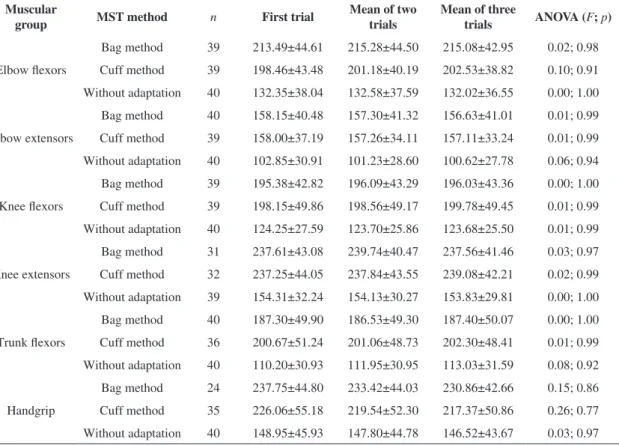
Table 2 gives the descriptive data of the strength measurements for all MST methods, as well as the ANOVA results regarding the comparisons between the various sources of outcomes. The values provided by the different sources of outcomes were similar for all evaluated methods (0.01<F≤0.26;
0.77≤p≤1.00). Furthermore, the sample size varied
according to the assessed muscular group and the
MST method. As shown by the n value, the
non-adapted sphygmomanometer was able to provide
measurements of all assessed muscular groups.
For the bag adaptation, 27 strength measurements were lost, whereas for the he cuff adaptation, 19 measures were lost, with the greatest losses arising from the evaluations of the knee extensors and handgrip muscles. These losses were due to
equipment limitations in reading the strength values
of very strong individuals and to the dificulty of the evaluator in stabilizing the body segment during the measurement of the knee extensor muscles of one of
the participants.
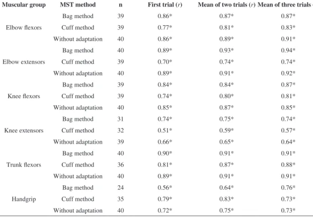
For all muscular groups and sources of
outcomes, significant and positive correlations were observed between the measurements obtained with the portable dynamometers and the three MST methods (Table 3). The magnitudes of the correlations were classiied as moderate to very high (0.51≤r≤0.94; p
≤0.003). In general, the non-adapted sphygmomanometer and the bag adaptation
showed correlations that were similarly classiied and of greater magnitude, compared with those of the cuff
adaptation to most muscular groups.
Discussion
To our knowledge, the present study was the first to investigate the best MST method for
the assessment of muscular strength in healthy
individuals. In addition, it aimed to determine whether the sphygmomanometer without any type of adaptation was an appropriate method for assessing
strength and to identify the best source of outcome
values when using the MST. All MST methods demonstrated signiicant and suitable correlations with the portable dynamometer for all sources of outcomes. However, some distinct characteristics between the methods should be considered.
Among the studies that evaluated the associations between the measurements obtained with the MST and portable dynamometry, which is considered the gold
standard for the assessment of isometric strength2,
signiicant and high correlations (0.75≤r≤0.98) were
found only for the elbow lexors of healthy individuals
using the cuff adaptation9, handgrip muscles of elderly individuals25, and healthy adults using the bag7,26 and cuff27 adaptations, as well as the anterior
trunk lexors of healthy individuals and individuals with low back pain using the bag adaptation11. Most previous studies have investigated the correlations
between the MST and portable dynamometry measures of UL muscular groups, while no study has evaluated these associations for the LL muscles or made use of the non-adapted sphygmomanometer.
Two studies used the bag and cuff adaptations for the assessment of strength of individuals with
rheumatoid arthritis and found no significant
differences between the measurements provided by the two methods5,10. However, the authors showed
differences between the two MST adaptations and reported that the bag adaptation is more elastic, requires more air in the pre-inlation of the equipment, has a larger contact area with the skin of the participant and reaches higher pressure values when an external force is applied5,10. In the present study,
the measurements obtained with the bag adaptation showed higher values than those obtained with the cuff adaptation only for the elbow lexors and the handgrip muscles, with the other measurements showing similarities between the two methods or lower values with the bag adaptation (knee lexors and anterior trunk flexors). The measurements obtained with the non-adapted sphygmomanometer were lower than those registered with other methods for all assessed muscular groups, possibly due to its larger contact area, which may have provided lower pressure values, when subjected to external forces.
The MST investigation using the non-adapted
sphygmomanometer aimed to raise the potential
clinical use of the equipment, because it does not Table 2. Descriptive statistics (means ± SD) of the measures of muscular strength with the three Modiied Sphygmomanometer Test (MST, mm Hg) methods and the ANOVA results regarding the comparisons between the various sources of outcome scores for all employed methods.
Muscular
group MST method n First trial
Mean of two trials
Mean of three
trials ANOVA (F; p)
Elbow lexors
Bag method 39 213.49±44.61 215.28±44.50 215.08±42.95 0.02; 0.98 Cuff method 39 198.46±43.48 201.18±40.19 202.53±38.82 0.10; 0.91 Without adaptation 40 132.35±38.04 132.58±37.59 132.02±36.55 0.00; 1.00
Elbow extensors
Bag method 40 158.15±40.48 157.30±41.32 156.63±41.01 0.01; 0.99 Cuff method 39 158.00±37.19 157.26±34.11 157.11±33.24 0.01; 0.99 Without adaptation 40 102.85±30.91 101.23±28.60 100.62±27.78 0.06; 0.94
Knee lexors
Bag method 39 195.38±42.82 196.09±43.29 196.03±43.36 0.00; 1.00 Cuff method 39 198.15±49.86 198.56±49.17 199.78±49.45 0.01; 0.99 Without adaptation 40 124.25±27.59 123.70±25.86 123.68±25.50 0.01; 0.99
Knee extensors
Bag method 31 237.61±43.08 239.74±40.47 237.56±41.46 0.03; 0.97 Cuff method 32 237.25±44.05 237.84±43.55 239.08±42.21 0.02; 0.99 Without adaptation 39 154.31±32.24 154.13±30.27 153.83±29.81 0.00; 1.00
Trunk lexors
Bag method 40 187.30±49.90 186.53±49.30 187.40±50.07 0.00; 1.00 Cuff method 36 200.67±51.24 201.06±48.73 202.30±48.41 0.01; 0.99 Without adaptation 40 110.20±30.93 111.95±30.95 113.03±31.59 0.08; 0.92
Handgrip
Bag method 24 237.75±44.80 233.42±44.03 230.86±42.66 0.15; 0.86 Cuff method 35 226.06±55.18 219.54±52.30 217.37±50.86 0.26; 0.77 Without adaptation 40 148.95±45.93 147.80±44.78 146.52±43.67 0.03; 0.97
require any extra cost or any time demand from
the professional to perform certain adaptations.
Moreover, this was the only method capable of
providing measures of strength for all assessed
muscular groups and individuals, with the exception of the knee extensors of a male participant, whose strength could not be evaluated by examiner 1, even with the dynamometer, due to the dificulty of manual stabilization of the segment. This problem most likely occurred due to a particular limitation of the examiner, who was not able to exert the necessary force to adequately stabilize the segment when this muscle group exerted its maximum isometric strength.
This type of limitation has been described in the literature for the evaluation of the strength of major
muscular groups with portable dynamometry28,29.
One disadvantage of the use of the non-adapted sphygmomanometer was the greater dificulty in stabilizing the equipment, most likely due to its larger contact area (378 cm2) in relation to the assessed
segment and the examiner’s hand, requiring more
training for the evaluator to perform the test. When using such a method to evaluate strength, in addition to maintaining the lattened hand on the equipment, it is necessary that the examiner be able to stabilize the sphygmomanometer distally, avoiding even slight movements of the segments, as they might lead to
slippage of the equipment and the need to repeat the measurement. This method required a greater need
for training and more repetitions, because the loss of the necessary stabilization occurred more frequently.
The cuff adaptation is a simple, quick, and low cost
method that uses readily available materials and can
be easily used by professionals. Moreover, it is easily stabilized by the examiner, unlike the non-adapted sphygmomanometer. An important disadvantage of this method was that many individuals exceeded the readability of the equipment, limiting its use on
stronger individuals.
The bag adaptation is the most commonly employed method in the literature8, and studies reported that it is able to provide more consistent
Table 3. Correlation coeficients between the measures obtained with the three Modiied Sphygmomanometer Test (MST) methods and the portable dynamometer considering the various sources of outcome scores.
Muscular group MST method n First trial (r) Mean of two trials (r) Mean of three trials (r)
Elbow lexors
Bag method 39 0.86* 0.87* 0.87*
Cuff method 39 0.77* 0.81* 0.83*
Without adaptation 40 0.86* 0.89* 0.91*
Elbow extensors
Bag method 40 0.89* 0.93* 0.94*
Cuff method 39 0.70* 0.74* 0.74*
Without adaptation 40 0.89* 0.91* 0.92*
Knee lexors
Bag method 39 0.84* 0.84* 0.87*
Cuff method 39 0.74* 0.80* 0.81*
Without adaptation 40 0.85* 0.87* 0.85*
Knee extensors
Bag method 31 0.74* 0.75* 0.74*
Cuff method 32 0.51* 0.59* 0.57*
Without adaptation 39 0.66* 0.65* 0.64*
Trunk lexors
Bag method 40 0.90* 0.91* 0.91*
Cuff method 36 0.81* 0.87* 0.88*
Without adaptation 40 0.89* 0.91* 0.91*
Handgrip
Bag method 24 0.56* 0.64* 0.76*
Cuff method 35 0.79* 0.83* 0.73*
Without adaptation 40 0.72* 0.75* 0.73*
measurements5,10, compared with the cuff adaptation.
Among the three methods, this adaptation showed to be the easiest to be trained and stabilized. Although it requires the fabrication of a cotton bag (average cost of R$15.00), this bag facilitates the stabilization of the equipment and the containment of the inlatable part. However, this adaptation showed a lower ability to
assess strength of stronger individuals compared to
the other two MST methods.
Regarding the sources of outcome values, some studies investigated the best way to assess strength in healthy individuals with portable dynamometers30,31.
Bohannon and Saunders31 investigated whether the
values and test-retest reliability of three sources of measurements (irst trial, highest value, and mean of three trials) to assess the strength of the elbow lexor muscles of healthy individuals were similar and reported no differences. Coldham et al.30 investigated
whether there was a difference in the test-retest
reliability of the same sources of outcomes for the evaluation of handgrip strength of healthy individuals
and individuals with various health conditions that affected their hands. They found that there were no
differences and also reported that only one repetition
was necessary. Furthermore, these authors also
reported that a greater number of repetitions is more
time-consuming31 and can lead to fatigue31 and pain30.
Taking into account the results of the present study
and the disadvantages of obtaining a higher number of
repetitions, only one repetition, after familiarization,
could be performed for the assessment of strength in
healthy individuals with the MST, regardless of the
chosen method.
As previously mentioned, there were no systematic
errors or inconsistencies of the measurements
in successive calibrations, suggesting that the
equipment can be used for repeated measurements
of strength. Because the manufacturer recommends
a full calibration of the sphygmomanometer
every two years (depending upon the frequency of use) in an authorized service32, we suggest that
this recommendation should be followed when
the equipment is used to measure strength. The calibration performed in the present study does not
need to be performed within clinical settings.
To ensure the internal validity of the present
study, the reading and the recording of all strength measurements were conducted by an assistant examiner, whereas within clinical contexts, the
same examiner is responsible for all stages of the
evaluation. Future studies should investigate the
consistency of the measurements when the same examiner is responsible for implementing the test
and reading and recording the values.
The MST shows great potential for the clinical evaluation of strength because it is portable, inexpensive, and showed signiicant correlations with the portable dynamometer measures for all
of the three investigated methods. The different
characteristics should be considered when selecting the best method. Some characteristics favor the choice of a particular method. A detailed description of these characteristics was performed, so that the MST method can be selected to prioritize the characteristics that the examiner considers most important. For all the three investigated methods, the use of only one repetition, after familiarization, was shown to be
adequate.
Acknowledgments
The authors thank the Minas Gerais State Research Foundation (Fundação de Amparo à Pesquisa do Estado de Minas Gerais - FAPEMIG), the Brazilian Federal Agency for the Support and Evaluation of Graduate Education (Conselho Nacional de Desenvolvimento Científico e Tecnológico - CAPES), the National Council for Scientific and Technological Development (Conselho Nacional de Desenvolvimento Cientíico e Tecnológico - CNPq) and the Dean’s Ofice for Research Studies at the Federal University of Minas Gerais (Pró-reitoria de Pesquisa da Universidade Federal de Minas Gerais - PRPq/UFMG) for the inancial support.
References
1. Conable KM, Rosner AL. A narrative review of manual muscle testing and implications for muscle testing research. J Chiropr Med. 2011;10(3):157-65. PMid:22014904 PMCid:PMC3259988.
2. Stark T, Walker B, Phillips J, Fejer R, Beck R. Hand-held Dynamometry correlation with the gold standard isokinetic dynamometry: a systematic review. Phys Med Rehabil. 2011;3(5):472-9.
4. Hébert LJ, Maltais DB, Lepage C, Saulnier J, Crête M, Perron M. Isometric muscle strength in youth assessed by hand-held dynamometry: a feasibility, reliability, and validity study. Pediatr Phys Ther. 2011;23(3):289-99. PMid:21829128. http://dx.doi. org/10.1097/PEP.0b013e318227ccff
5. Helewa A, Goldsmith CH, Smythe HA. The modiied sphygmomanometer - an instrument to measure muscle strength: a validation study. J Chronic Dis. 1981;34(7):353-61. http://dx.doi.org/10.1016/0021-9681(81)90073-4 6. Kaegi C, Thibault M, Giroux F, Bourbonnais D. The
interrater reliability of force measurements using a modified sphygmomanometer in elderly. Phys Ther. 1998;78(10):1095-1103. PMid:9781703.
7. Lucareli PRG, Lima MO, Lima FPS, Gimenes RO, Lucareli JGA, Garbelotti Junior SA, et al. Comparison of methods of measurement of the inger lexor muscles’ strength through dynamometry and modiied manual sphygmomanometer. Einstein. 2010;8(2 -Pt 1):205-8. 8. Souza LAC, Martins JC, Teixeira-Salmela LF, Godoy M, Aguiar LT, Faria CDCM. Evaluation of muscular strength with the modiied sphygmomanometer test: a review of the literature. Fisioter Mov. 2013;26(2):437-52. http://dx.doi. org/10.1590/S0103-51502013000200021 9. Bohannon RW, Lusardi MM. Modiied sphygmomanometer versus strain gauge hand-held dynamometer. Arch Phys Med Rehabil. 1991;72(11):911-14. http://dx.doi. org/10.1016/0003-9993(91)90010-G
10. Helewa A, Goldsmith CH, Smythe HA. Patient, observer and instrument variation in the measurement of strength of shoulder abductor muscles in patients with rheumatoid arthritis using a modified sphygmomanometer. J Rheumatol. 1986;13(6):1044-9. PMid:3560090. 11. Helewa A, Goldsmith CH, Smythe HA. Measuring
abdominal muscle weakness in patients with low back pain and matched controls: a comparison of 3 devices. J Rheumatol. 1993;20(9):1539-43. PMid:8164211. 12. Munro BH. Statistical methods for health care research.
5th ed. Philadelphia: Lippincott Williams & Wilkins; 2005. 13. Faria CDCM, Teixeira-Salmela LF, Neto MG, Rodrigues-de-Paula F. Performance-based tests in subjects with stroke: outcomes scores, reliability and measurement errors. Clin Rehabil. 2012;26(5):460-9. PMid:22008883. http://dx.doi.org/10.1177/0269215511423849
14. Centers for Disease Control and Prevention. Physical activity trends - United States, 1990-1998. MMWR Morb Mortal Wkly Rep. 2001;50(9):166-9. PMid:11393487. 15. Yancosek KE, Mullineaux D. Stability of handwriting
performance following injury-induced hand-dominance transfer in adults: a pilot study. J Rehabil Res Dev. 2011;48(1):59-68. http://dx.doi.org/10.1682/ JRRD.2010.04.0074
16. Carregaro RL, Cunha RR, Cardoso JR, Pinto RS, Bottaro M. Effects of different methods of antagonist muscles pre-activation on knee extensors neuromuscular responses.
Rev Bras Fisioter. 2011;15(6):452-9. PMid:22031273. http://dx.doi.org/10.1590/S1413-35552011005000028 17. Portney LG, Watkins MP. Foundations of clinical research:
applications to practice. 3rd ed. New Jersey: Prentice-Hall; 2009.
18. Kendall FP, McCreary EK, Provance PG. Muscles Testing and Function. 5th ed. Philadelphia: Lippincott Williams & Wilkins; 2007.
19. Reis M, Arantes P. Assessment of hand grip strength - validity and reliability of the saehan dynamometer. Fisioter Pesqui. 2011;18(2):176-81. http:// dx.doi.org/10.1590/S1809-29502011000200013 20. Bohannon RW. Reference values for extremity muscle strength obtained by hand-held dynamometry from adults aged 20 to 79 years. Arch Phys Med Rehabil. 1997;78(1):26-32. http://dx.doi.org/10.1016/S0003-9993(97)90005-8 21. Amaral JF, Mancini M, Novo-Júnior JM. Comparation
of three hand dynamometers in relation to the accuracy and precision of the measurements. Rev Bras Fisioter. 2012;16(3):216-24. PMid:22801514. http://dx.doi. org/10.1590/S1413-35552012000300007
22. Martins JC, Souza LAC, Teixeira-Salmela LF, Aguiar LT, Lara EM, Faria CDCM. Assessment of muscle strength in stroke subjects with portable dynamometry: literature review. Fisioter Mov. In press, 2014.
23. Bohannon RW. Recovery and correlates of trunk muscle strength after stroke. Int J Rehabil Res. 1995;18(2):162-67. PMid:7665262. http://dx.doi. org/10.1097/00004356-199506000-00010
24. Bohannon RW. Test-retest reliability of hand-held dynamometry during a single session of strength assessment. Phys Ther. 1986;66(2):206-09. PMid:3945674. 25. Rice C, Cunningham D, Paterson D, Rechnitzer P.
Strength in an elderly population. Arch Phys Med Rehabil. 1989;70(5):391-7. PMid:2719543.
26. Balogun J, Acomolafe C, Amusa L. Reproducibility and criterion-related validity of the modified sphygmomanometer for isometric testing of grip strength. Physiother Can. 1990;42(6):290-5.
27. Hamilton GF, McDonald C, Chenier TC. Measurement of grip strength: validity and reliability of the sphygmomanometer and Jamar grip dynamometer. J Orthop Sports Phys Ther. 1992;16(5):215-9. PMid:18796752. http://dx.doi.org/10.2519/jospt.1992.16.5.215
28. Martin HJ, Yule V, Dennison EM, Cooper C, Aihie Sayer A. Is hand-held dynamometry useful for the measurement of quadriceps strength in older people? A comparison with the gold standard Bodex dynamometry. Gerontology. 2006;52(3):154-9. PMid:16645295. http:// dx.doi.org/10.1159/000091824
Correspondence
Christina Danielli Coelho de Morais Faria Universidade Federal de Minas Gerais Departamento de Fisioterapia
Av. Antônio Carlos, 6627, Campus Pampulha CEP 31270-901, Belo Horizonte, MG, Brasil e-mail: [email protected]; [email protected] 30. Coldham F, Lewis J, Lee H. The Reliability of one vs. three
grip trials in symptomatic and asymptomatic subjects. J Hand Ther. 2006;19(3):318-26. PMid:16861131. http:// dx.doi.org/10.1197/j.jht.2006.04.002
31. Bohannon RW, Saunders N. Hand-held dynamometry: a single trial may be adequate for measuring muscle strength in healthy individuals. Physiother Can. 1990;42(1):6-9. 32. Welch Allyn Ltd. DuraShock™ Integrated Aneroid
![Table 1. Subjects’ demographic and physical characteristics (n=40). Variables Results Age (years): mean ± SD; range [min–max] 22.98±2.26 20-28 Body mass index (kg/m 2 ): mean ± SD 21.52±3.00 Gender Men: number (%) 18 (45%) Women: number (%) 22 (55%)](https://thumb-eu.123doks.com/thumbv2/123dok_br/19016376.469513/3.765.71.379.137.389/table-subjects-demographic-physical-characteristics-variables-results-gender.webp)