Review
Printed in Brazil - ©2014 Sociedade Brasileira de Química 0103 - 5053 $6.00+0.00
*e-mail: [email protected]
Clinical Applications and Methemoglobinemia Induced by Dapsone
Fábio R. Oliveira, Mariely C. Pessoa, Rosyana F. V. Albuquerque, Taysa R. Schalcher andMarta C. Monteiro*
Graduate Program in Phamaceutical Sciences, Faculty of Pharmacy, Federal University of Pará (UFPA), Avenida Augusto Correa, 01, Guamá, 66075-110 Belém-PA, Brazil
Dapsona é uma sulfona sintética que é utilizada como um antibiótico em seres humanos e animais para prevenir e tratar doenças, incluindo hanseníase, tuberculose, malária, e pneumonia por Pneumocystis carinii e encefalites por Toxoplasma gondii em pacientes com síndrome da imunodeficiência adquirida (AIDS), bem como em doenças anti-inflamatórias como dermatite herpetiforme. No entanto, este fármaco também está associado com vários efeitos adversos, incluindo a hemólise relacionada com a dose, metemoglobinemia, psicose, neuropatia periférica, agranulocitose, anemia aplástica, síndrome de hipersensibilidade, síndrome de sulfona, e outros. Destes efeitos, a metemoglobinemia é o mais comum efeito adverso da dapsona, que leva a anemia funcional e hipóxia celular com sintomas de cianose, dores de cabeça, fadiga, taquicardia, fraqueza e tonturas. Assim, esta revisão sumariza informações relevantes sobre a estrutura, mecanismo de ação, indicação clínica, e reações adversas de dapsona.
Dapsone is a synthetic sulfone that is used as an antibiotic in humans and animals to prevent and treat diseases including leprosy, tuberculosis,malaria, and Pneumocystis carinii pneumonia and Toxoplasma gondii encephalitis in acquired immune deficiency syndrome (AIDS) patients as well as in anti-inflammatory conditions, such as dermatitis herpetiformis. However, this drug is also associated with several adverse effects, including dose-related hemolysis, methemoglobinemia, psychosis, peripheral neuropathy, agranulocytosis, aplastic anemia, hypersensitivity syndrome, sulfone syndrome, and others. Of these effects, methemoglobinemia is the most common side effect of dapsone, which leads to functional anemia and cellular hypoxia with symptoms of cyanosis, headache, fatigue, tachycardia, weakness, and dizziness. Thus, this review summarizes relevant information on the structure, mechanism of action, clinical indication, and adverse reactions of dapsone.
Keywords: dapsone, methemoglobinemia, 4,4’-diamino-diphenylsulfone, dapsone hydroxylamine
1. Introduction
Dapsone was synthesized a century ago (1908) and has been used for treatment of several pathologies, including infectious processes such as leprosy, malaria, and
Pneumocystis carinii pneumonia and Toxoplasma gondii
encephalitis in acquired immune deficiency syndrome (AIDS) patients as well as in anti-inflammatory conditions, dermatitis herpetiformis and linear immunoglobulin A (IgA) disease.1 However, this drug may lead to adverse effects
such as dose-related hemolysis and methemoglobinemia.2
Therefore, this review summarizes relevant information on the structure, mechanism of action, clinical indication, and adverse reactions of dapsone.
2. Molecular Structure and Mechanisms of Antimicrobial Action
Dapsone (4,4’-diamino-diphenylsulfone, DDS) is an aromatic amine that belongs to the family of sulfones, which also includes diadiphenyl sulfone, sulfonyl dianiline, and disulfone.3 Sulfone is a very weak Lewis base with
no readily dissociable hydrogen ion (H+), which has the
structure of the simplest of the sulfones: a sulfonyl group (a sulfur atom bound to two oxygen atoms by double bonds) linked to two carbon atoms, as illustrated in Figure 1. Dapsone is a white, odorless crystalline powder that darkens upon exposure to light but remains chemically unaltered. It is poorly water-soluble but easily soluble in alcohol.4
essential for their pharmacological activity but also may be responsible for its toxicity.5,6
Eric Fromm and J. Whitmann were the first to describe the synthesis of sulfones, including dapsone, from
p-nitrosothiophenol in 1908;7 however, the synthesis of
dapsone from p-chloronitrobenzene was described in 1945.8
Therapeutic properties of this drug remained unknown until 1943, where the first in vivo studies showing the antimicrobial effect of dapsone in streptococcal infections were reported by Buttle et al..9 That same year, Faget et al.10
reported that a dapsone derivative, promin, can be useful for the treatment of leprosy. Therefore, in 1948, this drug was used as standard treatment for leprosy.11 In 1976,
during a workshop on leprosy chemotherapy, the World Health Organization (WHO) recommended the use of dapsone, rifampicin, and clofazimine as a leprosy multidrug therapy,12,13 and, currently, dapsone has been employed as
a broad-spectrum antimicrobial agent.
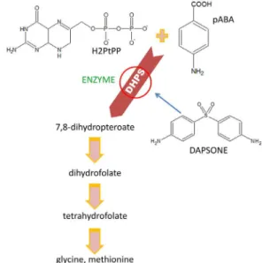
The use of dapsone as an antibiotic is due to its structural analogy of sulfa drugs, including sulfonamides and sulfones, to the substrate p-aminobenzoic acid (pABA). These drugs act as competitive antagonists of the enzyme 6-hydroxymethyl-7,8-dihydropteroate synthase (DHPS) that is responsible for the conversion of pABA to folate, a purine precursor, thereby interfering in the production of DNA necessary for cell division.14-21 In this
regard, DHPS catalyzes the formation of a C–N bond, joining pABA with 6-hydroxymethyl-7,8-dihydropterin-pyrophosphate (H2PtPP) to form 6-hydroxymethyl-7,8-dihydropterin-pyrophosphate and 6-hydroxymethyl-7,8-dihydropteroate. It is essential to
produce reduced folate cofactors by a de novo biosynthetic pathway present in prokaryotes and in some eukaryotes, such as protozoa, yeast, and plants, but it is absent in mammals.17,22-25 Ho et al.26 reported that DDS was found to
be approximately nine times more effective as a competitive inhibitor of DHPS than sulfonamides. These authors also suggested that this remarkable inhibitory activity of DDS can be attributed to its symmetry, which generates a single molecule with two identical planes quite similar to pABA, and this arrangement may facilitate the binding of DDS to the active site of DHPS. In addition, Bell and Roblin27
have reported that a structural similarity between the sulfonyl group present in sulfones and sulfonamides and the carbon dioxide (CO2) of pABA may be important to the
antibacterial activity of these drugs. Thus, they suggested that high potency of DDS depended upon the negativity of the sulfone (SO2) group and of steric factors that remain
to be explained.
Structure-activity relationships and in vivo studies have shown that dapsone is a symmetrical molecule and that the SO2 group is essential for the pharmacological activity of this
drug but is also responsible for its observed hemotoxicity.28
Our group has shown through geometric properties of the structure of dapsone that the symmetry of the SO2 moiety
is essential for the electron distribution between the two
para-related aniline moieties. Molecular symmetry is the most significant aspect for the nucleophilicity of dapsone, and the symmetric conformational isomer has lower energy than the asymmetric conformational isomer, showing the amine influence of conformation with participation of the SO2 moiety by resonance effect on both aniline rings.
Moreover, the SO2 moiety may stabilize the radical formed
during oxidation through conjugation (resonance effect), and the aniline ring is the nucleophilic moiety with possible biological properties through redox mechanisms, mainly electron transfer or oxidation for formation of the dapsone hydroxylamine (DDS-NHOH) metabolite.29,30
3. Clinical Experience and Recommended Use
Dapsone is an effective drug that is used as an antibiotic in humans and animals to prevent and treat diseases.31
Moreover, DDS is a synthetic sulfone with bactericidal and bacteriostatic activity against Mycobacterium leprae and
M. ulcerans, in high concentrations (≥ 8 mg mL–1), is also
effective against M. tuberculosis including the multi-drug resistant (MDR) strains M. intracellulare, M. kansasii,
M. fortuitum, and the Mycobacterium aviumcomplex
(MAC).31-36 In combination with pyrimethamine, dapsone
has been used for malaria prophylaxis in chloroquine-resistant Plasmodium falciparum and P. vivax strains,
displaying activity against all multiplying stages in the plasmodium life cycle.37,38 Combinations, such
as chlorproguanil-dapsone and artesunate-dapsone-proguanil, can be used in treatment and prophylaxis of malaria caused by P. falciparum and antimalarial action of schizonticides.39-41 Furthermore, dapsone also may
be used as a systemic adjuvant when it is combined with oral corticosteroids in the treatment of pemphigus vulgaris42 or in combination with pyrimethamine, in the
treatment and prevention of opportunistic infections in AIDS patients, such as Pneumocystis carinii pneumonia and Toxoplasma gondii encephalitis, or for the treatment of Kaposi’s sarcoma.43-51
Besides use of dapsone as an antimicrobial agent, this drug may also be used as an anti-inflammatory agent. Since this activity is unrelated to its antibacterial action, it may, therefore, be used in non-infectious inflammatory diseases such as dermatitis herpetiformis, IgA dermatitis, rheumatoid arthritis, acne conglobata, chronic urticaria, vasculitis, leukocyte bullous systemic lupus erythematosus, and spider bite and urticarial vasculitis syndromes.4,20,49,52-56
Accordingly, dapsone alone or with concurrent systemic corticosteroids can also be used as efficacy therapy in other dermatologic conditions, such as mucous membrane pemphigoid, pemphigus vulgaris, bullous pemphigoid,57
pyoderma gangrenosum (PG),58-60 subcorneal pustular
dermatosis (SPD)60-62 and Behcet’s disease.63,64
In these forms of dermatitis, the anti-inflammatory activity of dapsone is associated with polymorphonuclear leukocytes (PMNs) during the inflammatory process, such as cellular migration and apoptosis.56,65 In general, in vitro
studies showed that dapsone stimulates neutrophil motility, and clinical studies reported that DDS mediated stimulation of PMN migration.60,66 In this regard, the therapeutic
response of dapsone in PG resulted from inhibition of neutrophil chemotaxis and reduction of oxygen intermediates, thereby suppressing the inflammation,58,59
while for SPD and Behcet’s disease, dapsone can inhibit the neutrophil cytotoxicity and neutrophil chemotaxis and alter the glycosaminoglycans, diluting inflammatory mediators.60,61,63,64 Moreover, the inhibition of neutrophil
chemotaxis induced by dapsone can be explained due to the drug’s interference with the activation or function of the G-protein that initiates the signal transduction cascade common to chemotactic stimuli.67
In urticaria, dapsone may inhibit the neutrophils’ functions due to its antimicrobial and anti-inflammatory effects by a mechanism that involves the down-regulation of leukotriene B4 and interference with the expression of the cluster of differentiation 11B (CD11B) molecule.68,69
Other studies indicate that dapsone may inhibit
mitogen-stimulated transformation of lymphocytes, the alternate pathway of complementary activation, by decreasing levels of complement 3 (C3) protein and C3 pro-activator and interfering with the myeloperoxidase-peroxidase-halide-mediated cytotoxic system within neutrophils.70 In
addition, in vitro studies showed that dapsone may prevent myeloperoxidase- and eosinophil peroxidase-mediated tissue injury at sites where the peroxidase enzymes are secreted and diluted into the neutral pH environment of the tissue interstitial space. Dapsone may inhibit tissue proteases because it is known to oxidize glutathione (GSH); this is important because reduced GSH is required for most proteolytic enzymes to function.71,72
4. Pharmacokinetic Properties
Dapsone is administered orally, and about 80-85% is absorbed slowly from the gastrointestinal tract and uniformly distributed to all tissues. However, it tends to accumulate mainly in the skin, especially in the muscles, liver, and kidneys. Traces of this sulfone can be found up to three weeks after end of the treatment. This drug can cross both blood-brain and placental barriers and can be found in breast milk. Dapsone and its metabolic derivatives can be conjugated with glucuronic acid in the liver and excreted mainly by kidneys and approximately 10% as bile.4,56,65,73-76 In addition, about 70% of dapsone is bonded
to plasma proteins with plasma concentrations ranging from 0.4-1.2 mg L-1 24 hours after ingestion of 100 mg of the
drug, and the maximum plasma concentration is reached within 4-8 hours.75,77-80 Blood levels stabilize approximately
7-10 days after initiation of therapy.81 Dapsone shows a
large half-life ranging from 24-36 hours and can be found in the organism after 35 days following the end of treatment.4
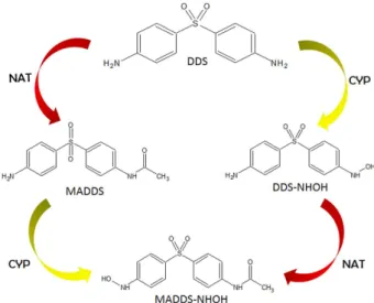
After absorption, DDS is transported through the portal circulation and is metabolized in the liver by two distinct routes: Nitrogen (N)-acetylation and N-hydroxylation (Figure 2). Dapsone acetylation is polymorphic and occurs via action of the liver cytosolic enzyme N-acetyltransferase (NAT), which is present in hepatic cells and in red blood cells.4,12N-acetylation is the major route of biotransformation
of drugs containing arylamine or anilines (Ar-NH2).82 The
aromatic amine is catalyzed by NAT enzymes present in two forms in humans (NAT1 and NAT2). These enzymes detoxify by converting the aromatic amines into amides, which are less toxic metabolites, producing the
N-hydroxy metabolites, including monoacetyl dapsone (MADDS), formed by acetylation of the amine group (NH2), and diacetyl-diaminosulfone (DADS), formed by
The N-hydroxylation that occurs during the oxidative clearance of dapsone and its acetylated derivative is mainly mediated by hepatic cytochrome P450 (CYP)84-86
and glucuronidation, conjugation of glucuronic acid and uridine diphosphate (UDP) through the action of the enzyme UDP-glucuronosyltransferase (UGT).3,28 N-hydroxylated
metabolites include dapsone hydroxylamine (DDS-NHOH) and monoacetyl dapsone hydroxylamine (MADDS-NHOH). These result from a process that occurs mainly by action of a cytochrome P450 isoform, such as CYP3A4, CYP2E1, CYP2C9, and CYP2C19.20,29,86,87 Recently, Schalcher et al.86
showed by molecular modeling the interaction mechanism and possible binding of DDS with the active site of enzyme CYP2C19. However, other CYP isoforms are involved in the metabolism of DDS, such as CYP2E and CYP3A.84,88-90
After oral administration, a constant MADDS/DDS is established, which tends to increase the rates of acetylation. In this regard, Coleman and Tingle91 reported that MADDS
is not primarily responsible for the toxicity of DDS, unless the other amino group is N-hydroxylated. Dapsone and its metabolites are conjugated in the liver and excreted in the form of mono-N-glucuronide and mono-N-sulfamate and, in lower concentrations, excreted in saliva and breast milk.3,92 Its elimination rate after a single dose is 50% during
the first 24 hours.79,80
5. Adverse Effects
Adverse reactions related to the use of dapsone range from digestive problems such as nausea, vomiting, and stomatitis.93 The occurrences of toxic hepatitis, cholestatic
jaundice, cutaneous photosensitivity reactions, psychosis,
and a syndrome that became known as the “sulfone syndrome” (fever, malaise, jaundice, exfoliative dermatitis or morbilliform rash, hepatic dysfunction, lymphadenopathy, methemoglobinemia, hemolytic anemia and lymphocytosis with atypical lymphocytes) are unusual.54,93 The occurrence
of liver disease associated with DDS is about 5% when administered alone, but the incidence of liver damage by DDS increased to 40% when the drugs were co-administered with other drugs such as trimethoprim for AIDS patients.94
The most frequent adverse reactions caused by dapsone is methemoglobinemia production and hemolysis (Figure 3),3,95-97
showing inclusions resulting from the changes in hemoglobin called Heinz bodies.96-100 The recommended dose of dapsone
is 50-100 mg per day (the dose should not exceed 300 mg per
day), but the most serious side effects are observed when the dose is > 100 mg per day101 and agranulocytosis
occurs.102 Dapsone taken orally in higher doses can
generally cause nausea, vomiting, and epigastric pain.103
Agranulocytosis is not a dose-dependent side effect of dapsone, with an unknown mechanism of development initiating after 4-12 weeks of treatment. It gradually progresses with symptoms of fever, swelling of the lymph nodes, inflammation and ulcers of the oral cavity, pharynx, and esophagus, increased susceptibility to sepsis, and death throughout the disease.100,104 Another adverse effect of
DDS therapy is called dapsone hypersensitivity syndrome (DHS), caused by an idiosyncratic reaction105,106 related
to metabolized products of DDS, as DDS-NHOH, which produces a great number of effects such as fever, rash, and internal organ involvement.107,108
The most frequent and well-reported adverse effects of dapsone were dose-limiting hemotoxic effects, such as hemolytic anemia and methemoglobinemia, which
Figure 2. Possible metabolic pathways of dapsone involving NAT and CYP enzymes. NAT: N-acetyl transferase; CYP: Cytochrome P450; DDS: 4,4’-diamino-diphenylsulfone; DDS-NHOH: dapsone hydroxylamine; MADDS: monoacetyl dapsone; MADDS-NHOH: monoacetyl dapsone hydroxylamine.
are associated with long-term administration of the drug at standard doses (100 mg per day), resulting in methemoglobinemia in about 15% of patients.109,110 The
hemotoxic effects of DDS increase in a dose-dependent manner, especially in infants and elderly patients.44,46,111-113
After DDS therapy is initiated at the > 300-mg per day dose, the patient should present symptoms of hemolytic anemia as a dose-dependent side effect that usually occurs in 3-4 weeks; the mechanism is unknown but is associated with a reduction in the lifespan of red blood cells101 The most common side effect of DDS treatment is
methemoglobinemia, which occurs when the concentration of methemoglobin (MetHb) in erythrocytes is more than 1%, leading to functional anemia and cellular hypoxia with symptoms of cyanosis at levels around 15% and headache, fatigue, tachycardia, weakness, and dizziness experienced in 30-40% of patients. Concentrations of approximately 60% may cause hypoxia leading to acidosis, paralysis, arrhythmias, coma, and convulsions, ultimately leading to death when concentrations reach 70-80% levels, due to the incapability of abnormal hemoglobin to carry oxygen or CO2.
114,115 N-hydroxy metabolites, DDS-NHOH and
MADDS-NHOH, have been investigated as the predominant hemolytic metabolites responsible for hemotoxic reactions, mainly methemoglobinemia.20,89,90,116,117 In addition, dapsone
is contraindicated in patients with low glucose-6-phosphate dehydrogenase (G6PD) levels, whereas gradually increasing the dose of dapsone (25-150 mg per day for three days) in patients with normal G6PD levels may lead to a degree of hemolytic anemia and associated symptoms.118
6. Methemoglobinemia
Methemoglobinemia is a clinical syndrome caused by increased blood concentration of MetHb.119 It occurs
naturally without any tissue damage due to regulation by the enzyme nicotinamide adenine dinucleotide phosphate (NADPH)-dependent methemoglobin reductase; however, it can occur in congenital chronic form owing to changes in the synthesis or metabolism of hemoglobin, as acquired from disease in which there is an increase in the rate of oxidation of hemoglobin to methemoglobin120-125 or
induced by agents with oxidizing action, such as DDS, sulfonamides, local anesthetics, methylene blue at high doses,20,30 primaquine, chloroquine, bupivacaine,
nitroglycerin, lidocaine, and nitroprusside, among others.126-128 The changes to the MetHb molecule include
the original ferrous (Fe2+) atom being oxidized to a ferric
(Fe3+) atom, leading to an allosteric change in the heme
portion of the oxidized hemoglobin molecule, decreasing its oxygen-binding capacity.120,121,123,124
According to Kinoshita et al.,129 the reduction of MetHb
occurs by two main mechanisms: the NADPH-dependent pathway system, represented by NADPH-dependent methemoglobin reductase, and NADH-cytochrome b5 reductase (also known as NADH-dependent methemoglobin reductase). The NADPH-methemoglobin reductase is capable of reducing MetHb formed under normal conditions, since under conditions of high oxidation MetHb reduction is responsible for NADH-cytochrome b5 reductase.130 The supply of NADH required for the
reduction of MetHb is from the anaerobic glycolysis of the Embdem-Meyerhof pathway from the glucose oxidation reaction that generates ATP and NADH, while for the other pathway the substrate is NADPH via the pentose-phosphate pathway from the activation of G6PD.
The most important pathway to reduce erythrocytes is that of NADH-cytochrome b5 reductase, influenced by the availability of NADH and cytochrome b5, a heme-protein present in the cytoplasm of cells with a primary role in the reduction of MetHb.131 In the case of MetHb formation,
40% up-regulation induced by oxidizing agents and some toxic compounds via NADH-cytochrome b5 reductase is compromised, thus patients with clinical manifestations, such as cyanosis and apnea, may be treated with methylene blue intravenously at 1-2 mg kg-1 body weight during a
5-minute period.22,125,132 This is due to the methylene blue
being a cofactor for the enzyme NADPH-methemoglobin reductase, thus the treatment results in the oxidation of the methylene blue by accepting an electron from NADPH in the presence of NADPH-methemoglobin reductase to leucomethylene blue, which acts as an artificial electron acceptor to MetHb, resulting in MetHb’s conversion back to hemoglobin.123,125,133,134 However, this pathway
is dependent on NADPH, and in patients with G6PD deficiency, methylene blue is ineffective and may still
induce hemolysis135 as well as symptoms including
dyspnea, chest pain, and persistent cyanosis.136
The methemoglobinemia occurs from the inability of MetHb to bind oxygen, causing a state of functional anemia, and increasing the binding affinity of the non-reduced ferrous heme for oxygen. This increase and spread of this process causes significant shortfalls in the supply of oxygen to the tissues, causing other important clinical manifestations, such as dyspnea, nausea, and tachycardia, when levels of MetHb reach up to 30%.133
Lethargy, stupor, and unconsciousness result from levels of approximately 50-70% of MetHb that can cause cardiac arrhythmias, circulatory failure, and central nervous system depression. Levels greater than 70% usually lead to death.54 Furthermore, it is possible to observe that in
the erythrocyte may occur, causing denaturation of the hemoglobin and precipitation of polypeptides that form insoluble aggregates, so-called Heinz bodies.137
Heinz bodies are characterized by the formation of hemichromes. These are the first products of denaturation and progressive globin dissociation, and in subsequent processes they take up another position in the molecule due to the change in the structure by denaturation of the
α and β chains, forming irreversible hemichromes that may also precipitate with hemin and globin (free heme group).137 The potential to induce methemoglobinemia
in erythrocytes is related to the oxidation-reduction cycle with oxyhemoglobin and oxygen molecules and reactive oxygen species (ROS)-producing MetHb,138 resulting in
subsequent processes involving other cellular biomolecules, including proteins of the cytoskeleton and membrane lipids (lipid peroxidation). The combination of these factors is responsible for the hemolysis observed in this pathology.139
Methemoglobinemia and anemia are the most common adverse reactions during treatment with DDS and its hydroxylated metabolite DDS-NHOH,106,138,140-146 and occur
even at therapeutic doses (dapsone 100 mg per day).147-149
Hansen et al.150 showed that the patients had symptomatic
methemoglobinemia during treatment with MetHb levels of 35 and 37%. In children presenting cases of acute intoxication with MetHb rates between 23.5-49.7%, clinical manifestations, such as cyanosis, tachycardia, vomiting, dyspnea, and agitation, were observed.151
Electronic properties, such as highest occupied molecular orbital (HOMO), lowest unoccupied molecular orbital (LUMO), ionization potential, molecular electrostatic potential (MEP), and spin densities were correlated to the redox properties of DDS and its derivatives on MetHb and shown that MetHb properties are linked to the aniline moiety at the para-position, and the lowest ionization potential is related with the increase of methemoglobinemia.30
The major clinical problem associated with the use of DDS is a decreased lifetime of human erythrocytes. This effect may be the cause of anemia, thus causing increased morbidity and mortality152,153 associated with
the formation of hydroxylamine metabolites with toxic effects on the red blood cells, producing high levels of MetHb.154 The hydroxylated metabolites of DDS
(DDS-NHOH and MADDS-(DDS-NHOH) have similar toxic effects on human erythrocytes,155-158 although Rasbridge and Scott159
claim that MADDS-NHOH was significantly more potent compared to DDS-NHOH when incubated with normal erythrocytes and G6PD deficiency.
Research assessing the damage of DDS and its metabolite confirm the significant increase in MetHb formation with both increased ROS by a redox cycle of
DDS-NHOH production and by reduction in the activity of enzymes involved in antioxidant defense58,138,144,158 with
dose-dependent effects.138
The probable mechanism of this process is that hemoglobin suffers oxidation of these compounds, leading to the formation of MetHb and a compound derived from DDS called nitrosobenzene, reduced by the NADH-methemoglobin reductase enzyme and back to GSH by DDS spreading the oxidation of Hb until GSH levels are depleted.20 The G6PD from the oxidation provides electrons
for NADH-methemoglobin reductase converting the Fe3+
-hemoglobin to Fe2+-hemoglobin, reducing MetHb.160
The development of hemolytic anemia induced by DDS-NHOH was correlated with inhibition of G6PD
in vitro in human erythrocytes. In the study, DDS-NHOH was able to decrease the half-life of G6PD-deficient erythrocytes by inducing twice the anemic process when compared with normal erythrocytes exposed to this metabolite.83,161 Therefore, G6PD-deficient cells show
increased susceptibility to oxidative damage since they are unable to reduce NADP+ to NADPH. The G6PD catalyzes
the oxidation reaction of G6P to 6-phosphogluconolactone in a reaction that specifically uses NADP+ as coenzyme,
which, after reduction, is converted into NADPH.
The NADPH cofactor is highly reductive; therefore, it is extremely important that erythrocytes maintain the reduced form of the GSH molecule, since it is necessary to the supply of glutathione reductase activity, which
converts oxidized GSH (GSSG) into reduced GSH.162
As erythrocytes lack mitochondria or organelles, the only source of NADPH is the catalytic action of G6PD through the pentose-phosphate pathway, which favors the antioxidant defense capacity of erythrocytes by the action of GSH, which is able to detoxify peroxide as well as keep the cysteine residues of hemoglobin and other proteins of red blood cells in the reduced state.163,164
Other mechanisms may be related to formation of anemia by use of DDS as those discussed by Coleman and Jacobus,20 in which oxidative denaturation in the erythrocyte
membrane acts by accelerating the processes of cell hemolysis as well as the induction of progressive changes in the erythrocytes. The latter occurs first via cytosolic domains of membrane proteins of erythrocytes through tyrosine phosphorylation of band 3 that is responsible for anionic exchange after the formation of MetHb,165-167
including the regulation of glycolysis and changes in morphology and other parameters in erythrocytes.168-172
New therapeutic alternatives are studied in order to minimize the effects of oxidative stress caused by administration of dapsone. According to Lima et al.,173
A, C, and E, zinc, magnesium, and selenium, have produced good results regarding the decrease of methemoglobinemia; studies with cimetidine,91 isosorbide,174N-acetylcysteine,
and arginine have shown that the search for new antidotes is valid and promising.
7. Conclusions
In conclusion, dapsone alone or combination therapies are effective to prevent and treat diseases, including leprosy, malaria and numerous dermatologic diseases. However, dapsone on high dosage can lead hemolytic toxicities, including methemoglobinemia and hemolytic anemia. Studies of structure-activity relationships have shown that dapsone is a symmetrical molecule and that the sulfone group is essential for the pharmacological activity of this drug but is also responsible for the observed hemotoxicity. In this review, it was shown that the process of MetHb formation can be associated mainly with DDS-NHOH to exert oxidative stress on human erythrocytes and it can result in the detoxification of the hydroxylamine by reduction to the amine.
Acknowledgement
We are grateful to the Conselho Nacional de Desenvolvimento Científico e Tecnológico (CNPq), FAPESPA, Federal University of Pará for granting financial support for this work. M. C. Monteiro was recipient of fellowships from CNPq.
Marta Chagas Monteiro received her PhD in Basic and Applied Immunology from the Faculty of Medicine of Ribeirão Preto in the University of São Paulo (USP). She is a Professor at the Faculty of Pharmacy, Federal University of Pará (UFPA, Brazil). Her research interests are development of drugs, drug toxicity and oxidative stress associated with neglected diseases, such as leishmaniasis, tuberculosis and leprosy, and neurodegenerative disease. Currently, she investigates the structure-activity relationships of several drugs, action mechanisms and toxicity of dapsone in leprosy patients in use of multidrug therapy. She has authored over 40 publications and two patents, four book chapters, and is a member of the editorial board of at least 8 journals, including World Journal of Translational Medicine, African Journal of Applied Microbiology Research, Wudpecker Journal of Pharmacy and Pharmacology, The Open Epidemiology Journal; and scientific journals referee for over 30 journals.
Fábio Rodrigues de Oliveira is MSc in Pharmaceutical Sciences at Federal University of Pará (UFPA, Brazil), working with natural products, oxidative stress and neglected diseases. Currently, he is a Professor at the Faculty of Pharmacy, Federal University of Pará (UFPA, Brazil) and worked in development of drug, drug toxicity and oxidative stress associated with neglected diseases, such as leishmaniasis, tuberculosis and leprosy, and neurodegenerative disease.
Mariely Cristine Pessoa is MSc in Pharmaceutical Sciences at Federal University of Pará (UFPA, Brazil), working with natural products, dapsone, oxidative stress and neglected diseases.
Rosyana de Fatima Vieira de Albuquerque is MSc in Pharmaceutical Sciences at Federal University of Pará (UFPA, Brazil), has experience in arbovirology and hemorrhagic fevers, neglected diseases, as leprosy, oxidative stress and organic synthesis.
Taysa Ribeiro Schalcher is MSc in Pharmaceutical Sciences at Federal University of Pará (UFPA, Brazil) and is specialist in hospital pharmacy, working with dapsone, oxidative stress and neglected diseases, hospital pharmacy, and public health.
References
1. Subramaniam, A.; Corallo, C.; Nagappan, R.; Anaesth. Intensive Care2010, 38, 1070.
2. Jopling, W. H.; Lepr. Rev. 1983, 54, 261.
3. Carrazza, M. Z. N.; Carrazza, F. R.; Oga, S.; Rev. Saude Publica
2000, 34, 396.
4. Ford, P. G.; Curr. Probl. Dermatol.2000, 12, 242. 5. Lang Jr., P. G.; J. Am. Acad. Dermatol.1979, 1, 479.
6. Mahmud, R.; Tingle, M. D.; Maggs, J. L.; Cronin, M. T.; Dearden, J. C.; Park, B. K.; Toxicology1997, 14, 1.
7. Fromm, E.; Wittmann, J.; Ber. Dtsch. Chem. Ges. 1908, 41, 2264. 8. Windholz, M.; The Merck Index, 9th ed.; Merck and Co.:
Rahway, NJ, 1976.
10. Faget, G. H.; Pogge, R. C.; Johansen, F. A.; Dinan, J. F.; Prejean, V. M.; Eccles, C. G.; Public Health Rep. 1943, 58, 1729. 11. Barr, J.; Pharm. Hist.2011, 53, 123.
12. Gelber, R. H.; Int. J. Lepr. Other Mycobact. Dis.1976, 44, 369. 13. World Health Organization (WHO); Chemotherapy of Leprosy
for Control Programmes, Technical Report Series 675; WHO: Geneva, 1982.
14. Woods, D. D.; Br. J. Exp. Pathol.1940, 21, 74. 15. Brown, G. M.; J. Biol. Chem.1962, 237, 536.
16. Rogers, E. F.; Clark, R. L.; Becker, H. J.; Pessolano, A. A.; Leanza, W. J.; McManus, E. C.; Andriuli, F. J.; Cuckler, A. C.; Proc. Soc. Exp. Biol. Med.1964, 117, 488.
17. Shiota, T.; Disraely, M. N.; Mccann, M. P.; J. Biol. Chem.1964, 239, 2259.
18. Bock, L.; Miller, G. H.; Schaper, K. J.; Seydel, J. K.; J. Med. Chem. 1974, 17, 23.
19. Wolverton, S. E.; J. Am. Acad. Dermatol.1992, 26, 661. 20. Coleman, M. D.; Jacobus, D. P.; Biochem. Pharmacol.1993,
45, 1027.
21. Farhi, D.; Bégon, E.; Wolkenstein, P.; Chosidow, O.; EMC - Dermatología2005, 39, 1.
22. Richey, D. P.; Brown, G. M.; J. Biol. Chem.1969, 25, 1582. 23. Baca, A. M.; Sirawaraporn, R.; Turley, S.; Sirawaraporn, W.;
Hol, W. G.; J. Mol. Biol. 2000,302, 1193.
24. Bermingham, A.; Derrick, J. P.; BioEssays2002, 24, 637. 25. Hawser, S.; Lociuro, S.; Islam, K.; Biochem. Pharmacol.2006,
30, 941.
26. Ho, R. I.; Corman, L.; Mores, S. A.; Schneider, H.; Antimicrob. Agents Chemother.1975, 7, 758.
27. Bell, P. H.; Roblin, R. O.; J. Am. Chem. Soc.1942, 64, 2905. 28. Tingle, M. D.; Mahmud, R.; Maggs, J. L.; Pirmohamed, M.;
Park, B. K.; J. Pharmacol. Exp. Ther.1997, 283, 817. 29. Mendes, A. P. S.; Schalcher, T. R.; Barros, T. G.; Almeida, E. D.;
Maia, C. S. F.; Barros, C. A. L.; Monteiro, M. C.; Borges, R. S.; J. Comput. Theor. Nanosci.2011, 8, 1.
30. Borges, R. S.; Vale, J. K. L.; Schalcher, T. R.; Almeida, E. D.; Maia, C. S. F.; Monteiro, M. C.; Orestes, E.; da Silva, A. B. F.; J. Comput. Theor. Nanosci.2013, 10, 1.
31. Hughes, W. T.; Clin. Infect. Dis. 1998, 27, 191. 32. Dhople, A. M.; J. Antimicrob. Chemother.2001, 47, 93. 33. Gonzalez, A. H.; Berlin, O. G.; Bruckner, D. A.; J. Antimicrob.
Chemother.1989, 24, 19.
34. Rastogi, N.; Goh, K. S.; Labrousse, V.; Eur. J. Clin. Microbiol. Infect. Dis.1993, 12, 954.
35. Abate, G.; Miorner, H.; Ahmed, O.; Hoffner, S. E.; Int. J. Tuberc. Lung Dis. 1998,2, 580.
36. Shen, G. H.; Wu, B. D.; Hu, S. T.; Lin, C. F.; Wu, K. M.; Chen, J. H.; Int. J. Antimicrob. Agents2010, 35, 400.
37. Bruce-Chwatt, L. J.; Br. Med. J.1982, 285, 674. 38. Chiodini, P. L.; J. Antimicrob. Chemother.1987, 20, 297. 39. Nzila, A. M.; Nduati, E.; Mberu, E. K.; Sibley, C. H.; Monks,
S. H.; Winstanley, P. A.; Watkins, W. M.; J. Infect. Dis.2000, 181, 2023.
40. Krudsood, S.; Imwong, M.; Wilairatana, P.; Pukrittayakamee, S.; Nonprasert, A.; Snounou, G.; White, N. J.; Looareesuwan, S.; Trans. R. Soc. Trop. Med. Hyg.2005, 99, 142.
41. Leslie, T.; Mayan, M. I.; Hasan, M. A.; Safi, M. H.; Klinkenberg, E.; Whitty, C. J.; Rowland, M.; JAMA, J. Am. Med. Assoc. 2007,297, 2201.
42. Bernabe, D. G.; Moraes, N. P.; Correia, C. M.; Furuse, C. F.; Crivelini, M. M.; Rev. Odontol. UNESP2005, 34, 49. 43. Poulsen, A.; Hultberg, B.; Thomsen, K.; Wantzin, G. L.; Lancet
1984, 10, 10.
44. Lee, B.; Medina, I.; Benowitz, N.; Jacob, P.; Wofsy, C.; Mills, J.; Ann. Intern. Med.1989, 110, 606.
45. Girard, P. M.; Landman, R.; Gaudebout, C.; Olivares, R.; Saimot, A. G.; Jelazko, P.; Gaudebout, C.; Certain, A.; Boué, F.; Bouvet, E.; N. Engl. J. Med.1993, 328, 1514.
46. Torres, R.; Barr, M.; Thorn, M.; Gregory, G.; Kiely, S.; Chanin, E.; Carlo, C.; Martin, M.; Thorton, J.; Am. J. Med.
1993, 95, 573.
47. Podzamczer, D.; Salazar, A.; Jiménez, J.; Consiglio, E.; Santín, M.; Casanova, A.; Rufí, G.; Gudiol, F.; Ann. Intern. Med.1995,122, 755.
48. El-Sadr, W. M.; Murphy, R. L.; Yurik, T. M.; Luskin-Hawk, R.; Cheung, T. W.; Balfour Jr., H. H.; Eng, R.; Hooton, T. M.; Kerkering, T. M.; Schutz, M.; van der Horst, C.; Hafner, R.; N. Engl. J. Med.1998, 339, 1889.
49. Zhu, Y. I.; Stiller, M. J.; J. Am. Acad. Dermatol.2001, 45, 420. 50. Yan, J.; Huang, B.; Liu, G.; Wu, B.; Huang, S.; Zheng, H.;
Shen, J.; Lun, Z. R.; Wang, Y.; Lu, F.; Acta Trop. 2013, 127, 236. 51. Veggi, L. M.; Pretto, L.; Ochoa, E. J.; Catania, V. A.; Luquita,
M. G.; Taborda, D. R.; Sánchez Pozzi, E. J.; Ikushiro, S.; Coleman, M. D.; Roma, M. G.; Mottino, A. D.; Life Sci.2008, 83, 155. 52. Grindulis, K. A.; MacConkey, B.; J. Rheumatol.1984, 11, 776. 53. Thuong-Nguyen, V.; Kadunce, D. P.; Hendrix, J. D; Gammon,
W. R.; Zone, J. J.; J. Invest. Dermatol.1993, 100, 349. 54. Coleman, M. D.; Gen. Pharmacol.1995, 26, 1461.
55. Chang, D. J.; Lamothe, M.; Stevens, R. M.; Sigal, L. H.; Semin. Arthritis Rheum. 1996, 25, 390.
56. Paniker, U.; Levine, N.; Dermatol. Clin.2001, 19, 79. 57. Wojnarowska, F.; Kirtschig, G.; Highet, A. S.; Venning, V. A.;
Khumalo, N. P.; Br. J. Dermatol. 2002, 147, 214.
58. Chow, R. K.; Ho, V. C.; J. Am. Acad. Dermatol.1996, 34, 1047. 59. Wollina, U.; Orphanet J. Rare Dis. 2007, 2, 19.
60. Gordon, R. A.; Mays, R.; Sambrano, B.; Mayo, T.; Lapolla, W.; Dermatol. Ther.2012, 25, 38.
61. Reed, J.; Wilkinson, J.; Clin. Dermatol.2000, 18, 301. 62. Cheng, S.; Edmonds, E.; Ben-Gashir, M.; Yu, R. C.; Clin. Exp.
Dermatol.2008, 33, 229.
63. Sharquie, K. E.; Najim, R. A.; Abu-Raghif, A. R.; J. Dermatol.
64. Alpsoy, E.; Clin. Exp. Rheumatol. 2005, 23, 532.
65. Wolf, R.; Tüzün, B.; Tüzün, Y.; Clin. Dermatol.2000, 18, 37. 66. Anderson, R.; Gatner, M. S.; van Rensburg, C. E.; Grabow, G.;
Imkamp, F. M.; Kok, S. K.; van Rensburg, A. J.; Antimicrob. Agents Chemother.1981,19, 495.
67. Debol, S.; Herron, M.; Nelson, R.; J. Leukocyte Biol.1997, 62, 827. 68. Boehm, I.; Bauer, R.; Bieber, T.; Allergy1999, 54, 765. 69. Wozel, G.; Blasum, C.; Winter, C.; Gerlach, B.; Inflammation
Res. 1997, 46, 420.
70. Sengupta, U.; Ghei, S. K.; Venkatesan, K.; Bharadwaj, V. P.; Int. J. Lepr. Other Mycobact. Dis. 1979, 47,167.
71. Bozeman, P. M.; Learn, D. B.; Thomas, E. L.; Biochem. Pharmacol. 1992, 44, 553.
72. Mier, P.; Van den Hurk, J.; Br. J. Dermatol.1975, 93, 471. 73. Peters, J. H.; Gordon, G. R.; Karat, A. B.; Am. J. Trop. Med.
Hyg. 1975, 24, 641.
74. Edstein, M. D.; Veenendaal, J. R.; Newman, K.; Hyslop, R.; Br. J. Clin. Pharmacol.1986, 22, 733.
75. Pieters, F. A.; Zuidema, J.; Br. J. Clin. Pharmacol. 1986, 22, 491. 76. Gatti, G.; Hossein, J.; Malena, M.; Cruciani, M.; Bassetti, M.;
J. Antimicrob. Chemother.1997, 40, 113.
77. Ellard, G. A.; Br. J. Pharmacol. Chemother.1966, 26, 212. 78. Shepard, C. C.; Int. J. Lepr. Other Mycobact. Dis.1976, 44,
135.
79. Ahmad, R. A.; Rogers, H. J.; Br. J. Clin. Pharmacol. 1980, 10, 519.
80. Ahmad, R. A.; Rogers, H. J.; Br. J. Clin. Pharmac.1981, 11, 101.
81. Lammktausta, K.; Kangas, L.; Lammintausta, R.; Int. J. Clin. Pharmacol. Biopharm. 1979, 17, 159.
82. Hanna, P. E.; Adv. Pharmacol. 1994, 27, 401.
83. Jollow, D. J.; Bradshaw, T. P.; McMillan, D. C.; Drug Metab. Rev.1995, 27, 107.
84. Gill, H. J.; Tingle, M. D.; Park, B. K.; Br. J. Clin. Pharmacol. 1995, 40, 531.
85. Wright, J. D.; Helsby, N. A.; Ward, S. A.; Br. J. Clin. Pharmacol.
1995, 39, 441.
86. Schalcher, T. R.; Borges, R. S.; Coleman, M. D.; Batista Júnior, J.; Salgado, C. G.; Vieira, J. L. F.; Romão, P. R. T.; Oliveira, F. R.; Monteiro, M. C.; PLoS One2014, 9, e85712.
87. Coleman, M. D.; Holden, L. J.; Environ. Toxicol. Pharmacol.
2004, 17, 55.
88. Watkins, P. B.; Pharmacogenetics1994, 4, 171.
89. Vage, C.; Svensson, C. K.; Drug Metab. Dispos.1994, 22, 572. 90. Mitra, A. K.; Thummel, K. E.; Kalhorn, T. F.; Kharasch, E. D.; Unadkat, J. D.; Slattery, J. T.; Clin. Pharmacol. Ther.1995, 58, 556.
91. Coleman, M. D.; Tingle, M. D.; Drug Dev. Res.1992, 25, 1. 92. Grebogi, I. H.; Tibola, A. P. O. V.; Barison, A.; Grandizoli, C. W.
P. S.; Ferraz, H. G.; Rodrigues, L. N. C.; J. Inclusion Phenom. Macrocyclic Chem.2012, 73, 467.
93. Sánchez-Saldaña, L.; Dermatología Peruana2008,18, 229. 94. Zimmerman, H. J. In Drug Hepatotoxicity, An Issue of Clinics
in Liver Disease, 2nd ed.; Zimmerman, H. J.; Pyrsopoulos, N., eds.; Lippincott Williams & Wilkins: Philadelphia, 1999, p. 428. 95. Ronald, F. R. In Cecil Textbook of Medicine, 17th ed.; Ronald,
F. R.; Wyngaarden, J. B.; Smith Jr., L. H., eds.; Saunders: Philadelphia, 1985, ch. 13.
96. Rimiolli, L. F.; Godoy, M. F.; Rev. Bras. Hematol. Hemoter.
2001, 26, 93.
97. Webster, S. H.; Blood1949, 4, 479.
98. Wilson, J. R.; Harris, J. W.; Ohio State Med. J.1977, 73, 557. 99. Trillo Jr., R. A; Aukburg, S.; Anesthesiology1992, 77, 594. 100. Coleman, M. D.; Toxicology2001, 162, 53.
101. Zuidema, J.; Hilbers-Modderman, E. S.; Merkus, F. W.; Clin. Pharmacokinet.1986, 11, 299.
102. Duhra, P.; Charles-Holmes, R.; Br. J. Dermatol.1991, 125, 172. 103. Supuran, C. T.; Casini, A.; Mastrolorenzo, A.; Scozzafava, A.;
Mini-Rev. Med. Chem.2004, 4, 625.
104. Kobe, Y.; Setoguchi, D.; Kitamura, N.; Journal of Medical Case Reports2011, 5, 107.
105. Orion, E.; Matz, H.; Wolf, R.; Clin. Dermatol.2005, 23, 182. 106. Sener, O.; Doganci, L.; Safali, M.; Besirbellioglu, B.; Bulucu, F.;
Pahsa, A.; J. Invest. Allergol. Clin. Immunol.2006, 16, 268. 107. Prussick, R.; Shear, N. H.; J. Am. Acad. Dermatol.1996, 35,
346.
108. http://www.intechopen.com/books/anemia/hemolysis-and-anemia-induced-by-dapsone-hydroxylamine accessed in July 2014.
109. Coleman, M. D.; Rhodes, L. E.; Scott, A. K.; Verbov, J. L.; Friedmann, P. S.; Breckenridge, A. M.; Park, B. K.; Br. J. Clin. Pharmacol.1992, 34, 244.
110. Singh, S.; Sethi, N.; Pandith, S.; Ramesh, G. S.; J. Anaesthesiol., Clin. Pharmacol.2014, 30, 86.
111. Cream, J. J.; Scott, G. L.; Br. J. Dermatol.1970, 82, 333. 112. Medina, I.; Mills, J.; Leoung, G.; Hopewell, P.; Lee, B.; Modin, G.;
Benowitz, N.; Wofsy, C.; N. Engl. J. Med.1990, 323, 776. 113. Beumont, M. G.; Graziani, A.; Ubel, P. A.; MacGregor, R. R.;
Am. J. Med. 1996, 100, 611.
114. Curry, S.; Ann. Emerg. Med.1982, 11, 214.
115. Zosel, A.; Rychter, K.; Leikin, J. B.; Am. J. Ther.2007, 14, 585. 116. Cucinell, S. A.; Israeli, Z. H.; Dayton, P. G.; Am. J. Trop. Med.
Hyg.1972, 21, 322.
117. Halmekoski, J.; Mattila, M. J.; Mustakallio, K. K.; Med. Biol.
1978, 56, 216.
118. Rogers 3rd, R. S.; Mehregan, D. A.; Semin. Dermatol.1988, 7, 201.
119. Udeh, C.; Bittikofer, J.; Sum-Ping, S. T.; J. Clin. Anesth.2001, 13, 128.
120. Falkenhahn, M.; Kannan, S.; O’Kane, M.; Br. J. Anaesth.2001, 86, 278.
122. Bayard, M.; Farrow, J.; Tudiver, F.; J. Am. Board Fam. Pract.
2004,17, 227.
123. Birchem, S. K.; J. Am. Osteopath. Assoc.2005, 105, 381. 124. Percy, M. J.; Lappin, T. R.; Br. J. Haematol.2008, 141, 298. 125. Turner, M. D.; Karlis, V.; Glickman, R. S.; Anesth. Prog.2007,
54, 115.
126. Greer, F. R.; Shannon, M.; Pediatrics2005, 116, 784. 127. Yang, J. J.; Lin, N.; Lv, R.; Sun, J.; Zhao, F.; Zhang, J.; Xu,
J. G.; Acta Anaesthesiol. Scand.2005, 49, 586.
128. Nascimento, T. S.; Pereira, R. O. L.; de Mello, H. L. D.; Costa, J.; Rev. Bras. Anestesiol.2008, 58, 651.
129. Kinoshita, A.; Nakayama, Y.; Kitayama, T.; Tomita, M.; FEBS J.2007, 174, 1449.
130. Wright, R. O.; Lewander, W. J.; Woolf, A. D.; Ann. Emerg. Med. 1999, 34, 646.
131. Jaffé, E. R.; Prog. Clin. Biol. Res.1981, 51, 133. 132. Gibson, Q.; Blood2002, 100, 3445.
133. Khan, N. A.; Kruse, J. A.; Am. J. Med. Sci. 1999, 318, 415. 134. Ashurst, J. V.; Wasson, M. N.; Hauger, W.; Fritz, W. T.; J. Am.
Osteopath. Assoc. 2010, 110, 16.
135. Ward, K. E.; McCarthy, M. W.; Ann. Pharmacother.1998, 32, 549.
136. Sills, M. R.; Zinkham, W. H.; Arch. Pediatr. Adolesc. Med.1994, 148, 306.
137. Winterbourn, C. C.; Semin. Hematol.1990, 27, 41.
138. Reilly, T. P.; Bellevue 3rd, F. H.; Woster, P. M.; Svensson, C. K.; Biochem. Pharmacol.1998, 55, 803.
139. Kanias, T.; Acker, J. P.; FEBSJ.2010, 277, 343.
140. Hjelm, M.; de Verdier, C. H.; Biochem. Pharmacol. 1965, 14, 1119.
141. Glader, B.; Conrad, M.; J. Lab. Clin. Med.1973,81, 267. 142. Fletcher, K. A.; Barton, P. F.; Kelly, J. A.; Biochem. Pharmacol.
1988, 37, 2683.
143. Tingle, M. D.; Coleman, M. D.; Park, B. K.; Br. J. Clin. Pharmacol.1990, 30, 829.
144. Ciccoli, L.; Ferrali, M.; Rossi, V.; Signorini, C.; Alessandrini, C.; Comporti, M.; Toxicol. Lett.1999, 29, 57.
145. Goulart, I. M. B.; Penna, G. O.; Cunha, G.; Rev. Soc. Bras. Med. Trop. 2002, 35, 365.
146. Ash-Bernal, R.; Wise, R.; Wright, S. M.; Medicine2004, 83, 265. 147. Queiroz, R. H. C.; Junior Melchior, E.; De Souza, A. M.;
Gouveia, E.; Barbosa, J. C.; de Carvalho, D.; Pharm. Acta Helv.
1997, 72, 209.
148. Dalpino, D.; Diltor, L. A. M.; Opromolla, V. A.; Hansen Int.
1998, 23, 146.
149. Halim, N. K.; Ogbeide, E.; East Afr. Med. J.2002, 79, 100. 150. Hansen, D. G.; Challoner, K. R.; Smith, D. E.; J. Emerg. Med.
1994, 12, 347.
151. Bucaretchi, F.; Miglioli, L.; Baracat, E. C. E.; Madureira, P. R.; De Capitani, E. M.; Vieira, R. J.; J. Pediatr. (Rio J.) 2000, 76, 290.
152. Gonazales, M. A.; Menendez, C.; Font, F.; Kahigwa, E.; Kimario, J.; Mshnda, H.; Bull. W. H. O.2000, 78, 97. 153. Wertheim, M. S.; Males, J. J.; Cook, S. D.; Tole, M. D.; Br. J.
Ophthalmol.2006, 90, 516.
154. Evelo, C. T.; Spooren, A. A.; Bisschops, R. A.; Baars, L. G.; Neis, J. M.; Blood Cells, Mol., Dis. 1998, 24, 280.
155. Israili, Z. H.; Cucinell, S. A.; Vaught, J.; Davis, E.; Lesser, J. M.; Dayton, P. G.; J. Pharmacol. Exp. Ther. 1973, 187,138. 156. Coleman, M. D.; Coleman, N. A.; Drug Saf.1996, 14, 394. 157. Bradshaw, T. P.; Mcmillan, D. C.; Crouch, R. K.; Jollow, D. J.;
Free Radicals Biol. Med.1997, 22, 1183.
158. Vage, C.; Saab, N.; Woster, P. M.; Svensson, C. K.; Toxicol. Appl. Pharmacol. 1994, 129, 309.
159. Rasbridge, M. R.; Scott, G. L.; Br. J. Haematol.1973, 24, 169. 160. Hall, A. H.; Kurig, K. W.; Rumack, B. H.; J. Med. Toxicol.1986,
1, 253.
161. Grossman, S. J.; Jollow, D. J.; J. Pharmacol. Exp. Ther. 1988, 244, 118.
162. Mehta, A.; Mason, P. J.; Vulliamy, T. J.; Baillière’s Clin. Haematol.2000,13, 21.
163. Ursini, M. V.; Parrella, A.; Rosa, G.; Salzano, S.; Martini, G.; Biochem. J.1997, 323, 801.
164. Salvemini, F.; Franzé, A.; Iervolino, A.; Filosa, S.; Salzano, S.; Ursini, M. V.; J. Biol. Chem.1999, 274, 2750.
165. Baggio, B.; Bordin, L.; Clari, G.; Gambero, G.; Moret, V.; Biochim. Biophys. Acta1993, 1148, 157.
166. Baggio, B.; Bordin, L.; Gambaro, G.; Piccoli, A.; Marzaro, G.; Clari, G.; Miner. Electrolyte Metab.1993b, 19, 17.
167. Bordin, L.; Fiore, C.; Donà, G.; Andrisani, A.; Ambrosini, G.; Faggian, D.; Plebani, M.; Clari, G.; Armanini, D.; Fertil. Steril.
2010, 94, 1616.
168. Low, P. S.; Rathinavelu, P.; Harrison, M. L.; J. Biol. Chem.
1993, 268, 14627.
169. Bordin, L.; Clari, G.; Moro, I.; Dalla Vecchia, F.; Moret, V.; Biochem. Biophys. Res. Commun.1995, 213, 249.
170. Musch, M. W.; Hubert, E. M.; Goldstein, L.; J. Biol. Chem.
1999, 274, 7923.
171. Bordin, L.; Fiore, C.; Bragadin, M.; Brunati, A. M.; Clari, G.; Acta Biochim. Biophys. Sin.2009, 41, 846.
172. Pantaleo, A.; Ferru, E.; Giribaldi, G.; Mannu, F.; Carta, F.; Matte, A.; de Franceschi, L.; Turrini, F.; Biochem. J.2009, 418, 359.
173. Lima, E. S.; Roland, I. A.; Maroja, M. F.; Marcon, J. L.; Rev. Inst. Med. Trop. São Paulo2007, 49, 211.
174. Inal, M. E.; Egüz, A. M.; Cell Biochem. Funct.2004, 22, 129. Submitted: April 7, 2014