Vitamin D and skin repair: a prospective, double-blind and
Vitamin D and skin repair: a prospective, double-blind and
Vitamin D and skin repair: a prospective, double-blind and
Vitamin D and skin repair: a prospective, double-blind and
Vitamin D and skin repair: a prospective, double-blind and
placebo controlled study in the healing of leg ulcers
placebo controlled study in the healing of leg ulcers
placebo controlled study in the healing of leg ulcers
placebo controlled study in the healing of leg ulcers
placebo controlled study in the healing of leg ulcers
Vitamina
Vitamina
Vitamina
Vitamina
Vitamina D
D
D
D
D e
ee
ee cicatrização
cicatrização
cicatrização
cicatrização
cicatrização de
de
de
de
de pele:
pele:
pele:
pele:
pele: estudo
estudo
estudo
estudo
estudo prospectivo,
prospectivo,
prospectivo,
prospectivo,
prospectivo, duplo-cego,
duplo-cego,
duplo-cego,
duplo-cego,
duplo-cego, placebo
placebo
placebo
placebo
placebo
controlado
controlado
controlado
controlado
controlado na
na
na
na cicatrização
na
cicatrização
cicatrização
cicatrização
cicatrização de
de
de úlceras
de
de
úlceras
úlceras
úlceras de
úlceras
de
de
de perna
de
perna
perna
perna
perna
Claudine Juliana Cristina Caznoch Burkiewicz1; Fernanda Ampesaan Guadagnin2; Thelma Laroka Skare3; Marcelo Mazza do Nascimento3;
Santiago Cirilo Nogueira Servin4; Gleim Dias de Souza4
A B S T R A C T A B S T R A C T A B S T R A C T A B S T R A C T A B S T R A C T
Objective: Objective: Objective: Objective:
Objective: To analyze the relation between vitamin D insufficiency and wound healing in patients with venous ulcers; to correlate vitamin D insufficiency with characteristics of the ulcer (size and pain) and to evaluate if reposition of vitamin D in these subjects speeds up the healing of the ulcer. Methods: Methods: Methods: Methods: Methods: Were selected 26 patients with leg ulcers, and 26 patients without matched for gender, age, systemic arterial hypertension and tobacco use. The venous ulcer group was divided in two subgroups: one that received placebo (nine patients) and other that received vitamin D 50.000 IU per week over two months (13 patients). Blood was collected for 25-OH-vitamin D dosage before and after the medication. In the ulcer group, demographic data, leg ulcer size as well as pain severity measured by analogical visual scale were obtained. Data was grouped in contingency and frequency tables and the test of Fisher and chi-squared were used for nominal variables and Mann-Whitney for numerical variables. The adopted significance was of 5%. Results:Results:Results:Results:Results: Was found vitamin D insufficiency in the great majority of the patients. The median level in the ulcer group was 17,05 ng/dl and 22,75 ng/dl in the group without ulcer (p=0,0182) Was not found any relation between the ulcer size without treatment and the level of vitamin D. After treatment in the patients that took vitamin D the median size of the ulcer changed from 25 cm2 to 18 cm2 and in the placebo group changed from 27 cm2 to 24,5 cm2 (respectively p=0,7051 and p=0,7877). Considering the
variability of the size of the ulcer in the treatment group versus placebo group, the median size was equal to -0,75 cm2 in the first
group and +4cm2 in the second (p=0,0676) Conclusion: Conclusion: Conclusion: Conclusion: Conclusion: Patients with leg ulcers have more vitamin D deficiency. No difference in
the ulcer characteristics was noted between those with and without vitamin D deficiency. There was a trend toward a better healing in those with vitamin D reposition.
Key words Key words Key words Key words
Key words: Venous insufficiency. Skin ulcer. Leg ulcer. Vitamin D deficiency. Wound healing.
Work performed at the Clinic of the Vascular Surgery Service, Evangelical University Hospital of Curitiba, Curitiba, Paraná State – PR, Brazil.
1. Master’s Degree Graduate, Post-Graduation Program in Principles of Surgery, Evangelical Faculty of Paraná / Evangelical University Hospital of Curitiba; 2. Medical School Graduate, Evangelical Faculty of Paraná / Evangelical University Hospital of Curitiba; 3. PhD, Permanent Professor, Post-Graduation Program in Principles of Surgery, Evangelical Faculty of Paraná / Evangelical University Hospital of Curitiba; 4. Graduate, Post-Graduation Program in Principles of Surgery, Evangelical Faculty of Paraná / Evangelical University Hospital of Curitiba.
INTRODUCTION
INTRODUCTION
INTRODUCTION
INTRODUCTION
INTRODUCTION
L
ack of vitamin D is a common finding in our population, especially among the elderly. This is due to a combination of decreased intake and absorption, associated with limited exposure to sunlight1. Vitamin D deficiency is also commonin other age groups. One study2 with 290 patients
hospitalized in a general clinical care showed that vitamin D deficiency occurred in 57% of individuals, 22% of whom had a severe deficit of this substance (<8 ng/dl).
Holick3 sets a value above 30 mg/dl for the
25-OH-vitamin D as a standard value; a value between 21-29 ng/dl as deficiency; below 20 ng/dl as insufficiency; and below 8 ng/dl as severe insufficiency.
Levels of vitamin D in the Brazilian population are similar to other countries. One study1 that measured
serum vitamin D in inpatients and outpatients showed 71.2% of vitamin D deficiency in the first group and 43.8% in the second, and that women had significantly lower values than men.
To measure the levels of vitamin D, 25-OH-vitamin D is used. This is not the active form of the 25-OH-vitamin, but is more stable4.
200 genes, including those responsible for regulating cellular proliferation, differentiation, apoptosis and angiogenesis3.
Distinct from the classical vitamin D effect on serum calcium and phosphorus, the local control (such as regulation of cells in various tissues, including epidermal keratinocytes) is done by modifying growth factors and cytokines5. The effect on keratinocytes can be either
inhibitory (via activation of TGF-â and suppression of IL-1, IL-6 or IL-8) or stimulatory, through increased production of components of the family of epidermal growth factor and platelet growth factor5. In this latter situation, vitamin D
can affect wound healing.
The keratinocyte is an excellent model to study cellular differentiation in vitro. According to Hosomi et al.6
and Matsumoto et al.7 the 1-25-OH-vitamin D induces
differentiation and suppresses the growth of keratinocyte cells. However, in other studies, mitogenic effects of this hormone on keratinocytes were also observed4. This
discrepancy may be related to the concentration of the hormone and its effects on the synthesis of several local factors involved in growth and differentiation of these cells8.9.
Hosomi et al.6 found that the keratinocyte
differentiation induced by 1á,25(OH)2D3 is mediated through vitamin D receptors, whose expression in the skin has been well established. These effects were demonstrated on cell growth in vivo and in vitro.
PDGF (platelet-derived growth factor) plays an important role in wound healing5. It stimulates the
proliferation of fibroblasts and muscle cells, promotes the synthesis of collagen and extracellular matrix and acts as an attraction factor for fibroblasts, monocytes and neutrophils. In the healing process, PDGF acts together with many growth factors. However, PDGF receptors are not found in normal epidermis or keratinocytes. Zhang et al.5
indicated that the production of PDGF is up-regulated by 1á,25(OH)2D3 at the same time that cell growth is suppressed by the hormone in culture of human keratinocytes. This result suggests that the increased production of PDGF is accompanied by an increase in the factors that inhibit cell growth, such as TGF-â, which acts directly on the keratinocyte growth inhibition5.
Leg ulcers are common in the adult population, causing significant social and economic impact due to their recurrent nature and the long interval between onset and healing10. If not properly managed, venous ulcers have high
failure treatment rates and recurrence. Despite the high prevalence and huge importance of venous ulcers, they are often neglected and inadequately treated11.
In 46.7% of patients, leg ulcer remains open for twelve months or less; in 39.2% they last between 2 and 10 years; and 14.2%, more than 10 years12. Thus, the
patient with leg ulcers often needs medical and other health professionals care; this condition leads to labor problems, including early retirement10. A study in patients with venous
ulcers with mean age of 57 years showed that 35% were
retired; 16.1% were out of work due to the ulcer; 2.5% were receiving sick pays; and 4.2% were unemployed12.
All these factors cause a big burden to the health and soci-al security systems and interfere with the patient’s qusoci-ality of life due to treatment costs, job loss and decreased pleasure in daily activities.
This work was done in order to: evaluate the prevalence of vitamin D deficiency in patients with leg ulcers; relate vitamin D deficiency with ulcer characteristics (size and reported pain); and assess whether the replacement of vitamin D in deficient individuals accelerates ulcer healing.
METHODS
METHODS
METHODS
METHODS
METHODS
This study is prospective, placebo-controlled and double-blind. It was performed in the Clinic of Vascular Surgery of the Evangelical University Hospital of Curitiba, from October 2009 to October 2011, and was approved by the Ethics Committee in Research of the local institution. All participants signed an informed consent.
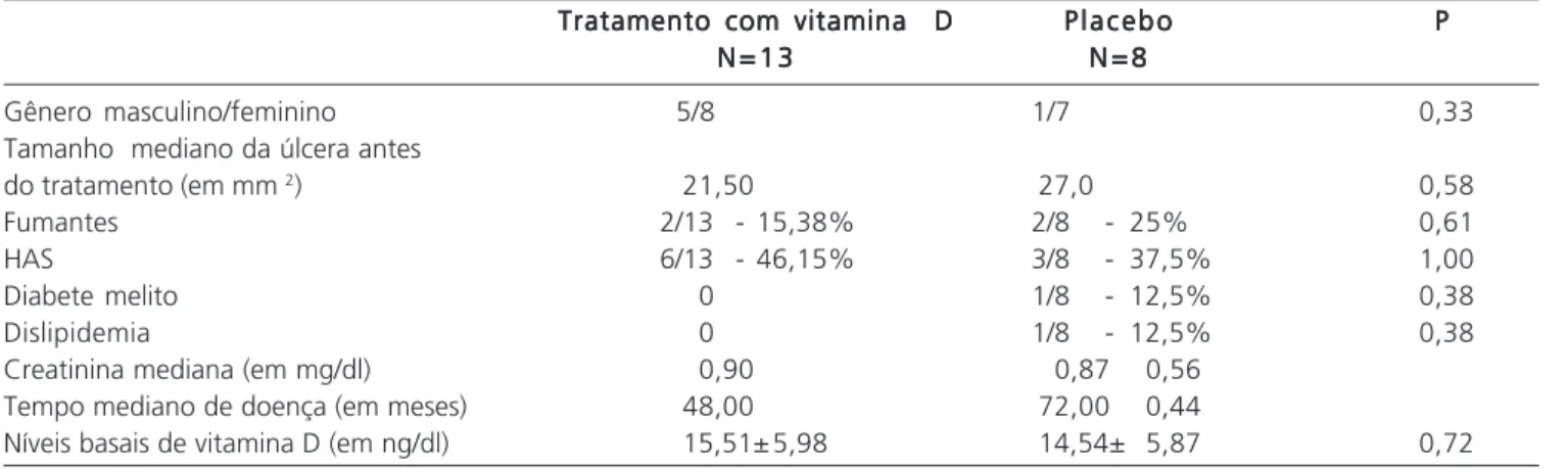
We studied patients of both genders with chronic venous leg ulcers randomly chosen in order of arrival for appointments. This group was called “ulcer group”. The ulcer group was subdivided into a group with vitamin D deficiency that took vitamin D (vitamin D group), another group with vitamin D deficiency that received placebo (placebo group) and a third group that did not receive any medicine, as the dosage of vitamin D was normal.
For controls we used patients from the outpatient rheumatology clinic in consultation for soft tissue involvement and here designated “group without ulcer”. Controls were matched with the ulcer group for gender, age and comorbidities. In the ulcer group we considered eligible for the study patients of both genders, with leg ulcers of venous origin, over 18 years and who agreed to participate in the trial. Exclusion criteria were pregnancy, presence of associated autoimmune disease, use of anticonvulsants (which alter the metabolism of vitamin D), concomitant osteomyelitis, skin tumors on venous ulcers and/or lymphedema, ulcers in legs of other cause than venous insufficiency, patients with renal insufficiency (serum creatinine greater than 1.5 mg/dl), uncontrolled hypertension (blood pressure greater than or equal to 140/100 mmHg on the day of inclusion), paresthesias of lower limbs and the need for surgical debridement
Patients from the ulcer group were examined in three situations, named visits 1, 2 and 3.
complete physical examination was performed and the size of the ulcerated area was recorded (obtained by measuring the vertical and longitudinal diameters to calculate the affected area (13). If more than one ulcer existed, the values
were added.
After application of the questionnaire, we collected blood from the patients, who returned in two weeks (visit 2) for the choice of the treatment group. If the vitamin D was normal, the patient was only observed. If the vitamin D was low, patients were allocated to a group receiving placebo or treatment with vitamin D. Replacement of vitamin D was made with 50,000 IU/week3. Placebo
and vitamin D were identified only by a numerical code and the capsules of the vitamin and placebo had similar appearance. Both patient and investigator were blinded as to the contents of the capsules, which were known only by a third physician that matched the sample.
Visit 3 was performed after eight weeks of treatment, and ulcer size and local pain measured by VAS were recorded. In that same visit, the medication code was broken and if the patient had received vitamin D, he/ she proceeded to a second serum vitamin D measurement to ensure that the drug was properly ingested and absorbed and that the patient had received a dose sufficient to correct the deficiency.
Vitamin D dosage was performed in venous blood, stored in dry tubes, protected from light and refrigerated at 2° to 8°C until testing. The analysis was performed by chemiluminescence, with equipment and brand Liaisom and with the kit DiaSorin, assuming as a normal value those above 30 mg/dl. Creatinine was simultaneously measured to confirm the absence of renal failure.
The data were analyzed using frequency and contingency tables. For measures of central tendency we used the mean and standard deviation or the median value. To study the association of nominal variables we used the Fisher’s and chi-square test and the Student t and Mann Whitney tests for numeric variables. The correlation study was done using the Spearman test. The significance adopted was 5%. To study the difference of ulcer size before and after treatment we performed a logarithm transformation (log10) due to variability and asymmetry of the values, to obtain a normal distribution. Calculations were made with the aid of GraphPad Prism version 5.00 for Windows, GraphPad Software, San Diego California USA, www.graphpad.com.
RESULTS
RESULTS
RESULTS
RESULTS
RESULTS
We studied 52 patients: 26 patients with leg ulcers with a mean age of 57.15 ± 11.36 years, among whom nine were male (34.61%) and 17 females (65.38%), had 1 to 360 months of disease duration (median of 30 months), ulcer size from 1 to 406 cm2 (mean of 26.5 cm2) and
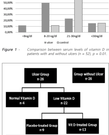
creatinine levels from 0.7 to 1.5 mg/dl (median 0.9 mg/dl). The other 26 patients formed the group without ulcer. This last group was formed by 4 (15.3%) males and 22 (84.6%) females (p=0.19) and had a mean age of 61.61±7.61 years (p=0.10) and mean serum creatinine of 0.83±0.24 mg/dl (p=0.17).The median serum vitamin D in the ulcer group was 17.05 ng/dl (5.64 to 42.00 ng/dL) and in the group without ulcer it ranged between 9.8 and 77.6 ng/dl (median: 22.75 ng/dl). The comparison of vitamin D levels between the two groups showed a p = 0.01 (Figure 1).
By studying the correlation between levels of vitamin D and characteristics of the ulcer we found no relation between the size of the ulcerated area and the levels of vitamin D levels (p = 0.48, R Speaman -0.144, 95% CI – 0.5129 to 0.268). Of the patients with ulcers who returned to visit 2, 22 (84.6%) had low levels of vitamin D and were divided into treatment groups with placebo (n = 9) and vitamin D (n = 13) (Figure 2).
The matching of data from the studied samples that were treated with placebo or vitamin D can be seen in table 1.
Taking into account the change in the area of the ulcer from the group taking vitamin D, which was
Figure 1 Figure 1 Figure 1 Figure 1
Figure 1 - Comparison between serum levels of vitamin D in patients with and without ulcers (n = 52), p = 0.01.
Figure 2 Figure 2 Figure 2 Figure 2
obtained by subtracting the after-treatment area of the ulcer from the pre-treatment one of all patients, we obtained the following values: (a) in the vitamin D treatment group
the area was between -41.0 to +8.75cm2 (median
-0.75cm2); (b) in the placebo group, the area ranged from
-8.0 to + 25cm2 (median 4cm2). From the analysis of medians,
there was greater variation in the first group (p = 0.06), with a tendency to statistical significance (Figure 3).
DISCUSSION
DISCUSSION
DISCUSSION
DISCUSSION
DISCUSSION
This study was motivated by the enormous amount of work on vitamin D published in recent years. In addition to bone metabolism, normal levels of vitamin D are essential for proper muscle function and low levels of this substance have been associated with reversible myopathy. Bischoff-Ferrari et al.14 correlate the level of
25-OH-vitamin D and muscle function of lower limbs in people over 60 years (n = 4100), which were selected by researchers from house to house. They measured the 25-OH-vitamin D levels and did walk and sit-stand tests with five repetitions. The researchers found a positive association between concentrations 25-OH-vitamin D and lower limb function. This correlation remained when they evaluated the following variables: age, gender, race, use of orthotics (cane or walker), body mass, number of comorbidities, month of evaluation and physical activity level.
As for the other functions of vitamin D, the evidence is controversial, suggesting that it may prevent diabetes mellitus type 1, hypertension, systemic sclerosis and many common cancers15.
In the skin, vitamin D binds to its hormone re-ceptor (VDR), increasing the secretion of cathelecidin16. The
importance of vitamin D and cathelicidin during wound healing turns vitamin D an attractive pharmacological target for the treatment of these situations. Also, defective expression of antimicrobial peptides has been observed in chronic ulcers and burns17.
Through the healing of venous ulcers, we tried to analyze the effect of vitamin D in the skin regeneration process. The contribution of the authors encourages a discussion on a still controversial field and the search for new therapeutic options for a very common situation in medical practice. Presently, venous ulcers have no effective therapeutic options and have a high treatment cost due to very slow healing and laboral incapacity. The study of vitamin D as a potential treatment can be a worthwhile innovation.
In selecting the sample for this study, we sought to exclude all possible confounding variables, such as: the use of anticonvulsants, which alter the metabolism of vitamin D, foot ulcers, which are influenced by footwear, and renal failure, due to vitamin D metabolism in the kidney. Due to the rigorous selection of patients, the sample size was relatively small. The measurement of vitamin D after treatment was of great importance, since it was found that it was effective, the initial values raising from 14.54±5.87ng/ dl to 40.80±13.06ng/dl.
The present study demonstrated that vitamin D deficiency was highly prevalent in all groups, in agreement
Tabela 1 Tabela 1 Tabela 1 Tabela 1
Tabela 1 - Pareamento da amostra com placebo (n=9) e vitamina D (n=13).
Tratamento com vitamina D Tratamento com vitamina D Tratamento com vitamina D Tratamento com vitamina D
Tratamento com vitamina D P l a c e b oP l a c e b oP l a c e b oP l a c e b oP l a c e b o PPPPP N = 1 3
N = 1 3 N = 1 3 N = 1 3
N = 1 3 N = 8N = 8N = 8N = 8N = 8
Gênero masculino/feminino 5/8 1/7 0,33
Tamanho mediano da úlcera antes
do tratamento (em mm 2) 21,50 27,0 0,58
Fumantes 2/13 - 15,38% 2/8 - 25% 0,61
HAS 6/13 - 46,15% 3/8 - 37,5% 1,00
Diabete melito 0 1/8 - 12,5% 0,38
Dislipidemia 0 1/8 - 12,5% 0,38
Creatinina mediana (em mg/dl) 0,90 0,87 0,56
Tempo mediano de doença (em meses) 48,00 72,00 0,44
Níveis basais de vitamina D (em ng/dl) 15,51± 5,98 14,54± 5,87 0,72
Figure 3 Figure 3 Figure 3
with the literature15,18. In the group without ulcer, the vast
majority of patients (88.41%) had vitamin D above 20ng/ dl. In the group with ulcers, of the 26 patients included, 63.53% had levels below 20ng/dl, which is compatible with severe deficiency of this substance. This difference may have multiple explanations: ulcer patients are instructed to rest indoors with legs elevated as part of the treatment; the appearance of the wounds, often with unpleasant odor, favors the use of more clothes, and many of these patients are away from work activities. Therefore they expose themselves to little sunlight.
Heilborn et al.8 studied leg ulcers and vitamin D
in interesting ways. They selected nine patients with venous ulcers of the lower limbs and performed biopsies of the edges of the wounds. They also selected healthy persons in which serial skin incisions were made in the abdominal region, and then proceeded to new incisions in the margin of the subsequent biopsies (in 5 minutes, 12 hours, two days, seven days and fourteen days after the first incision). For the ex vivo studies, they obtained human skin from plastic s and maintained them in culture media. The analysis of those materials showed that in the group of chronic leg ulcers many people were deficient of an antimicrobial peptide (hCAP18). This fact did not occur in healthy individuals and in ex vivo studies. This peptide is a molecule of the innate defense system and is important for skin integrity.
Other authors19 proved that treatment with
vitamin D increases the production of hCAP18 in human keratinocytes in vitro by binding to a vitamin D-responsive element in the promoter of the gene hCAP18. These observations imply that vitamin D is actually associated with skin regeneration.
Studying the ulcer group, those patients with normal vitamin D had ulcers with the same characteristics in relation to area and pain as measured by VAS when compared to those with low vitamin D. So we concluded that vitamin D deficiency may not be implicated in the cause of the ulcer, but it is a consequence. This fact does not mean that this deficiency cannot disturb the healing process. In our study, after treatment there was no statistically significant difference in the ulcer size when comparing groups treated with placebo and vitamin D. However, when analyzing the size variation of the wound after treatment, we found a trend towards statistical significance. It is possible that the small number of patients in our study has resulted in a type II statistical error.
In other diseases, such as psoriasis, vitamin D is topically applied with proven efficacy. This was shown in an epidemiological study with 11.631 patients with psoriasis, of which 59.7% were treated with topical application of vitamin D20.
A question raised in this work is whether the topical administration route would change the obtained results. In the literature there is no data about this idea.
As venous ulcers are diseases of long duration, another issue to be addressed is the observation time. In the present study patients were followed up to two months (just immediately after the normalization of vitamin D levels). The hypothesis that remains to be tested in other studies is if the monitoring was done for a longer time, or until ulcer closure, perhaps the result would be statistically significant. This is an area in which literature is very scarce and there is no data to support this idea.
Some studies suggest an association between pain and vitamin D deficiency4,21,22. Plotnikoff and Quigley22
studied 150 patients who arrived at the consultation center in Minneapolis, USA, with persistent, non-specific musculoskeletal pain. Of these, 100% had levels of vitamin D below 20 ng/ml. In Oslo, Norway, Knutsen et al.21 studied
this aspect in a different way. The authors included patients with musculoskeletal pain, headache and fatigue. A total of 572 individuals were chosen, of which 58% had levels of vitamin D below 20 ng/ml. In the present work there was no relation between the value of visual analogue scale for pain and serum vitamin D (p = 0.14), but it must be remembered that pain is a complex phenomenon, with influence of organic, psychological and social factors12,23
and it is possible that the appearance of the ulcer, with social and labor exclusion, influence more than the organic aspect.
The impaired wound healing of chronic ulcers is a clinical problem, and regardless of the underlying cau-se, such ulcers are characterized by chronic inflammation9,24,25. Persistent inflammation creates an
environment with high levels of proteases and cytokines. In addition, there is an imbalance in the proteolytic enzymes and their endogenous inhibitors26,27. All these
factors hinder wound healing28,29. Much progress has
occurred in relation to normal wound healing. However, the understanding of this fundamental process is still insufficient, and there is little therapeutic progress in the field of ulcers treatment8. Therefore, the role of vitamin D
R E S U M O R E S U M O R E S U M O R E S U M O R E S U M O
Objetivo: Objetivo: Objetivo: Objetivo:
Objetivo: Estudar a relação entre deficiência de vitamina D e cicatrização de pele em pacientes com úlceras de perna, relacionar esta deficiência com características da úlcera e avaliar se a reposição de vitamina D nos indivíduos deficientes acelera a cicatrização da úlcera. Métodos: Métodos: Métodos: Métodos: Métodos: Foram escolhidos aleatoriamente 26 pacientes com úlceras venosas de perna e 26 sem úlcera pareados para sexo, idade, HAS e tabagismo. Os grupos foram comparados com relação à dosagem sérica de vitamina D. O grupo úlcera foi dividido em dois subgrupos: um que tomou placebo e outro que recebeu vitamina D 50.000UI por semana durante dois meses. Foi realizada a dosagem da 25-OH-vitamina D e avaliados o tamanho da úlcera e a gravidade da dor, antes e após o tratamento. Resultados: Resultados: Resultados: Resultados: Resultados: A maioria dos pacientes apresentava níveis insuficientes de vitamina D. Não foi encontrada correlação entre o tamanho da úlcera sem tratamento e os níveis de vitamina D. Nos pacientes que receberam vitamina D, após o tratamento, o tamanho mediano da área da úlcera, diminui de 25cm2, para 18cm2 e no grupo placebo, de
27cm2 para 24,5cm2 (p=0,7051 e p=0,7877, respectivamente). Considerando-se a variabilidade da área da úlcera do grupo
vitamina D versus placebo, a mediana foi igual a -0,75cm2 no primeiro grupo e 4cm2 no segundo grupo (p=0,0676). Conclusão:Conclusão:Conclusão:Conclusão:Conclusão:
Pacientes com úlcera de perna têm mais deficiência de vitamina D que os sem. A deficiência de vitamina D não influiu nas características das lesões. A cicatrização nos pacientes com hipovitaminose D mostrou tendência para ser maior naqueles que receberam reposição vitamínica.
Descritores Descritores Descritores Descritores
Descritores: Insuficiência venosa. Úlcera cutânea. Úlcera da perna. Deficiência de vitamina D. Cicatrização.
REFERENCES
REFERENCES
REFERENCES
REFERENCES
REFERENCES
1. Saraiva GL, Cendoroglo MS, Ramos LR, Araújo LM, Vieira JG, Maeda SS, et al. Prevalência da deficiência, insuficiência de vita-mina D e hiperparatireoidismo secundário em idosos institucionalizasdos e moradores na comunidade da cidade de São Paulo, Brasil. Arq Bras Endocrinol Metabol. 2007;51(3):437-42. 2. Thomas MK, Lloyd-Jones DM, Thadhani RI, Shaw AC, Deraska DJ,
Kitch BT, et al. Hypovitaminosis D in medical inpatients. N Engl J Med. 1998;338(12):777-83.
3. Holick MF. Vitamin D deficiency. N Engl J Med. 2007;357(3):266-81.
4. Dawson-Hughes B, Heaney RP, Holick MF, Lips P, Meunier PJ, Vieth R. Estimates of optimal vitamin D status. Osteoporos Int. 2005;16(7):713-6.
5. Zhang JZ, Maruyama K, Ono I, Kaneko F. Production and secretion of platelet-derived growth factor AB by cultured human keratinocytes: regulatory effects of phorbol 12-myristate 13-acetate, etretinate, 1,25-dihydroxyvitamin D3, and several cytokines. J Dermatol. 1995;22(5):305-9.
6. Hosomi J, Hosoi J, Abe E, Suda T, Kuroki T. Regulation of terminal differentiation of cultured mouse epidermal cells by 1 alpha,25-dihydroxyvitamin D3. Endocrinology. 1983;113(6):1950-7. 7. Matsumoto K, Hashimoto K, Nishida Y, Hashiro M, Yoshikawa K.
Growth-inhibitory effects of 1,25-dihydroxyvitamin D3 on normal human keratinocytes cultured in serum-free medium. Biochem Biophys Res Commun. 1990;166(2):916-23.
8. Heilborn JD, Nilsson MF, Kratz G, Weber G, Sørensen O, Borregaard N, Ståhle-Bäckdahl M. The cathelicidin anti-microbial peptide LL-37 is involved in re-epithelialization of human skin wounds and is lacking in chronic ulcer epithelium. J Invest Dermatol. 2003;120(3):379-89.
9. Herrick SE, Sloan P, McGurk M, Freak L, McCollum CN, Ferguson MW. Sequential changes in histologic pattern and extracellular matrix deposition during the healing of chronic venous ulcers. Am J Pathol. 1992;141(5):1085-95.
10. Abbade LP, Lastória S, de Almeida Rollo H, Stolf HO. A sociodemographic, clinical study of patients with venous ulcer. Int J Dermatol. 2005;44(12):989-92.
11. Maffei FH, Magaldi C, Pinho SZ, Lastoria S, Pinho W, Yoshida WB, et al. Varicose veins and chronic venous insufficiency in Brazil: prevalence among 1755 inhabitants of a country town. Int J Epidemiol. 1986;15(2):210-7.
12. Callam MJ, Harper DR, Dale JJ, Ruckley CV. Chronic ulcer of the leg: clinical history. Br Med J. 1987;294(6584):1389-91.
13. Kantor J, Margolis DJ. Efficacy and prognostic value of simple wound measurements. Arch Dermatol. 1998;134(12):1571-4. 14. Bischoff-Ferrari HA, Dietrich T, Orav EJ, Hu FB, Zhang Y, Karlson
EW, et al. Higher 25-hydroxyvitamin D concentrations are associated with better lower-extremity function in both active and inactive persons aged > or =60 y. Am J Clin Nutr. 2004;80(3):752-8.
15. Holick MF. High prevalence of vitamin D inadequacy and implications for health. Mayo Clin Proc. 2006;81(3):353-73.
16. Schauber J, Gallo RL. Expanding the roles of antimicrobial peptides in skin: alarming and arming keratinocytes. J Invest Dermatol. 2007;127(3):510-2.
17. Zasloff M. Sunlight, vitamin D, and the innate immune defenses of the human skin. J Invest Dermatol. 2005;125(5):xvi-xvii.
18. Holick MF. Vitamin D status: measurement, interpretation, and clinical application. Ann Epidemiol. 2009;19(2):73-8.
19. Wang TT, Nestel FP, Bourdeau V, Nagai Y, Wang Q, Liao J, Tavera-Mendoza L, et al. Cutting edge: 1,25-dihydroxyvitamin D3 is a direct inducer of antimicrobial peptide gene expression. J Immunol. 2004 1;173(5):2909-12.
20. Takahashi H, Nakamura K, Kaneko F, Nakagawa H, Iizuka H; JAPANESE SOCIETY FOR PSORIASIS RESEARCH. Analysis of psoriasis patients registered with the Japanese Society for Psoriasis Research from 2002-2008. J Dermatol. 2011;38(12):1125-9.
21. Knutsen KV, Brekke M, Gjelstad S, Lagerløv P. Vitamin D status in patients with musculoskeletal pain, fatigue and headache: a cross-sectional descriptive study in a multi-ethnic general practice in Norway. Scand J Prim Health Care. 2010;28(3):166-71. 22. Plotnikoff GA, Quigley JM. Prevalence of severe hypovitaminosis
D in patients with persistent, nonspecific musculoskeletal pain. Mayo Clin Proc. 2003;78(12):1463-70.
23. Grey JE, Enoch S, Harding KG. Wound assessment. BMJ. 2006;332(7536):285-8.
24. Bollag WB. Differentiation of human keratinocytes requires the vitamin d receptor and its coactivators. J Invest Dermatol. 2007;127(4):748-50.
26. Callam MJ, Ruckley CV, Harper DR, Dale JJ. Chronic ulceration of the leg: extent of the problem and provision of care. Br Med J. 1985;290(6485):1855-6.
27. Gniadecki R. Stimulation versus inhibition of keratinocyte growth by 1,25-Dihydroxyvitamin D3: dependence on cell culture conditions. J Invest Dermatol. 1996;106(3):510-6.
28. Vieth R. What is the optimal vitamin D status for health? Prog Biophys Mol Biol. 2006;92(1):26-32.
29. Peters BS, dos Santos LC, Fisberg M, Wood RJ, Martini LA. Prevalence of vitamin D insufficiency in Brazilian adolescents. Ann Nutr Metab. 2009;54(1):15-21.
Received on 25/03/2012
Accepted for publication 30/05/2012 Conflict of interest: none
Source of funding: none How to cite this article: How to cite this article: How to cite this article: How to cite this article: How to cite this article:
Burkiewicz CJCC, Guadagnin FA, Nascimento MM, Skare TL, Dietz UA, Servin SCN, Souza GD. Vitamin d and skin repair: a prospective, double-blind and placebo controlled study of the role of vitamin d in the healing of leg ulcers. Rev Col Bras Cir. [periódico na Internet] 2012; 39(5). Disponível em URL: http://www.scielo.br/rcbc
Address for correspondence: Address for correspondence: Address for correspondence: Address for correspondence: Address for correspondence: