Cop
yright
© ABE&M t
odos os dir
eit
os r
eser
vados
.
Thyroid nodules and thyroid
cancer in Graves’ disease
Nódulos tiroidianos e câncer de tiroide na doença de Graves
Abbas Ali Tam1, Cafer Kaya1, Fevzi Balkan1, Mehmet Kılıç2, Reyhan Ersoy3, Bekir Çakır3
ABSTRACT
Objective: The frequency of thyroid nodules accompanying Graves’ disease and the risk of thyroid cancer in presence of accompanying nodules are controversial. The aim of this study was to evaluate the frequency of thyroid nodules and the risk of thyroid cancer in patients ope-rated because of graves’ disease. Subjects and methods: Five hundred and twenty-six patients in whom thyroidectomy was performed because of Graves’ disease between 2006 and 2013 were evaluated retrospectively. Patients who had received radioactive iodine treatment and external irradiation treatment in the neck region and who had had thyroid surgery previously were not included in the study. Results: While accompanying thyroid nodule was present in 177 (33.6%) of 526 Graves’ patients, thyroid nodule was absent in 349 (66.4%) patients. Forty-two (8%) patients had thyroid cancer. The rate of thyroid cancer was 5.4% (n = 19) in the Graves’ patients who had no nodule, whereas it was 13% (n = 23) in the patients who had nodule. The risk of thyroid cancer increased signiicantly in presence of nodule (p = 0.003). Three patients had recurrence. No patient had distant metastasis. No patient died during the follow-up period.
Conclusions: Especially Graves’ patients who have been decided to be followed up should be evaluated carefully during the follow-up in terms of thyroid cancer which may accompany. Arq Bras Endocrinol Metab. 2014;58(9):933-8
Keywords
Graves’ disease; thyroid nodules; thyroid cancer
RESUMO
Objetivo: A frequência da ocorrência de nódulos tiroidianos acompanhando a doença de Gra-ves e o risco de câncer de tiroide na presença desses nódulos é controversa. O objetivo deste estudo foi avaliar a frequência de nódulos tiroidianos e o risco de câncer de tiroide em pacien-tes operados por doença de Graves. Sujeitos e métodos: Quinhentos e vinte e seis pacientes anteriormente submetidos à tiroidectomia por doença de Graves entre 2006 e 2013 foram ava-liados retrospectivamente. Os pacientes que receberam tratamento com iodo radioativo e irra-diação externa da região do pescoço e que anteriormente passaram por cirurgia de tiroide não foram incluídos no estudo. Resultados: Enquanto os nódulos de tiroide se apresentaram em 177 (33,6%) dos 526 pacientes com doença de Graves, eles estiveram ausentes em 349 (66,4%) pacientes. Um total de 42 (8%) dos pacientes teve câncer de tiroide. A ocorrência de câncer de tiroide foi 5,4% (n = 19) nos pacientes com doença de Graves que não apresentaram nódulos, e 13% (n = 23) nos pacientes com nódulos. O risco de câncer de tiroide aumentou signiicati-vamente na presença de nódulos (p = 0,003). Três pacientes apresentaram recidivas. Nenhum paciente apresentou metástase distante e nenhum paciente veio a óbito durante o período de acompanhamento. Conclusões: Pacientes com doença de Graves devem ser avaliados cuida-dosamente no acompanhamento para a possível ocorrência de câncer de tiroide. Arq Bras Endocrinol Metab. 2014;58(9):933-8
Descritores
Doença de Graves; nódulos tiroidianos; câncer de tiroide
1 Ataturk Training and Research
Hospital, Department of Endocrinology and Metabolism, Ankara, Turkey
2 Yıldırım Beyazıt University,
Department of General Surgery, Ankara, Turkey
3 Yıldırım Beyazıt University,
Department of Endocrinology and Metabolism, Ankara, Turkey
Correspondence to:
Abbas Ali Tam
Ataturk Training and Research Hospital
06800 – Ankara, Turkey [email protected] Received on June/30/2014 Accepted on Aug/21/2014
Cop
yright
© ABE&M t
odos os dir
eit
os r
eser
vados
.
INTRODUCTION
T
hyroid nodules are frequently found in patients with Graves’ disease, though the frequency varies depending on the method used (1-3). The prevalence of palpable thyroid nodule is 3-fold higher compared to the general population. In epidemiological studies, a higher thyroid nodule prevalence is found when thy-roid ultrasonography is used for evaluation of thythy-roid morphology (2).In addition, thyroid nodule develops in approxi-mately half of the patients with Graves’ disease during the follow-up (4). There is an increased risk of thyroid cancer in presence of these nodules (3).
While the rate of malignancy is approximately 5% in palpable thyroid nodules in the general population, it varies between 2.3% and 45.8% in patients with Graves’ disease (2).
Moreover, there has been much controversy regard-ing biological behaviour of cancers in Graves’ patients. While some authors have reported that thyroid cancers have a more agressive course (5,6), some others have reported the contrary (7,8). The aim of this study was to investigate the frequency of thyroid nodule in pa-tients operated because of Graves’ disease and the fre-quency of thyroid cancer in patients with and without nodule.
SUBJECTS AND METHODS
Five hundred and twenty-six patients who underwent thyroidectomy because of Graves’ disease between 2006 and 2013 were included in the study. The diag-nosis of Graves’ disease was made with increased se-rum triiodothyronine (T3) and thyroxine (T4) levels and decreased thyroid stimulating hormone (TSH) levels and diffuse uptake on thyroid scintigraphy in patients who had a history and signs of hyperthyroi-dism. In most cases, the diagnosis was supported with increased thyroid stimulating antibody (TRAb) levels. Thyroid ultrasonography was performed in all patients. The patients were divided into two groups as the group with nodule and the group without nodule according to presence of thyroid nodule. Fine needle aspiration biopsy (FNAB) was performed in 172 of the patients who had thyroid nodule. Patients who had received radioactive iodine treatment and external irradiation treatment in the neck region and who had had thy-roid surgery previously were not included in the study.
The patients were treated with propylthiouracil or me-thimazole before surgery. The patients who required urgent surgery received Lugol solution for 7-10 days before surgery. The indications for surgery in Graves’ patients included failure of antithyroid drug treatment and/or development of side effects due to these drugs, goitre which caused to compression symptoms in the trachea or esophagus, severe opthalmopathy, suspicious malignancy on FNAB and request of the patient. Total or near total thyroidectomy was performed in all patients.
RESULTS
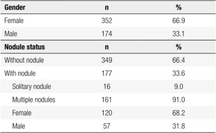
Five hundred and twenty-six patients 352 (66.9%) of whom were female and 174 (33.1%) of whom were male were included in the study. The mean age of the patients was 41.2 ± 12.4 years (range 17-69 years). While thyroid nodule was present in 177 (33.6%) pa-tients, 349 (66.4%) had no thyroid nodule. Among the patients who had thyroid nodule, 16 had solitary nodu-le and 161 had multipnodu-le nodunodu-les. The mean nodunodu-le di-ameter was 27.17 ± 14.10 mm (range 7.30-68.0 mm). One hundred and twenty (68.2%) of the patients who had thyroid nodule were female, whereas 57 (31.8%) were male and there was no statistically signiicant dif-ference (p > 0.05) (Table 1). According to the lar gest nodule diameter, the thyroid nodule diameter was higher than 1 cm in 171 of 177 patients.
Table 1. Clinical characteristics of patients
Gender n %
Female 352 66.9
Male 174 33.1
Nodule status n %
Without nodule 349 66.4
With nodule 177 33.6
Solitary nodule 16 9.0
Multiple nodules 161 91.0
Female 120 68.2
Male 57 31.8
Cop
yright
© ABE&M t
odos os dir
eit
os r
eser
vados
.
and in 5.4% (n = 19) of 349 patients who had no thy-roid nodule. This difference was statistically signiicant (p = 0.003). The risk of cancer increased markedly in presence of thyroid nodule. In our study, thyroid can-cer was present in 2 (12.5%) of 16 patients who had a solitary nodule and in 21 (13%) of 161 patients who had multiple thyroid nodules.
A total of 17 patients had variant thyroid cancer 6 of whom had classical variant, 3 of whom had oncocytic variant and 2 of whom had tall cell variant. The mean age of 42 patients who were found to have cancer was 43.1 ± 12.4 years (range 20-69 years). The mean age was 44.8 ± 12.1 years (range 22-69 years) in women who had thyroid cancer and 40.2 ± 12.8 years (range 20-61 years) in men who had thyroid cancer. This dif-ference was not statistically signiicant (p > 0,05). In the patients who were found to have thyroid cancer, the mean follow-up time between the diagnosis of Graves’ disease and surgery was 3.95 ± 1.61 years (range 1-8 years) and the mean follow-up time after the diagno-sis of thyroid cancer was 2.87 ± 1.61 years (range 0.5-7.50 years). Anti-Tg and Anti TPO were measured in nearly all patients with cancer and they were found to be positive in 52.5% of the patients and 48.8% of the patients, respectively (Table 2). FNAB was performed in 172 patients who had thyroid nodule and suspicious malignancy was present in 2 of them.
occured in 3 (7.1%). The tumor was multicentric in 5 patients. Cancer was present in both lobes in 5 patients. Three of 3 patients who had lymph node involvement, 3 of 6 patients who had thyroid capsule invasion and 2 of 4 patients who had invasion into the surrounding tissues had papillary thyroid cancer and the rest had mi-cropapillary thyroid cancer (Table 3).
Table 2. Clinical characteristics of cancer patients
Characteristic n %
Gender Male 16 38.1
Female 26 61.9
Anti-Tg Positive 21 52.5
Negative 19 47.5
Anti-TPO Positive 20 48.8
Negative 21 51.2
Age (years) 43.1 ± 12.4
Male 40.2 ± 12.8
Female 44.8 ± 12.1
Disease perioda (years) 3.95 ± 1.61
Cancer periodb (years) 2.87 ± 1.61
Data is expressed as mean ± SD; with the range presented in parenthesis a: the mean time between Graves’ disease and operation. b: the mean follow-up time after the diagnosis of thyroid cancer.
Lymph node involvement was present in 3 (7.1%) of 42 patients who had thyroid cancer, thryoid capcule invasion was present in 6 (14.3%), invasion into the sur-rounding tissues was present in 4 (9.5%) and recurrence
Table 3. Clinical characteristics of cancer patients
n %
Lymph node metastasis 3 7.1
PTC 3 100.0
MPTC 0 0.0
Mulicentricity 5 11.9
PTC 2 40.0
MPTC 3 60.0
Thyroid capsule invasion 6 14.3
PTC 3 50.0
MPTC 3 50.0
Surrounding tissue invasion 4 9.5
PTC 2 50.0
MPTC 2 50.0
Bilaterality 5 11.9
PTC 4 80.0
MPTC 1 20.0
Recurrence 3 7.1
PTC 2 66.6
MPTC 1 33.4
PTC: papillary thyroid cancer; MPTC: micropapillary thyroid cancer.
Recurrence occured in only one of 33 micropap-illary thyroid cancers and in 2 of 9 papmicropap-illary thyroid cancers. According to TNM staging, 37 patients had stage 1. Thirty-two of 33 micropapillary thyroid can-cers were stage 1 and one was stage 2. Five of 9 papil-lary thyroid cancers were stage 1, 1 was stage 2, 2 were stage 4a and 1 was stage 4b (Table 4).
There was no statistically signiicant difference be-tween the patients with thyroid cancer who showed and did not show lymph node invasion, thyroid capsule invasion, invasion into the surrounding tissues and re-currence in terms of age, TRAb titer and disease time (p > 0.05).
Cop
yright
© ABE&M t
odos os dir
eit
os r
eser
vados
.
who had nodule. Thus, the cancer focus was found in the parenchyma in 31 (73.8%) of 42 patients indepen-dent of accompanyment of thyroid nodule.Twenty-six cancers in the thyroid parancime were micropapillary. Mortality was not observed in any patient during the follow-up. Radioactive iodine treatment was given to 29 patients.
The data were assessed using SPSS 15.0 statistical package program. Chi-square and Fisher’s exact test were used in assessment of the categorical data and Stu-dent’s T-test and one-way variance analysis were used in assessment of the numerical data. p value of < 0,05 was considered statistically signiicant.
Kim and cols. detected thyroid nodule by palpation in 9.4% of the patients (23/245) and by thyroid ultra-sonography in 35.1% of the patients (6/245) in a pro-spective study which they conducted with 245 Graves patients. Fifty percent of these patients (43/86) had solitary nodules and 50% (43/86) had multiple no-dules (11).
Our center is a reference center and we initially per-form thyroid ultrasonography in all thyroid patients re-ferring to our department. In our series, we detected thyroid nodule in a total of 177 (33.6%) patients by thyroid ultrasonography; 16 of these patient had soli-tary nodule and 161 had multiple nodules. According to the size of the dominant nodule, 171 patients had a nodule larger than 1 cm.
Presence of these nodules increases the risk of thy-roid cancer. However, the risk of differentiated thythy-roid cancer in Graves patients remains as a controversial is-sue. The malignancy rate in these nodules varies be-tween 10% and 46% (10). This rate is approximately 5% in the general population (12). While the annual inci-dence of clinical thyroid cancer in the general euthyroid population has been reported to be 0.5-8/100.000, it is 175/100.000 in Graves disease (13).
Recently, Ren and cols. detected thyroid nodule in 22.7% of Graves patients (96/423) by ultrasonography. Twenty ive of these patients (26%) had solitary nod-ule and 71 (74%) had multiple nodnod-ules. In the same study, the total incidence of thyroid cancer was 13.7% (58/423). While thyroid nodule accompanied to 46 of 58 patients, thyroid nodule was absent in 12 (3,6%) patients and the tumor was detected incidentally (14).
In our study, the rate of thyroid cancer in Graves’ patients was 8% (42/526). One of the important points in our series was the high cancer rate in the parenchyma of the patients with Graves’ disease. There was a can-cer focus in the thyroid parenchyma in 31 (73.8%) of 42 patients. This inding is notable, because nearly all cancers reported in series of Graves’ patients with co-existing carcinoma are in the thyroid nodule (1). Er-bil and cols. (1) found that 67% (n = 12) of thyroid cancers found incidentally in Graves’ patients were in the parenchyma outside the nodule. Ren and cols. (14) reported this rate to be 3.6% (n = 12). This shows that malignancy is not always related with nodule (12). If these patients were treated only with antithyroid and/ or radioactive iodine, they would not have received an appropriate treatment because of accompanying thy-roid cancer (15).
Table 4. TNM staging in cancer patients
TNM n %
Stage 1 37 88.0
Stage 2 2 4.8
Stage 4A 2 4.8
Stage 4B 1 2.4
Total 42 100.0
DISCUSSION
Thyroid nodule is observed with a higher rate in pa-tients with Graves’ disease compared to the general population. The prevalence of these nodules varies depending on the method used (examination, thy-roid scintigraphy, thythy-roid ultrasonography or combi-nations of these). The prevalence is lower when only clinical examination and scintigraphy are performed compared to ultrasonography (9). Palpable thyroid nodules are observed with a rate of 5% in the general population and with a rate of approximately 15% in Graves’ disease (1,10). When more sensitive echo-graphic imaging is used, thyroid nodules are found more frequently in Graves’ disease compared to the general population (10).
Cantalamessa and cols. found thyroid nodule with a diameter of 8 mm or larger in 33.6% of Graves’ pa-tients. When smaller lesions were included, the igure reached to 40.6% (4).
Cop
yright
© ABE&M t
odos os dir
eit
os r
eser
vados
.
Thyroid cancers found in Graves’ patients mostly have papillary origin (16). Many carcinomas related with Graves’ disease are small and are found inciden-tally during surgery or postoperative pathological ex-amination (17). In our study, all cancers detected in our study had papillary origin. Seventy-eight point six percent of these (33/42) were micropapillary thyroid cancer.
The pathogenic relationship between Graves’ disea-se and thyroid cancer could not be understood clearly. Thyrotropin receptor antibodies TRAbs may possibly play a role in the initiation and progression of thyroid cancer. TRAbs and TSH activate the same intracel-lular pathway and both of them have mitogenic and antiapoptotic effects on thyroid follicular cells. TRAbs stimulate angiogenesis, which has a critical role in the growth and development of the tumor in the thyroid, by upregulating vascular endothelial growth factor and placental growth factor (2,10,13,18).
Biological behavior and optimal management of differentiated thyroid cancers accompanying Graves’ disease are controversial (18). It has been reported that patients who have undergone thyroidectomy because of Graves’ disease and who have small thyroid cancer (a diameter of 1 cm or smaller) have excellent prognosis and longer disease-free survival compared to patients who have small thyroid cancer without Graves’ disease (19). Most of the patients in our series had small thy-roid cancer. Thirty-seven of 42 patients had a TNM stage of 1. Thirty-two of 33 patients with micropapil-lary thyroid cancer had a TNM stage of 1 and recur-rence was observed in only one of these patients. In addition, no patient died during the follow-up.
Currently, antithyroid drugs, radioactive iodine and surgery are used as current treatment methods in treatment of Graves’ disease. Each method has its own side effects and success and failure rates. The advantage of surgical treatment is that it improves thyrotoxico-sis faster compared to the other methods and provides effective treatment (20). The recommended operation for Graves’ disease is total thyroidectomy. Less exten-sive surgery (subtotal thyroidectomy) carries a consi-derable risk of recurrent thyrotoxicosis (up to 30% of the patients) and revisional thyroid surgery is related with a high complication risk (21). Therefore, total or near total thyroidectomy was performed in all patients in our series. Radioactive iodine treatment following surgery was given to 29 of our patients who were found to have thyroid carcinoma.
Conclusively, the risk of cancer is considerably in-creased in Graves’ patients in presence of accompanying nodule. Our study included the patients in whom sur-gery was performed because of Graves’ disease. Patients who were given radioactive iodine or medical treatment for treatment of Graves’ disease were not included in our study. Studies including these patients are needed to demonstrate the frequency of thyroid nodules and the risk of cancer clearly in Graves’ patients. The risk of cancer in the thyroid parenchyma in Graves’ patients should not be ignored. Long-term treatment and fol-low-up of these patients in whom surgery has not been performed should be pursued meticulously not only for providing and maintaining the euthryoid state, but also because of increased risk of thyroid cancer.
Disclosure: no potential conlict of interest relevant to this article was reported.
REFERENCES
1. Erbil Y, Barbaros U, Ozbey N, Kapran Y, Tükenmez M, Bozbora A, et al. Graves’ disease, with and without nodules, and the risk of thyroid carcinoma. J Laryngol Otol. 2008;122:291-5.
2. Beliore A, Russo D, Vigneri R, Filetti S. Graves’ disease, thyroid nodules and thyroid cancer. Clin Endocrinol. 2001;55:711-8. 3. Kraimps JL, Bouin-Pineau MH, Mathonnet M, De Calan L,
Ron-ceray J, Visset J, et al. Multicenter study of thyroid nodules in patients with Graves’s disease. Br J Surg. 2000;87:1111-3. 4. Cantalamessa L, Baldini M, Orsatti A, Meroni L, Amodei V,
Casta-gnone D. Thyroid nodules in graves disease and the risk of thy-roid carcinoma. Arch Intern Med. 1999;159:1705-8.
5. Beliore A, Garofalo MR, Giuffrida D, Runello F, Filetti S, Fiumara A, et al. Increased aggressiveness of thyroid cancer in patients with Graves’ disease. J Clin Endocrinol Metab. 1990;70:830-5. 6. Pellegriti G, Beliore A, Giuffrida D, Lupo L, Vigneri R. Outcome of
differentiated thyroid cancer in Graves’ patients. J Clin Endocri-nol Metab. 1998;83:2805-9.
7. Pacini F, Elisei R, Di Coscio GC, Anelli S, Macchia E, Concetti R, et al. Thyroid carcinoma in thyrotoxic patients treated by surgery. J Endocrinol Invest. 1988;11:107-12.
8. Hales IB, McElduff A, Crummer P, Clifton-Bligh P, Delbridge L, Hoschl R, et al. Does Graves’ disease or thyrotoxicosis af-fect the prognosis of thyroid cancer. J Clin Endocrinol Metab. 1992;75:886-9.
9. Mishra A, Mishra SK. Thyroid nodules in Graves’ disease: impli-cations in an endemically iodine deicient area. J Postgrad Med. 2001;47:244-7.
10. Pellegriti G, Mannarino C, Russo M, Terranova R, Marturano I, Vi-gneri R, et al. Increased mortality in patients with differentiated thyroid cancer associated with Graves’ disease. J Clin Endocrinol Metab. 2013;98:1014-21.
11. Kim WB, Han SM, Kim TY, Nam-Goong IS, Gong G, Lee HK, et al. Ultrasonographic screening for detection of thyroid cancer in patients with Grave’s disease. Clin Endocrinol. 2004;60:719-25. 12. Lima PC, Moura Neto A, Tambascia MA, Zantut Wittmann DE.
Cop
yright
© ABE&M t
odos os dir
eit
os r
eser
vados
.
13. Pazaitou-Panayiotou K, Michalakis K, Paschke R. Thyroid cancer in patients with hyperthyroidism. Horm Metab Res. 2012;44:255-62. 14. Ren M, Wu MC, Shang CZ, Wang XY, Zhang JL, Cheng H, et al. Predictive factors of thyroid cancer in patients with Graves’ disea se. World J Surg. 2014;38:80-7.
15. Weber KJ, Solorzano CC, Lee JK, Gaffud MJ, Prinz RA. Thyroidec-tomy remains an effective treatment option for Graves’ disease. Am J Surg. 2006;191:400-5.
16. Boostrom S, Richards ML. Total thyroidectomy is the preferred treatment for patients with Graves’ disease and a thyroid nodule. Otolaryngol Head Neck Surg. 2007;136:278-81.
17. Chao TC, Lin JD, Chen MF. Surgical treatment of thyroid cancers with concurrent Graves disease. Ann Surg Oncol. 2004;11:407-12.
18. Lee J, Nam KH, Chung WY, Soh EY, Park CS. Clinicopathologic features and treatment outcomes in differentiated thyroid carci-noma patients with concurrent Graves’ disease. J Korean Med Sci. 2008;23:796-801.
19. Kikuchi S, Noguchi S, Yamashita H, Uchino S, Kawamoto H. Prog-nosis of small thyroid cancer in patients with Graves’ disease. Br J Surg. 2006;93:434-9.
20. Phitayakorn R, Morales-Garcia D, Wanderer J, Lubitz CC, Gaz RD, Stephen AE, et al. Surgery for Graves’ disease: a 25-year perspec-tive. Am J Surg. 2013;206:669-73.