357
Corresponding author: Dra. Divina das Dôres de Paula Cardoso.
e-mail: [email protected]
Received 16 December 2015
Accepted 5 April 2016
Detection of antibodies to Oropouche virus in
non-human primates in Goiânia City, Goiás
Marize Moreira Gibrail
[1], Fabíola Souza Fiaccadori
[1], Menira Souza
[1],
Tâmera Nunes Vieira Almeida
[1],
Jannifer Oliveira Chiang
[2], Lívia Caricio Martins
[2],
Milene Silveira Ferreira
[2]and Divina das Dôres de Paula Cardoso
[1][1]. Departamento de Microbiologia, Imunologia, Parasitologia e Patologia Geral, Instituto de Patologia Tropical e Saúde Pública, Universidade Federal de Goiás, Goiânia, Goiás, Brasil. [2]. Seção de Arbovirologia e Febres Hemorrágicas, Instituto Evandro Chagas, Ananindeua, Pará, Brasil.
Abstract
Introduction: Arboviruses are associated with human disease, and non-human primates (NHPs) are important primary hosts. This study shows the detection of antibodies to Oropouche virus (OROV) in NHPs either living in urban parks or acclimatized at the Wild Animal Screening Center, Goiânia city. Methods: Fifty blood samples were analyzed by hemagglutination-inhibition and neutralization assays. Results: Two monkeys (Alouatta caraya) had antibodies to OROV by both techniques. Conclusions:
This is the irst report demonstrating the detection of OROV antibodies in Goiás State and may represent the introduction/
circulation of OROV in the region and a potential risk to the human population.
Keywords: Non- Human Primates. Oropouche virus. Arbovirus.
Oropouche virus (OROV) belongs to the genus Orthobunyavirus and the family Bunyaviridae, which also comprises the genera Hantavirus, Nairovirus, Phlebovirus, and Tospovirus. Orthobunyaviruses include many human pathogens such as Caraparu, Catu, Guaroa, Marituba, and Tacaiuma(1).
OROV, like other orthobunyaviruses, is a single stranded, negative-sense, three-segmented virus. The segments, small (S), medium (M) and large (L), encode the nucleocapsid (N) protein, the surface (Gn and Gc) glycoproteins and the viral ribonucleic acid (RNA)-dependent RNA polymerase, respectively(2) (3). In
addition, in some orthobunyaviruses, the S and M segments can also encode the non-structural proteins NSs and NSm, respectively(3). Phylogenetic analysis of the OROV genome has
identiied four virus genotypes (I, II, III and IV), all of which
occur in Brazil(4).
OROV is an important agent responsible for at least 30 epidemics in the Amazon region of Brazil, which resulted in approximately 500,000 human cases of infection. Oropouche fever is characterized by an acute febrile illness with sudden onset. Infected patients report headaches, chills, myalgia, arthralgia, retroocular pain, and, in a few cases, it may be accompanied by aseptic meningitis or meningoencephalitis(5).
OROV is maintained in nature in two distinct cycles: urban and sylvatic. In urban areas, the midge, Culicoides paraensis
is considered the primary vector and humans are the vertebrate hosts, while in the sylvatic cycle its primary vector remains unknown. A single strain of OROV was recovered from a pool containing the mosquito Aedes serratus in the Brazilian Amazon, and in Trinidad, another isolate was obtained from the mosquito Coquillettidia venezuelensis. Sloths, non-human primates (NHPs), and wild birds are considered to play a role in the maintenance cycle of this virus(2) (3) (5) (6).
Worldwide, 496 species of NHPs have been described. The NHPs from Brazil belong to the suborder Platyrrhini,
that includes ive families: Atelidae, Cebidae, Callitrichidae,
Aotidae and Pitheciidae. The family Atelidae, includes the Alouatta caraya species; Cebidae, includes, Cebus libidinosus species; and Callitrichidae, includes the Callithrix penicillata species (7). These species have been associated with yellow fever
epidemics in Brazil(8) and are found endemically in parks and
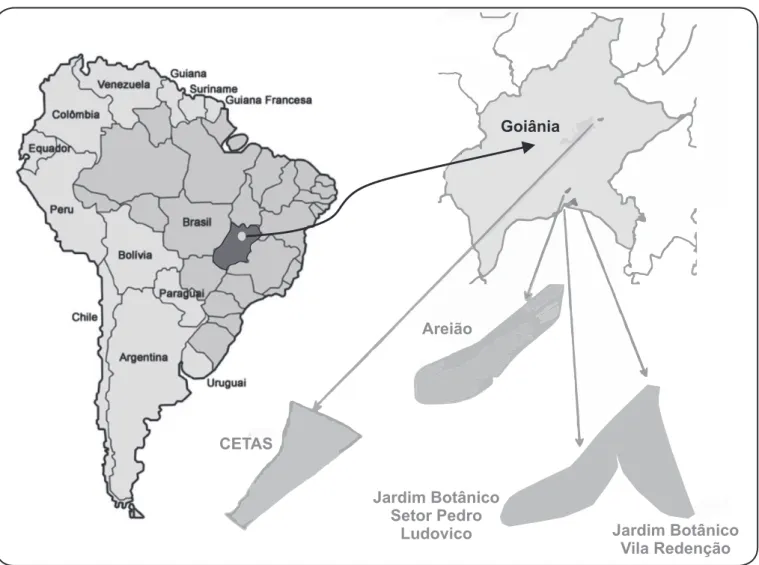
areas of vegetation in the municipality of Goiânia, in the State of Goiás, Central Brazil (Figure 1).
Among the 50 NHPs studied, 27 Cebus libidinosus individuals were living in three urban parks in Goiânia. The other 23 animals were acclimatized in the Wild Animal Screening Center [Centro de Triagem de Animais Silvestres/ Instituto Brasileiro do Meio Ambiente e dos Recursos Naturais Renováveis (CETAS/IBAMA)]. Of these 23 animals, 15 were Cebus libidinosus, ive were Alouatta caraya, and three were Callithrix penicillata. The animal samples from the Goiânia urban parks were collected in 2014, while those of acclimated
animals at CETAS/IBAMA were collected in 2011 and 2013.
Rev Soc Bras Med Trop 49(3):357-360, May-June, 2016 doi: 10.1590/0037-8682-0425-2015
358
Gibrail MM et al .- Oropouche virus antibodies in non-human primates
Goiânia
CETAS
Areião
Jardim Botânico Setor Pedro
Ludovico Jardim Botânico
Vila Redenção
FIGURE 1 - Location of non-human primate capture and blood sampling sites. Shown are Goiânia City, Goiás: Parque Areião, Parque Jardim Botânico Setor Pedro Ludovico, Parque Jardim Botânico Vila Redenção, and CETAS-IBAMA. Adapted from SIGGO (Sistema de Informações Geográicas de Goiânia). CETAS: Centro de Triagem de Animais Silvestres; IBAMA: Instituto Brasileiro do Meio Ambiente e dos Recursos Naturais Renováveis.
The animals were captured by attraction to a trap/cage
tomahawk and they were chemically restrained by intramuscular
administration of 2.0mg/kg tiletamine-zolazepam (Zoletil®).
Physiological parameters were monitored until the end of sedation. The capture of each animal was performed only
once, and conirmed on the basis of a microchip reading. From
each animal, a 3-10ml blood sample was collected by femoral or brachial venipuncture depending on the species(8).
All sera samples were initially screened by an hemagglutination inhibition (HI) assay as described by Clarke and Casals et al.(9) and
adapted to a microplate format by Shope(10). Positive samples by
HI, with a titer equal to or greater than 1:40, were conirmed by
a separate neutralization test (NT) as described by Beaty et al.(11)
Serological tests were performed in the Laboratório de Referência para Arboviroses e Febres Hemorrágicas of the Instituto Evandro Chagas, Ananindeua, Pará.
For the HI assay, serum samples were treated (100% acetone and 0.85% NaCl), hydrated with borate buffer saline (BBS; 1.5 M NaCl, 0.5M H3BO3, 1.0M NaOH, pH 9.0) and adsorbed
with goose (Anser cinereus) erythrocytes. The test was performed
in two steps; in the irst step, each serum sample (1:20) was tested
against 23 viral antigens (4 hemagglutinating units (HU) of each) prepared from intracerebral inoculation (IC) in newborn mice and centered on three different viral families, namely Togaviridae (eastern equine encephalitis, western equine encephalitis, Mayaro, and Mucambo viruses), Bunyaviridae (Caraparu, Catu, Guaroa, Maguari, Oropouche, Tacaiuma, Belem, and Icoaraci viruses) and Flaviviridae (yellow fever, Bussuquara, Cacipacore, Ilheus, Saint Louis encephalitis, Rocio, and dengue 1-4 viruses). After 1-hour incubation of the serum-antigen complex a suspension of goose erythrocytes was added.
359
Rev Soc Bras Med Trop 49(3):357-360, May-June, 2016
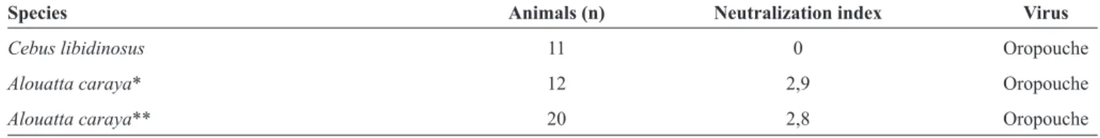
TABLE 1 - Seropositivity of non-human primates as tested by serum neutralizing test for arboviruses observed in Goiânia City, Goiás.
Species Animals (n) Neutralization index Virus
Cebus libidinosus 11 0 Oropouche
Alouatta caraya* 12 2,9 Oropouche
Alouatta caraya** 20 2,8 Oropouche
*Animal with unknown provenance. **Animals from the City of Goiânia.
The NT assay was carried out using newborn mice (1-3 days). For each serum sample, 18 suckling mice were used. Three dilutions of each antigen were used. Serum samples were mixed
with each of the speciic antigens and incubated at 37°C for 60 minutes. Mice were then inoculated with 20µL of virus/antigen
mixture by an IC route. For each reaction, negative and positive control sera were included. The assay readout was obtained by evaluating each animals survival within an inoculated group (6 animals were inoculated per dilution of virus). The antibody titer was considered as the average value of dilutions for each serum sample in which death occurred in more than 50% of inoculated animals [lethal dose (LD50)]. The titer was deined as the logarithmic neutralization index (LNI), using log 10, as described by Reed & Muench(12). The sample was considered
positive when its LNI was equal to or greater than 1.7. Of the total NHPs studied, three animals had antibodies for OROV with a titer greater than 1:40 by the HI test. One of the animals was Cebus libidinosus and two were Alouatta caraya. When submitted to the neutralization test, the two samples of Alouatta caraya were conirmed as positive for OROV (Table 1).
In the period 2007-2008, a yellow fever epizootic circulation in NHPs inhabitants living in urban parks in Goiânia was registered, where the death of 61 animals from the species Alouatta caraya, Cebuslibidinosus and Callithrix penicillata were recorded. In addition, human infections were also diagnosed(8), resulting in an outbreak of the disease.
Arboviruses have geographic distributions covering all continents. However, except for in temperate countries, maintenance cycles are interrupted owing to low temperatures(6). Brazil has
characteristics that favor the spread of these viruses: indeed, a third of the country is covered by tropical forest with is large degree of biodiversity and high density of vectors and vertebrate hosts than can easily co-circulate, enhancing the possibility of arbovirus maintenance and subsequent spread into urban and suburban areas(13).
This is the irst study conducted in the State of Goiás aimed
at investigating the occurrence of arboviruses in a population of NHPs living in urban conservation areas. In the present study, antibodies positive for antigens of OROV were detected in two Alouatta caraya, both of which had been acclimatized at the
CETAS/IBAMA. The other sample, from Cebus libidinosus,
which was irst found to be positive by HI, was negative by
NT. This could have been due to re-assortment, or even cross reaction, with other Orthobunyavirus species(14).
OROV is an arbovirus of great importance to public health owing to the increased morbidity of Oropouche fever
in humans, with clinical presentations ranging from febrile illness to aseptic meningitis(5). It has been also responsible for
explosive epidemics in urban areas of large cities and small
villages of the Amazon region, since its irst outbreak in 1960(4).
The occurrence of this virus in humans has also been seen in Brazil and other South American countries(4), in concert with its
occurrence in NHPs, as observed at Arinos, Minas Gerais State, Brazil (Callithrix genus)(2). In the State of Goiás, to our knowledge,
there have been no reports of the occurrence of Oropouche fever in humans. Therefore, the detection of antibodies for this virus in NHPs is important considering the possibility of dissemination among these animals, and the potential for introduction among humans living in populated areas. The presence in an NHP population may therefore present a risk to the human population.
In fact, it is noteworthy that most of the animals from the CETAS/
IBAMA were captured in urban parks of Goiânia, a city whose people have close contact with parks where animals live. It is not known whether Culicoides paraensis, the urban OROV vector, is found in the State of Goiás, but other Culicoides species have been described in this State that have the potential to become OROV vectors(15). Finally, the conirmation of immunity to OROV in NHPs
living in parks in Goiânia City should be considered as an important warning sign of ongoing virus circulation, and further studies should be conducted to implement continuous monitoring of these sentinel animals as an early warning of potential viral outbreaks.
ETHICAL CONSIDERATIONS
The study was approved by the Ethics Committee for Animal Use [Comissão de Ética no Uso de Animais (CEUA)] of the Universidade Federal de Goiás (Process 32/2014) and the Information System on Biodiversity [Sistema de Informação em Biodiversidade (SISBIO)] (Process 24629).
Acknowledgments
We would like to thank Doctor Pedro Fernando da Costa Vasconcelos for collaboration in this study.
Conlict of interest
The authors declare that there is no conlict of interest.
Financial Support
Pedro Fernando da Costa Vasconcelos is supported by Conselho Nacional de Desenvolvimento Científico e Tecnológico (CNPq) grants (processes
360
REFERENCES
1. International Committee on Taxonomy of Viruses (Internet). Virology division: IUMS; 2013 (Cited 2015 January 28); Available
at: http://ictvonline.org/virusTaxonomy.asp.
2. Nunes MRT, Martins LC, Rodrigues SG, Chiang JO, Azevedo RSS, Travassos da Rosa APA, et al. Oropouche virus isolation, Southeast Brazil. Emerg Infect Dis 2005; 11:1610-1613.
3. Shi X, Kohl A, Vincent HJ, Ping Li L, McLees A, Elliot RM. Requirement of the N-Terminal Region of Orthobunyavirus Nonstructural Protein NSm for Virus Assembly and Morphogenesis. J Virol 2006; 80:8089-8099.
4. Vasconcelos HB, Nunes MRT, Casseb LMN, Carvalho VL, Silva EVP, Silva M, et al. Molecular epidemiology of Oropouche virus, Brazil. Emerg Infect Dis 2011; 17:800-806.
5. Bastos MS, Figueiredo LTM, Naveca FG, Monte RL, Lessa
M, Figueiredo RMP, et al. Identiication of Oropouche Orthobunyavirus in the cerebrospinal luid of three patients in the
Amazonas, Brazil. Am J Trop Med Hyg 2012; 86:732-735.
6. Casseb AR, Casseb LMN, Silva SP, Vasconcelos PFC. Arbovírus:
Importante zoonose na Amazônia brasileira. Vet e Zootec 2013;
20:9-21.
7. Primate Specialist Group. (Internet) IUCN/SSC; 2014 (Cited 2015
July 8); Available at: http://www.primate-sg.org/
8. Ministério da Saúde. Secretaria de Vigilância em Saúde. Boletim da Secretaria de Vigilância em Saúde. Situação da Febre Amarela
Silvestre no Brasil, 2007 e 2008. (Internet). Brasília: MS; 2008
(Cited 2008 March 25); Available at: http://portal.saude.gov.br/saude/.
9. Clarke DH, Casals J. Technique for hemagglutination and hemagglutination inhibition with arthropod-borne viruses. Am J Trop Med Hyg 1958; 7:561-573.
10. Shope RE. The use of a micro-hemagglutination inhibition test to follow antibody response after arthropod-borne virus infection in a community of forest animals. An Microbiol 1963; 11:167-171.
11. Beaty BJ, Calisher CH, Shope RE. Arboviruses. In: Schmidt NJ, Emmons EW, editors. Diagnostic procedures for viral, rickettsial and chlamydial infections. 6th edition. Washington: American
Public Health Association; 1989. p. 797-855.
12. Reed LJ, Muench H. A simple method of estimating ifty per cent endpoints. Am J Epidemiol 1938; 27: 493-497.
13. Figueiredo LTM. Emergent arboviruses in Brazil. Rev Soc Bras Med Trop 2007; 40:224-229.
14. Aguilar PV, Barrett AD, Saeed MF, Watts DM, Russell CG, Ampuero JS, et al. Iquitos Virus: a novel reassortant Orthobynyavirus associated with human illness in Peru. PLos Negl Trop Dis 2011; 5:e1315:1-10.
15. Naves HAM, Carvalho MESV, Costa JA, Oliveira RA. Frequência domiciliar de Culicidae in zona urbana de Goiânia-Goiás-Brasil. Rev Pat Trop 1996; 1:43-49.