INTRODUCTION
T
he surgical techniques to approach liver bleeding in-clude local compression, cauterization, bandages, su-tures, resections and drainage1,2. In complex liver lesions accompanied by hemodynamic instability, laparotomy is indicated for bleeding control with eventual Pringle ma-neuver2-4, ligation of affected vessels and ducts, as de-scribed by Patcher2, and even damage control surgery5.The development of a wide variety of hemo-static agents and tissue adhesives that occurred in re-cent years6 offers surgeons the opportunity to use these products in order to achieve quicker and easier bleeding control. The seriousness and the difficulty in managing certain cases of liver trauma motivate the search for new therapeutic alternatives, especially for bleeding control. The efficiency of the new hemostatic lead to the hypoth-esis to test the efficacy of collagen adhesives associated with fibrinogen and thrombin, compared with the con-ventional suture in the treatment of experimental trau-matic liver injury.
METHODS
This experimental study was conducted in the Surgical Technique Laboratory of the Faculdade de Medicina de Jundiaí, Jundiaí-SP, and was ap-proved by the Ethics Committee for Animal Use with number 81/110.
We included 30 adult, male, Wistar rats, with a mean age of 3.55 months, weighing on average 442,80g (342g-527g). The animals were randomly divided into three groups, A, B and C, ten subjects in each.
All rats received premedication with atropine at a dose of 0.05 mg/kg subcutaneously in the dorsal region and acepromazine (Acepran® 1% – Univet, São Paulo) 1mg/kg by the same route. After 15 minutes of application of premedication, they received an associa-tion of tiletamine and zolazepan (Zoletil® 50 – Virbac, São Paulo) 20mg/kg intramuscularly. We initiated the operative procedure after full action of the anesthetic drugs, monitored by loss of corneal and eyelid reflexes and limbs flexion.
The collagen, fibrinogen and thrombin biological adhesive is
effective in treating experimental liver injuries
O adesivo biológico de colágeno, fibrinogênio e trombina é eficaz no
tratamen-to de lesões hepáticas experimentais
FrederiCo MiChelinode oliveira1; MarCus viníCius h. de Carvalho1; evaldo MarChi1; Clóvis antônio lopes pinto2.
A B S T R A C T
Objective: to evaluate the effectiveness of an collagen-based adhesive associated with fibrinogen and thrombin in experimental liver injuries in rats. Methods: we randomly divided 30 Wistar rats into three groups: A, B and C. All underwent a standard liver traumatic injury. In group A, the lesion was treated with the adhesive; in group B, with conventional, absorbable suture; group C received no treatment. We analyzed the time of hemostasis, mortality, occurrence of adhesions and any histological changes. Results: there was no statistical difference in relation to mortality (p=0.5820). The adhesive treated group showed the lowest hemostasis times (p=0.0573, odds ratio 13.5) and lower incidence of adhesions (p=0.0119). The histological alterations of the Groups A and B were similar, with foreign body granuloma formation separating the adhesive material and the hepatic stroma suture. Conclusion: the collagen adhesive associated with fibrinogen and thrombin was effective in treating experimental hepatic injury, providing a lower incidence of adhesions between the liver and surrounding structures.
Keywords: Wounds and Injuries. Liver. Hemostatics. Thrombin. Tissue Adhesives.
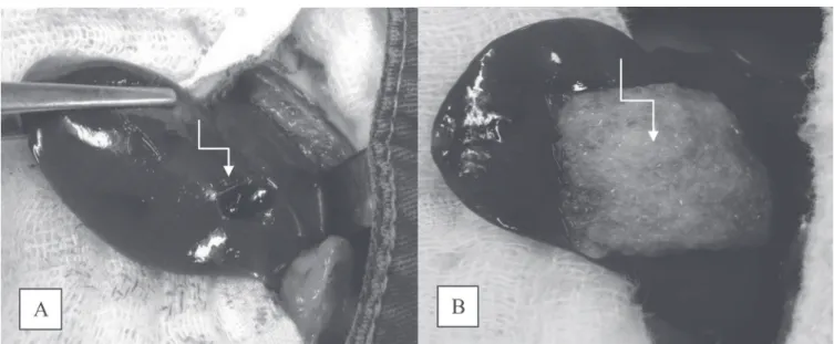
All rats underwent laparotomy under asep-tic technique, started from the xiphoid, approximate-ly 3cm long. After opening the abdominal wall, we positioned a small orthostatic retractor and identified the liver, the organ chosen to perform the standard-ized injury with a biopsy surgical instrument (Punch Keyes® – ABC Surgical Instruments, Brazil) 5mm in diameter, introduced 5mm in depth into the paren-chyma (Figure 1A).
From then on, we treated the animals accord-ing to the group to which they belonged. In Group A, after one minute of bleeding we performed treatment of injury using the surgical collagen adhesive associat-ed to fibrinogen and thrombin (Tachosil® – Nycomassociat-ed, Austria), previously activated in 0.9% saline (Figure 1B), with subsequent cleaning of the cavity and abdominal wall closure. In Group B, one minute after bleeding, we performed treatment of the injury with parenchymal liver suture using 3-0 polyglactin-910 (Vicryl® – Ethicon, USA) and subsequent cleaning of the cavity and the abdominal wall closure. In Group C, control group, we did not carry out any treatment of the hepatic injury, and only closed the abdominal wall.
In the experiments in groups A and B were re-corded the hemostasis times for further analysis. Postop-eratively, all rats received analgesia with dipyrone drops added to water and diet with appropriate chow at will. After eight weeks, the surviving rats were euthanized in a carbon dioxide chamber, with immediate necropsy for
observation of intra-abdominal conditions and removal of the liver for histological analysis.
The study variables were the time to hemosta-sis, the occurrence of deaths, the occurrence of adhe-sions and any histological changes.
The hemostasis time was the time required to control bleeding are noted in the groups A and B. In group C, we did not record the time to hemo-stasis, immediately closing the abdominal wall after the liver injury. In the study design, we opted not to interfere in any way in the hemostasis of the induced injuries of the control group. We feared that, during the bleeding observation to note the time of hemo-stasis, if the bleeding was heavy the researcher might fell motivated to interfere with gauze compression or absorbing the blood with gauze. Attitudes like these would interfere with the results, with a tendency to decrease adhesions.
adhesions, we removed the rats livers and placed them in 10% formalin with subsequent preparation of slides with hematoxylin-eosin and picrosirius for microscopic analysis.
Statistical analysis was performed with the pre-sentation of absolute (n) and relative (%) frequency dis-tribution tables for all variables.
We analyzed the variables death, hemostasis time and the occurrence of adhesions with the Fisher’s exact test. For the qualitative death variable, we made the comparison using the Fisher’s exact test because the conditions of application of the chi-square test were not met. For the variable time of hemostasis, we compared the occurrence of the shorter time, which was two min-utes between the two groups (adhesive and suture), us-ing the Fisher’s exact test, because it is a qualitative vari-able; we also calculated the odds ratio with its respective confidence interval. The significance level for the statisti-cal tests was 5%.
RESULTS
Hemostasis Time
The overall average was 3.5 minutes, ranging between two and ten minutes. In Group A, the average
time was 2.4 minutes, with the shortest time two, and the longest, five. In Group B, the average time was 4.2 minutes, ranging from two to ten minutes.
The distribution of the occurrence of hemosta-sis time of each group is shown in Figure 2.
When we grouped and analyzed the results with time equal to two minutes and longer than two minutes, in groups A and B (Table 1) we obtained a borderline significance between them by the Fisher ex-act test (p=0.0573). The odds ratio was 13.5 (range 1.20 to 15.2), which means that the animals of group B are 13.5 times more likely to have greater hemosta-sis time than two minutes. Therefore, this data shows statistical significance.
Death
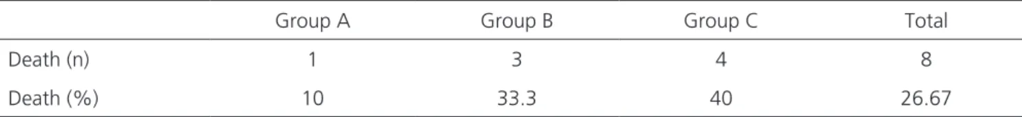
Group A showed mortality of 10% (1/10 an-imals), group B had mortality of 33.3% (3/10 animals) and group C, 40% (4/10 animals). Overall mortality was 26.67% (8/30 animals). Table 2 and Figure 3 show the distribution of the number of deaths in each group.
The Fisher’s exact test did not identify differ-ence with statistical significance when comparing Group A with Group B (p=0.5820), Group A with Group C (p=0.3034) and Group B with Group C (p=1.0000).
Table 1. Distribution of hemostasis time equal to two minutes and greater than two minutes in groups A and B in absolute numbers and percentages (in parentheses)
2 minutes > 2 minutes Total
Group A 6 (60%) 4 (40%) 10 (100%)
Group B 1 (10%) 9 (90%) 10 (100%)
Total 7 (35%) 13 (65%) 20 (100%)
Hemostasis time – animals of Group A versus Group B – Fisher exact test, p = 0.0573, Odds Ratio = 13.5.
Figure 2. Distribution of the lesions repair times between groups A and B. Vertically, the number of rats, and horizontally, the time of hemostasis
and six with Grade I. Group B had two rats with Grade I adhesions, three with Grade II and two with Grade III. The C group had one mouse with Grade I adhesions, four with Grade II and one with Grade III. No rat showed Grade IV adhesions.
Table 3 shows the distribution of the degree of adhesions in each study group.
When analysed the adhesions variable, we found that Group A had a lower incidence than Group B, with statistical significance (p=0.0119 – Fisher’s ex-act test). A similar result occurred when comparing group A with group C (p=0.0069). When comparing Groups B and C, we found no statistically significant difference (p=1.0000).
Histological Changes
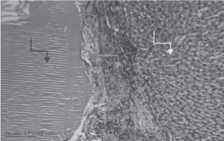
Histological changes found in the slides of the rats’ livers of Group A were reaction to the foreign body with formation of histiocytes palisades, separating amorphous material (adhesive) from stromal liver cells (Figure 4) and plasma cell infiltrate and bilirubin extrav-asation due to ductal injury. We also observed intense collagen deposition (Figure 5), with dense fibrosis. His-tological changes found in the slides of Group B rats’ livers were foreign body, granuloma-type inflammatory reaction around the suture fragments, with giant cells
tion of inflammatory tissue.
DISCUSSION
The induced liver injuries tried to reproduce intermediate lesions that correspond to grade III le-sions when compared to liver trauma classification of the American Association for the Surgery of Trauma (AAST)1,3,8.
For the choice of tissue adhesive, we looked for a product that could take advantage of the prop-erties of bleeding, barrier offered by mechanical hemostatic agents, associated with direct action on blood clotting, offered by active hemostatic agents. Thus, the choice fell on a combination of products al-ready on the market, represented by the combination of collagen associated to fibrinogen and thrombin9-13. This is a totally biological product, without synthetic components. This adhesive was evaluated in clinical studies as support to hemostasis in different kinds of surgery, most often in elective situations, especially on parenchymatous organs, showing effectiveness in controlling bleeding9-13.
Frilling, in 2005, reported the adhesive superior-ity compared with the argon beam during liver resection with respect to homeostasis time12. We obtained similar
Table 3. Distribution of Adhesions in Groups A, B and C.
ADHESIONS
Group A Group B Group C
Grade zero 3 0 0
Grade I 6 2 1
Grade II 0 3 4
Grade III 0 2 1
findings when evaluating the injury repair time with the use of adhesive compared with conventional suturing. The shorter hemostasis time obtained reflects the easy han-dling and effectiveness of the material in controlling bleed-ing, a fact already identified with the use of collagen alone, as demonstrated by Mantovani et al.14, or when combined with fibrinogen and thrombin, as shown by experimen-tal studies using dogs9 and pigs10. It is noteworthy that in some rats treated with injury suture, the extended time to achieve hemostasis was due to the difficulty of manipula-tion of the liver tissue, which was very frail.
Like the collagen, fibrinogen and thrombin adhesive, other hemostatic agents are also cited as ef-fective in the control of various types of bleeding. In 1990, De la Garza and Rumsey showed effectiveness in controlling bleeding with the use of fibrin glue in two patients suffering from liver trauma15. In the same year, Ochsner et al. used this product in 26 patients suffer-ing from liver and splenic injuries, also with effective bleeding control16.
Several experimental studies show the effec-tiveness of fibrin adhesive in controlling hepatic hemor-rhage in dogs17, pigs18,19, rats20 and rabbits21, with good adhesion to the injured liver, little local inflammatory re-action and few complications. In our study we obtained similar findings to those of the cited works.
The occurrence of adhesions, which can be classified as a complication of surgical treatment, was statistically lower in the group treated with the adhesive compared with the group treated with suture (p=0.0119).
This may be due to the animals treated with suture pre-senting major bleeding and bruising at the site of injury, resulting in greater inflammatory reaction and conse-quent adhesion.
Frena and Martin13, in 2006, found the ab-sence of biliary fistulas with the use of this product in elective hepatectomies in humans, which also occurred in our study, even when dealing with liver trauma, which increases the chance of this complication.
The mortality of the group treated with the adhesive (10%) showed no statistically significant dif-ference from the group treated with suture (33.3% / p = 0.5820) and the control group (40% / p=0.3034). In a study of 1,000 patients suffering from liver trauma led by Feliciano et al. between 1979 and 1984, the mortality rate found was 10%22, and in another study, conducted by Saaiq et al. in Islamabad, Pakistan, be-tween 2003 and 2010, mortality was 9.73%23. Thus, mortality with the adhesive experimental use is similar to those found in liver trauma treatments convention-ally performed in humans.
The presence of foreign body inflammatory reaction found in the histological analysis of the rats’ livers treated with the collagen adhesive associated with fibrinogen and thrombin was similar to changes found in studies using fibrin glue in rats24, fibrin glue in rabbits21 and polyglycolic acid mesh in pigs25. We did not observe histological findings suggestive of liver tissue necrosis or vacuolar degeneration, as described with the use of cyanoacrylate26, or the presence of Figure 4. Photomicrograph of histological section stained with
hema-toxylin-eosin, showing a Group A rat liver. The black arrow points to the adhesive amorphous material; the green arrow points to the foreign body, granuloma-type inflammatory process area, with histiocytes distributed in palisade, sepa-rating the adhesive material from the liver stroma.
development of less invasive therapies such as angiogra-phy with embolization28,29, decreases the need for surgery to control liver bleeding. However, in situations of he-modynamic instability or with associated trauma to other organs, particularly in hollow viscus, surgical treatment is often mandatory1,3,8,22,27,28. The liver operative approach can be a complex procedure, requiring great skill and ex-perience of the surgeon2. This study showed that a
col-adhesive associated with fibrinogen and thrombin was effective in experimental hepatic injury, opening new per-spectives for use in liver injuries in humans.
ACKNOWLEDGEMENT
We thank Professor Siani Sirlei Morais for the statistical analysis of this study.
R E S U M O
Objetivo: avaliar a eficácia de um adesivo a base de colágeno associado ao fibrinogênio e trombina, no trauma hepático experimental em ratos. Métodos: toram incluídos no estudo 30 ratos Wistar, igualmente divididos aleatoriamente em três grupos: A, B e C. Todos foram submetidos à lesão traumática hepática padronizada. No grupo A, a lesão foi tratada com o adesivo, no grupo B, com sutura convencional com fio absorvível, e no grupo C, não houve tratamento da lesão. Foram analisados o tempo de hemostasia, mortalidade, ocorrência de aderências e eventuais alterações histológicas. Resultados: os resultados mostraram que não houve diferença estatística em relação à mor-talidade (p=0,5820). O grupo tratado com adesivo apresentou os menores tempos de hemostasia (p=0,0573 e odds ratio 13,5) e menor ocorrência de aderências (p=0,0119). Microscopicamente as alterações histológicas dos grupos A e B foram semelhantes, com a formação de granuloma de corpo estranho separando o material do adesivo e do fio de sutura do estroma hepático. Conclusão: o adesivo de coláge-no associado ao fibricoláge-nogênio e trombina foi eficaz coláge-no tratamento do trauma hepático experimental, proporcionado mecoláge-nor ocorrência de aderências entre o fígado e as estruturas vizinhas.
Descritores: Ferimentos e Lesões. Fígado. Hemostáticos. Trombina. Adesivos Teciduais.
REFERENCES
1. Piper GL, Peitzman AB. Current manage-ment of hepatic trauma. Surg Clin North Am. 2010;90(4):775-85.
2. Feliciano DV, Pachter HL. Hepatic trauma revisit-ed. Curr Probl Surg. 1989;26(7):453-524.
3. Moore EE. Edgar J. Poth Lecture. Critical deci-sions in the manegement of hepatic trauma. Am J Surg. 1984;148(6):712-6.
4. Pringle JH. V. Notes on the arrest of hepat-ic hemorrhage due to trauma. Ann Surg. 1908;48(4):541-9.
5. Weber DG, Bendinelli C, Balogh ZJ. Damage con-trol surgery for abdominal emergencies. Br J Surg. 2014;101(1):e109-18.
6. Achneck HE, Sileshi B, Jamiolkowski RM, Alba-la DM, Shapiro ML, Lawson JH. A comprehen-sive review of topical hemostatic agents: effi-cacy and recommendations for use. Ann Surg. 2010;251(2):217-28.
7. Mazuji MK, Kalambaheti K, Pawar B. Preventive of adhesions with polyvinylpyrrolidone. Prelimi-nary report. Arch Surg. 1964;89:1011-5.
8. Rasslan S, Monteiro RP. Tratamento não-op-eratório do trauma hepático. Rev Col Bras Cir. 1999;26(6):379-87.
10. Grottke O, Braunschweig T, Daheim N, Coburn M, Grieb G, Rossaint R, et al. Effect of TachoSil in a coag-ulopathic pig model with blunt liver injury. J Surg Res. 2011;171(1):234-9.
11. Erdogan D, van Gulik TM. Evolution of fibrin-ogen-coated collagen patch for use as a topical hemostatic agent. J Biomed Mater Res B Appl Bio-mater. 2008;85(1):272-8.
12. Frilling A, Stavrou GA, Mischinger HJ, de Hemp-tinne B, Rokkjaer M, Klempnauer J, et al. Effec-tiveness of a new carrier-bound fibrin sealant ver-sus argon beamer as haemostatic agent during liver resection: a randomised prospective trial. Langenbecks Arch Surg. 2005;390(2):114-20. 13. Frena A, Martin F. How to improve bilio-stasis in
liver surgery. Chir Ital. 2006;58(6):793-5.
14. Mantovani M, Vidal BC, Concon Filho A. Tamponamento das lesões hepáticas trans-fixantes com colágeno tipo I. Acta cir bras. 1998;13(2):80-5.
15. de la Garza JL, Rumsey E Jr. Fibrin glue and he-mostasis in liver trauma: a case report. J Trauma. 1990;30(4):512-3.
16. Ochsner MG, Maniscalco-Theberge ME, Champi-on HR. Fibrin glue as a hemostatic agent in hepatic and splenic trauma. J Trauma. 1990;30(7):884-7. 17. Kram HB, Reuben BI, Fleming AW, Shoemaker
WC. Use of fibrin glue in hepatic trauma. J Trauma. 1988;28(8):1195-201.
18. Feinstein AJ, Varela JE, Cohn SM, Compton RP, McKenney MG. Fibrin glue eliminates the need for packing after complex liver injuries. Yale J Biol Med. 2001;74(5):315-21.
19. Delgado AV, Kheirabadi BS, Fruchterman TM, Scherer M, Cortez D, Wade CE, et al. A novel biologic hemostatic dressing (fibrin patch) re-duces blood loss and resuscitation volume and improves survival in hypothermic, coagulopath-ic swine with grade V liver injury. J Trauma. 2008;64(1):75-80.
20. Jakob H, Campbell CD, Stemberger A, Wried-Lüb-be I, Blümel G, Replogle RL. Combined applica-tion of heterologous collagen and fibrin sealant for liver injuries. J Surg Res. 1984;36(6):571-7. 21. Taha MO, De Rosa K, Fagundes DJ. The role of
biological adhesive and suture material on rabbit hepatic injury. Acta Cir Bras. 2006;21(5):310-4. 22. Feliciano DV, Mattox KL, Jordan GL Jr, Burch JM,
Bitondo CG, Cruse PA. Management of 1000 consecutive cases of hepatic trauma (1979-1984). Ann Surg. 1986;204(4):438-45.
23. Saaiq M, Niaz-ud-Din, Zubain M, Shah SA. Pre-sentation and outcome of surgically managed lives trauma: experience at a tertiary care teach-ing hospital. J Pak Med Assoc. 2013;63(4):436-9. 24. Fontes CER, Taha MO, Fagundes DJ, Ferreira MV,
Prado Filho OR, Mardegan MJ. Estudo compar-ativo do uso de cola de fibrina e cianoacrilato em ferimento de fígado de rato. Acta Cir Bras. 2004;19(1):37-42.
25. Bakker FC, Wille F, Patka P, Haarman HJ. Sur-gical treatment of liver injury with an absorb-able mesh: an experimental study. J Trauma. 1995;38(6):891-4.
26. Silveira LMG, Matera A, Cortopassi SRG, Ferrigno CRA, Xavier JG, Cunha F. Comparação entre os efeitos da associação gelatina-resorcina-formaldeí-do e gelatina-resorcina-formaldeí-do n-butil-2-cianoacrilato na síntese gelatina-resorcina-formaldeí-do parên-quima hepático de coelhos. Braz J Vet Res Anim Sci. 2005;42(4):284-90.
27. Ahmed N, Vernick. Management of liv-er trauma in adults. J Emliv-erg Trauma Shock. 2011;4(1):114-9.
28. Bouras AF, Truant S, Pruvot FR. Manage-ment of blunt hepatic trauma. J Visc Surg. 2010;147(6):e351-8.
29. Misselbeck TS, Teicher EJ, Cipolle MD, Pasquale MD, Shah KT, Dangleben DA, et al. Hepatic angio-embolization in trauma patients: indications and complications. J Trauma. 2009;67(4):769-73.
Received: 10/10/2015
Accepted for publication: 17/03/2016 Conflict of interest: none.
Source of funding: none.
Mailing address: