ISSN 1549-3636
© 2009 Science Publications
Corresponding Author: Jasni Mohamad Zain, Department of Graphics and Multimedia Technology, Faculty of Computer System and Software Engineering, University Malaysia Pahang, Lebuhraya Tun Razak, 26300, Gambang Kuantan, Pahang, Malaysia
Clinical Assessment of Watermarked Medical Images
1
Jasni Mohamad Zain,
2Abdul Rani M. Fauzi and
3Azian A. Aziz
1
Department of Graphics and Multimedia Technology,
Faculty of Computer System and Software Engineering, University Malaysia Pahang,
Lebuhraya Tun Razak, 26300, Gambang Kuantan, Pahang, Malaysia
2
Department of Internal Medicine, International Islamic University Malaysia,
P.O. Box 141, 25710, Kuantan, Pahang Malaysia, Malaysia
3
Department of Radiology, International Islamic University Malaysia,
P.O. Box 141, 25710, Kuantan, Pahang Malaysia
Abstract: Problem statement: Digital watermarking provides security to medical images.
Watermarking in Region Of Interest (ROI) however distorts medical images but it is known that the resulting loss of fidelity is visually imperceptible. Approach: Clinical assessment will objectively evaluate the distortion on medical images to see whether or not medical diagnosis is altered. We used 75 medical images consisting of x-rays, ultrasound and CT scans. Digital watermarking was inserted in ROI and ROI/Region Of Non Interest (RONI) in all of them. Three assessors were randomly assigned 225 images, each receiving 75, a mixture of watermarked and non watermarked images. Results: Chi square test was used and p<0.05 was considered significant. There was no significant difference between original images and those watermarked in ROI or ROI/RONI. There was no comment on image quality in all the images assessed. Conclusion/Recommendations: Digital watermarking does not alter medical diagnosis when assessed by clinical radiologists. The quality of the watermarked images was also unchanged.
Key words: Digital watermarking, clinical assessment, medical images, region of interest, region of non interest
INTRODUCTION
Nowadays it is becoming easier and easier to tamper with digital image in ways that are difficult to detect. The implication of this is colossal especially when it involves medical images used in patient care or the tamper was done to fabricate image based evidence[1].
Image authentication techniques are usually based on two kinds of tools, digital signature and watermarking[2]. A digital signature is non-repudiation, encrypted version of the message digest extracted from the data. It is usually stored as a separate file, which can be attached to the data to prove integrity and originality[3].
Watermarking techniques consider the image as a communication channel. The embedded watermark, usually imperceptible, may contain either a specific producer ID or some content-related codes that are used for authentication. Digital watermarking[1,4] offers a promising alternative to digital signatures in image authentication applications. The use of watermarks
instead of digital signatures typically records additional functionality by exploiting inherent properties of image content.
The main advantage of digital watermarking is that the authentication information is directly embedded into the image data. As a result, the authentication information survives even when the host image undergoes format conversions. The digital watermark’s capability for isolating manipulated image regions is another advantage. This functionality is known as the tamper localization property. It is worth mentioning that both digital signatures and authentication watermarks are useful only for establishing the sources of the image and detecting manipulations occurring after the signature/watermark has been inserted.
applications, where the need for authentication is often paramount, there are typically stringent constraints on data fidelity that prohibit any permanent signal distortion in the watermarking process. For instance, artifacts in a patient’s diagnostic image may cause errors in diagnosis and treatment with possible life-threatening consequences.
Evaluation of the quality of watermarked medical image remains an important issue, by both objective and subjective means[4]. Image quality has two implications: Fidelity and intelligibility. The former describes how the reconstructed image differs from the original one, with Peak-Signal-to-Noise Ratio (PSNR) as a typical example and the latter shows the ability through which the image can offer information to people, with classification-accuracy. Whether an objective measure on image quality is efficient or not, depends strongly on its accordance with subjective measure[5]. Most methods for watermarking data have been evaluated on the basis of minimizing an objective distortion measure such as PSNR at a given amount of watermark[6,7]. However, higher PSNR does not always mean better quality in the watermarked image because PSNR is not necessarily a subjective measure of the quality.
Clinicians are somewhat reserved to new technology even when they were expected to benefit most from them. A survey from a rural area of Scotland involving doctors and other health care professionals revealed a rather low uptake in Information Communication Technology (ICT) citing concern over the impact on patient care as one of the reasons[9]. New medical technology demands meticulous scrutiny primarily to assess efficacy on care and equally crucial the issue of cost and the benefits accrued[10]. A hierarchy of evaluation for new technology has been proposed highlighting the need to scrutinize new medical technology meticulously[11].
Our study should be viewed in this context. We perform this clinical evaluation to subjectively assess watermarked medical images. To the best of our knowledge, this is the first clinical evaluation involving clinicians to study this technique. The process of watermarking in Region of Interest (ROI) and region of Non-Interest (RONI) as suggested by Coatrieux et al.[8] is also discussed.
MATERIALS AND METHODS
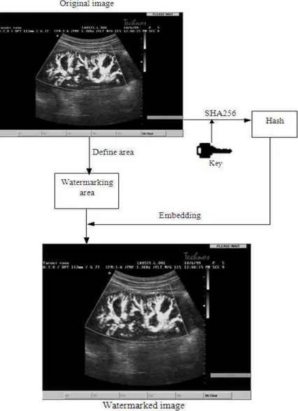
Watermark in Region of Non-Interest (RONI): We
used method in[12] to embed the hash (256 bits) of the original image in the RONI of each image. Figure 1 shows the definition used for ROI and Fig. 2 shows the general method used to embed watermark in the RONI.
Fig. 1: ROI is defined inside the rectangle
Watermark in ROI and RONI: Another watermarking
technique was used to embed watermark in both ROI and RONI. This technique allows us to detect and localize tamper.
We propose a block of size 8×8 for better accuracy of localization, although the scheme will allow user to choose how accurate they want. We need to prepare a one to one block mapping sequence A→B→C→D→...→A for watermarking embedding,
where symbol denotes an individual block. The intensity feature of block A will be embedded in block B and the intensity feature of block B will be embedded in block C. Voyatzis and Pitas[13] presented a two dimensional, discrete Torus automorphism for creating a unique and random mapping of the pixels within an image. We use a 1D transformation based on[13] to get a one-to-one mapping:
B = [(k×B) mod Nb]+1 (1)
Where:
B = B, kє[1,Nb]
k = A secret key (prime number)
N = The total number of blocks in the image
The generation algorithm of the block-mapping sequence is as follows:
• Divide the image into non-overlapping of 8x8 pixels • Assign a unique integer Bє{1,2,3,…,Nb} to each
block from left to right and top to bottom, where Nb = (M/8)×(N/8)
• Randomly pick a prime number kє[1, Nb]
• For each block number B, apply Eq. 1 to obtain B, the number of its mapping block
Fig. 2: RONI embedding method
Table 1: Mapping of blocks with k = 23, 26 and Nb = 40
k B 1 2 3 4 5 6 21 22 23 24 23 B 24 7 30 13 36 19 4 27 10 23 26 B 27 13 39 25 11 37 27 13 39 25
Note that the secret key, k, must be a prime in order to obtain a one to one mapping: otherwise, the period is less than N and a one to many mapping may occur. Table 1 shows some parts of the mapping sequence generated with Nb = 40, k = 23 (prime) and 26
(not prime) respectively. In Table 1, Bstarts to repeat at B = 21 when k = 26, which is not a prime.
Embedding: For each block B of 8×8 pixels, we
further divide it into 4 sub-blocks of 4×4 pixels. The watermark in each sub-block is a 3 tuple (v, p, r), where
both v and p are 1-bit authentication watermark and r is a 7 bit recovery watermark for the corresponding sub-block within sub-block A mapped to B. The following algorithm describes how the 3 tuple watermark of each sub-block is generated and embedded:
• Set the LSB of each pixel within the block to zero and compute the average intensity of the block and each of its four sub-blocks, denoted by avg_B and avg_Bs, respectively
• Generate the authentication watermark, v, of each sub-block as:
s
1ifavg _ B avg _ B, v
0 otherwise
≥
=
Fig. 3: Watermark positioned in the LSB of 3×3 block
Fig. 4: Watermark distribution for the whole image with PSNR = 54.15 dB
• Generate the parity check bit, p, of each sub-block as:
1 if numis odd, p
0 otherwise
=
(3)
where num is the total number of 1s in the seven MSBs of avg_Bs
• From the mapping sequence generated in the preparation step, obtain block A whose recovery information will be stored in block B
• Compute the average intensity of each corresponding sub-block As within A and denote it
avg_As
• Obtain the recovery intensity, r, of As by taking 7
MSB in avg_As. Seven bits is used as we are using
one bit for watermarking
• Embed the 3-tupel watermark (v, p, r), 9 bits in all, onto shown in Fig. 4, where r1 is the MSB, e.g., if the intensity of As is 155, r1, r2, r3, r4, r5, r6 and r7 is 1, 0, 0, 1,1, 0 and 1 respectively
(a)
(b)
(c)
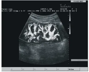
Fig. 5: (a) original image (b) RONI watermarked image (c) RONI and ROI watermarked image
example of an original image, the same image with watermark embedded in RONI and watermark embedded in the whole image (ROI/RONI).
Clinical evaluation: This section will describe our clinical evaluation. Seventy-five images (21 x-rays, 27 CT films and 27 ultrasound images) from our hospital teaching bank were watermarked twice, in Region Of Non-Interest (RONI) and in ROI/RONI, the whole image, making 225 images altogether.
Three assessors whom were all consultant radiologists were randomly given a selection of both Watermarked (WM) and non-watermarked (nWM) images. Each received 75 images, 25 nWM images or the original and 50 WM images. WM images were approximately equal proportion of RONI and ROI/RONI.
They were asked to give a clinical diagnosis to each image aided by a short clinical stem. The stem was constructed with the aid of a clinician to closely resemble the actual clinical information inserted in any imaging request form. Figure 6 is one actual example of the question. The radiologists were asked to answer the questions in the ways they themselves have been accustomed to in practice to simulate the actual reporting in day to day clinical situations. There was no restriction on time. At the end of each question, the radiologist was invited to comment on the quality of the image if he or she deemed justified.
Fig. 6: A sample question used during assessment
The assessors’ responses were then compared with the original diagnoses and an independent radiologist assessed all vague responses. If a clinical diagnosis was not possible, the assessor had to choose a reason or state her or his own reason. We used Chi Square test statistics to test the difference between the three groups and p<0.05 was considered significant.
RESULTS
The three assessors evaluated 225 images. Each received 25 and 50 WM (ROI and ROI/RONI) images. Table 2 and 3 listed the image types and their diagnoses used in the study respectively. Table 4 gave the number of Correct Responses (CR) from each assessor. Chi Square test showed no significant difference between the three groups.
Table 2: Types of Images used in the study
X-ray (n = 21) CT films (n = 27) Ultrasound (n = 27)
Head 0 6 0
Chest 19 16 0 Abdomen 0 4 14 Pelvic 0 1 8 Deep veins 0 0 4 Ankle 1 0 0
Calf 0 0 0
Thigh 0 0 1 Shoulder 1 0 0
Table 3: All the diagnoses of images used in the study
Table 4: Images types (N = 225) given to each Assessor (A) and the Correct Responses (CR)
A1 A2 A3 Total (CR) ROI + RONI 23 27 25 75 (CR) (22) (25) (23) (70) RONI 27 23 25 75 (CR) (26) (23) (24) (73) nWM 25 25 25 75 (CR) (23) (25) (24) (72) Total images 75 75 75 225 (Total CR) (70) (72) (71)
There were five wrong responses from assessors1, 3 from assessor 2 and 4 from the last assessor. These were all judged incorrect responses when assessed by the independent radiologist. In addition, image quality was not implicated in all wrong responses. Overall, there was also no comment on image quality from all the assessors. Chi Square test on CR from the three groups (nWM, RONI and ROI) was not statistically significant (p = 0.5).
DISCUSSION
Digital watermarking is an exciting new technology. Watermarking capabilities to detect and localize tamper make it a powerful tool in security[2]. Healthcare givers however are inherently slow at adapting to new technology. The survey from Scotland provided some insight into this with the concern on the impact of a new technology on patient care being one of the causing factors[9]. Although this study was only looking at the usage of ICT among health staffs in general practitioners’ clinics but the customary scrutiny imposed on new technology point to a similar prevalent general view[10].
The arrival of a new technology to the medical field has to go through very rigorous scrutiny not forgetting the obvious concern from health managers of the cost implications. A new technology must also be relevant at the point of the delivery of care that is measured in terms of the benefits that they accrue to patients. Each new technology must be able to answer successively relevant questions, each designed to safeguard what essentially is the question of patient’s benefits and quality care[11]. The technology of digital watermarking on medical images must therefore be scrutinized in similar manner.
To the best of our knowledge there has never been a similar study done before. We had employed practicing radiologists simulated under normal clinical situations to assess watermarked images in both RONI and ROI/RONI. The questions were framed by a clinician to resemble the actual day to day clinical scenarios to enable the assessors to judge under normal
conditions with normal restrictions placed on issuing imaging reports. We have also added a subjective section to enable the assessor to express their thoughts on the quality of the images. As pointed earlier, the result was reassuring.
CONCLUSION
From this study, we concluded that watermarking medical images did not alter clinical diagnoses. It was also evident that the area where watermarking was embedded was immaterial as both sites; ROI and RONI gave similar results when they were clinically assessed. We noted the previous suggestion by Coatrieux et al.[8] to preserve ROI to safeguard diagnostic zone but our study has shown that ROI could also be an area for watermarking.
In addition, there was no difference in image quality when visually assessed by medical radiologists. We are therefore confident that digital watermarking in safe in terms of preserving image quality. Both physicians and radiologist should be reassured that this technique of digital watermarking ensures image quality preservation and therefore clinical diagnoses can be made with high reliability.
REFERENCES
1. Cox, I., M. Miller and J. Bloom 2001. Digital Watermarking. 1st Edn., Morgan Kaufman, San Francisco, CA., ISBN:1558607145, pp: 576. 2. Coatrieux, G., H. Maitre, B. Sankur, Y. Rolland
and R. Collerec, 2000. Relevance of watermarking in medical imaging. Proceeding of the IEEE EMBS Conference on Information Technology Applications in Biomedicine, Nov. 9-10, IEEE Xplore Press, Arlington, VA., USA., pp: 250-255. DOI:10.1109/ITAB.2000.892396
3. Cho, Y., B. Ahn, J.S. Kim, I.Y. Kim and S.I. Kim, 2001. A study for watermark methods appropriate to medical images. J. Digit. Imag., 14: 184-186. http://www.ncbi.nlm.nih.gov/pubmed/11442090 4. Langelaar, G.C., I. Setyawan and R.L. Lagendijk,
2000. Watermarking digital image and video data. IEEE Sign. Process. Mag., 17: 20-46. DOI: 10.1109/79.879337
5. Eskicioglu, A.M. and P.S. Fisher, 1995. Image quality measures and their performance. IEEE Trans. Commun., 43: 2959-2965. DOI: 10.1109/26.477498
7. Karunasekera, S.A. and N.G. Kingsbury, 1995. A distortion measure for blocking artifacts in images based on human visual sensitivity. IEEE Trans. Image Process., 4: 713-724. DOI: 10.1109/5.286196
8. Coatrieux, G., H. Maitre and B. Sankur, 2001. Strict intergrity control of biomedical images. Proceeding of the SPIE Conference on Security and Watermarking of Multimedia Contents III, Jan. 22-25, San Jose CA., pp: 229-240. http://cat.inist.fr/?aModele=afficheN&cpsidt=1405 0744
9. Richards, H., G. King, M. Reid, S. Selvaraj and I. McNicol et al., 2005. Remote working: Survey of attitudes to eHealth of doctors and nurses in rural general practices in the United Kingdom.
Fam. Pract., 22: 2-7.
http://www.ncbi.nlm.nih.gov/pubmed/15642724 10. Hillman, B.J., 1994. New imaging technology and
cost containment. Am. J. Roentgenol., 162: 718-740. http://cat.inist.fr/?aModele=afficheN&cpsidt=3952 827
11. Fryback, D.G. and J.R. Thornbury, 1991. Efficacy of diagnostic imaging. Med. Dec. Mak., 11: 88-94. DOI: 10.1177/0272989X9101100203
12. Zain, J.M., L.P. Baldwin and M. Clarke, 2004. Reversible watermarking for authentication of DICOM images. Proceeding of the IEEE 26th Annual International Conference on Engineering in Medicine and Biology Society, Sept. 1-5, IEEE Xplore Press, USA., pp: 3237-3240. http://ieeexplore.ieee.org/xpl/freeabs_all.jsp?arnum ber=1403911