André Luiz da Costa MICHELOTTO(a)
Cacio MOURA-NETTO(b)
Angela Toshie ARAKI(b)
Eduardo AKISUE(c)
Gilson Blitzkow SYDNEY(d)
(a)Universidade Tuiuti do Paraná – UTP, School
of Dentistry, Curitiba, PR, Brazil.
(b)Universidade Cruzeiro do Sul, School of
Dentistry, Graduate Program in Dentistry, São Paulo, SP, Brazil.
(c)Universidade Santa Cecilia – Unisanta,
School of Dentistry, Santos, SP, Brazil.
(d)Universidade Federal do Paraná, School of
Dentistry, Curitiba, PR, Brazil.
Penetration of a resin-based filling
material into lateral root canals and
quality of obturation by different
techniques
Abstract: The aim of this study was to evaluate the penetration of a resin/polyester polymer-based material (Resilon Real Seal; SybronEndo Corp., Orange, USA) into simulated lateral canals, and the quality of obturations by different techniques. A total of 30 standardized simulated canals were divided into three groups according to the technique of obturation used: MS (McSpadden), SB (SystemB/Obtura II),
and LC (Lateral Condensation). To analyze the penetration of the illing
material, the simulated canals were digitalized and the images were analyzed using the Leica QWIN Pro v2.3 software. The data of the middle and apical thirds were separately submitted to analysis of variance (ANOVA), followed by the Tukey’s test for the comparison
of the techniques. Results showed a signiicant difference (p < 0.05)
between groups (LC < SB) in the middle third, and a signiicant
difference (p < 0.05) between groups (LC < SB and MS < SB) in the apical third. To analyze the quality of the obturations, the canals were radiographed and evaluated by three examiners. The Kappa test on interexaminer agreement and the nonparametric Kruskal-Wallis test
indicated no signiicant difference between illing techniques. It was
concluded that Resilon achieves greater levels of penetration when associated with thermoplastic obturation techniques.
Keywords: Root Canal Filling Materials; Root Canal Obturation; Endodontics.
Introduction
The goal of endodontic therapy is to obtain a root canal illing that
allows tridimensional sealing of the root canal system, using a nonirri-tant material to support periapical healing.1 The root canal system has a
very complex morphology with many irregularities including ins, deltas,
and accessory and lateral canals. Lateral canals are reported in 27.4-45% of all teeth,2,3 and majority are located in the apical third of the roots.3,4
Reports have shown developing or persistent periapical pathology around
untreated and/or unilled lateral canals.5,6 Thus, illing these areas appears
to be very important to improve the success rate of endodontic therapy. Obturation techniques involving gutta-percha plasticization have
shown the greatest eficiency in illing root canal systems.7,8 An
alterna-Declaration of Interests: The authors certify that they have no commercial or associative interest that represents a conflict of interest in connection with the manuscript.
Corresponding Author:
Cacio Moura-Netto E-mail: [email protected]
DOI: 10.1590/1807-3107BOR-2015.vol29.0010
Submitted: May 09, 2014
tive is the Resilon cone (Resilon Research, LLC, North Branford, USA), which is a resin/polyester polymer-based obturation material associated with a hydro-philic resin-based sealer (Real Seal, SybronEndo Corp., Orange, USA). This obturation system was introduced with the concept of monoblock formation between the cone and sealer. The hydrophilic prop-erty of this methacrylate resin-based sealer allows deeper penetration into dentinal tubules, which may increase its sealing ability.9
According to the manufacturer, the polymers in Resilon (Resilon Research, LLC) provide the mate-rial with thermoplastic properties allowing it to be applied using thermoplastic techniques,10,11,12 because
its melting point is the same as gutta-percha (60°C).13
This could be an advantage because several studies
showed superior sealing ability and illing material
adaptation with a thermoplastic technique.9,14
Thus, the aim of this study was to evaluate the penetration of Resilon (Real Seal, SybronEndo Corp., Orange, USA) into simulated lateral canals, and the
quality of the illings obtained using different tech
-niques: Lateral Condensation (LC), System B/Obtura II (SB), and McSpadden (MS).
Methodology
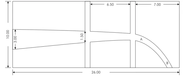
This study used 30 simulated curved canals (angle: approximately 30°; length: 24 mm), prepared at the Prosthesis Laboratory, Dental School, Universidade
Federal do Paraná (UFPR, Curitiba, Brazil) ready for obturation. Each sample contained four lateral canals (diameter: 0.3 mm; length: 7.0 mm), two located in the middle and two in the apical third (Figure 1). The specimens were divided into three groups
accord-ing to the illaccord-ing technique: MS, SB, and LC Groups.
Filling protocol
The filling material used was the Real Seal system (SybronEndo Corp., Orange, USA). For all groups, a 0.04-taper Resilon cone was fitted to the working length (1 mm prior to canal exit). After primer application, the sealer was inserted into the canal using the Resilon cone, until the sealer covered the entire extension of the cone. These procedures were performed according to the manufacturer’s instructions.
Group MS (McSpadden)
A thermo compactor (SybronEndo Corp.) with a larger tip (#60), attached to a contra-angle handpiece
and a low-speed motor, was inserted into the illed
canal. The compactor was activated at 15,000 rpm and inserted into the canal with smooth in and out movements (amplitude: 3 mm). The compactor reached 20 mm, at the beginning point of curvature. At this point, the compactor was gently removed from the canal using the same in and out movement.Once thermo-compaction was performed, a cold plugger
26.00
1
0
.0
0
1
.5
0
6.50 7.00
A
B
3
.0
0
was used for vertical condensation of the illing mate -rial, to achieve better adaptation to the canal walls.
Group SB (System B/Obtura II)
The System B device (SybronEndo Corp.) using Resilon pellets (Real Seal, SybronEndo Corp.) was
calibrated to 150°C. After the root canal was illed
with the sealer and the solid material (as described above), the #2 condenser was activated and introduced until the same depth established for the MS group (20.0 mm). At this point, heating was stopped and the condenser remained under pressure for 10 seconds. Furthermore, heating was rapidly activated and the condenser was removed from the interior of the canal. Condensation of the remaining thermoplas-ticized material was performed in the apical third with a cold plugger. Filling of the middle and cervi-cal thirds was completed with successive stages of injection of thermoplasticized Resilon using Obtura II (Obtura Corp., Fenton, USA) and cold vertical con-densation using cold pluggers.
Group LC (Lateral Condensation)
The master Resilon cone was itted into the root
canal with the sealer. Furthermore, spaces were
cre-ated using a C inger spreader (Dentsply Maillefer,
Tulsa, USA) to introduce MF accessory cones until
the canal is illed. Obturation was performed with
a heated plugger, the excess of gutta-percha was removed, and a cold plugger was used for vertical
condensation of the illing material.
Evaluation of penetration of the filling material
The penetration of the illing material into lateral
canals was evaluated using images of the simulated canals taken with a stereomicroscope (Leica Micro- systems, Bannockburn, USA), and analyzed using the Leica QWIN Pro software (Leica Microsystems). The parameters were the total length of the lateral
root canals and the extent of penetration of the ill
-ing material. The data were submitted to analysis of variance (ANOVA) and Tukey’s test (p < 0.05). Statis-tical analysis was performed with SPSS 13.0 software (SPSS Inc., Chicago, USA).
Radiographic Quality of Obturation
The quality of root canal filling was scored on the following modified three-point scale: (1) a good score for homogenous filling obturating the entire prepared root canal, well adapted to the canal wall, with only a few minor areas of
rela-tive radiolucency (diameter: ≤ 0.25 mm; without
porosity); (2) a satisfactory score for imperfect
fill-ing with irregularities of ≤ 1 mm, where the fillfill-ing may be slightly shorter (diameter: ≤ 0.5 mm) than
the working length (few porosities); and, (3) a bad score for inadequately filled canals with irregu-larities (diameter: > 1 mm), where the filling diam-eter may be > 0.5 mm shorter than the working length (several porosities). Three trained exam-iners performed the evaluations simultaneously. Examiner reliability was assessed by reevaluation of all specimens on a separate occasion. In case of disagreement, the ultimate decision was reached by consensus. The collected data were submitted for statistical analysis using the Kappa correla-tion test for interexaminer comparison, and the Kruskal-Wallis nonparametric test for score anal-ysis (p < 0.05). Statistical analysis was performed with SPSS 13.0 software (SPSS Inc., Chicago, USA).
Results
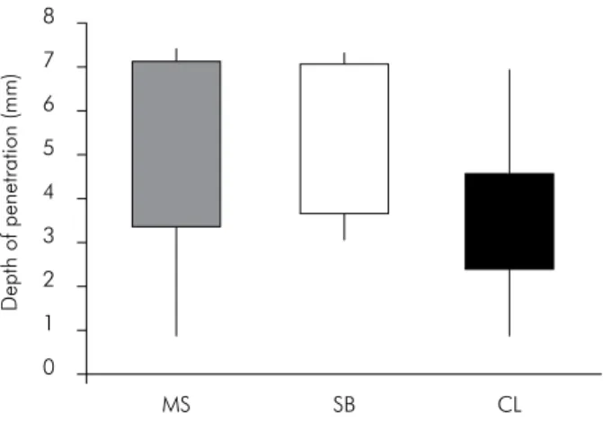
The obturation technique had a signiicant impact
on the illing quality of lateral root canals. In the mid
-dle third, illing penetration into lateral canals was shallower in the LC Group than in the SB and MS
Groups (Figure 2; p < 0.05). On the other hand, there
was no significant difference between the SB and MS groups. In the apical third, greater penetration
was obtained for the SB Group than the MS and LC
groups (Figure 3; p < 0.05). But there was no signiicant
difference between the MS and LC groups (p > 0.05).
The quality of the illings was analyzed by three
experienced endodontists using radiographs of the simulated root canals. Interexaminer agreement analysis produced a kappa value of 0.75 (75%), indi-cating a high correlation between the three exam-iners. Accordingly, a nonparametric Kruskal-Wallis
test was performed, and indicated no signiicant dif
Discussion
The purpose of this study was to compare the efficiency of three obturation techniques for the penetration of the Resilon Real Seal (SybronEndo
Corp.) illing material into lateral canals. Standard
-ized experiments were conducted with simulated root canals to avoid the variables between natural teeth.7,8,15,16 These root canals had lateral canals in the
middle and apical thirds, visible in the buccolingual
direction, to evaluate the penetration depth of illing
material by radiographic analysis.
The thermoplastic properties of Resilon are attrib-uted to the addition of polycaprolactone, which has a melting point of 60°C,12 and is indicated for
ther-moplastic obturation techniques.8,14,17 Thermoplastic
Figure 2. Impact of the obturation technique on filling material penetration (mm) into lateral canals of the middle third region.
D
e
pth
o
f
pe
ne
tratio
n
(mm)
8
7
6
5
4
3
2
1
0
MS SB CL
Figure 3. Impact of the obturation technique on filling material penetration (mm) into lateral canals of the apical third region.
D
e
pth
o
f
pe
ne
tratio
n
(mm)
8
7
6
5
4
3
2
1
0
MS SB CL
Resilon was found superior to conventional gutta-percha for plasticization.17,18
The present study demonstrates that the relative
eficiency of the obturation techniques depends on the
position of the lateral root canals. In the middle third,
the illing material penetrated deeper by thermoplas
-tic obturation (SB or MS) than by LC. In the apical third, superior obturation was obtained by SB than
by MS or LC. In fact, plasticization of illing material by MS was more eficient in the middle third than in
the apical third of the root. This discrepancy is con-sistent with the fact that the thermo compactor was inserted up to the beginning of the curvature. After
this point, the illing material had a poorer plastici
-zation; therefore, penetration into the apical lateral
canals was lower. This inding suggests that the supe
-rior eficacy of MS is limited to the straight portion of the root canal. Beyond the compactor tip, the illing
material is poorly plasticized, as previously reported.7
The thermo compactor of MS only remained activated
long enough to initiate illing material plasticization. A longer activation time could improve the lowability of illing material but could also increase the porosity
of the obturation. 7 Thus, SB is more effective for the
plasticization of illing material in the apical third.
Karr et al.19 and Tanomaru-Filho et al.20 reported
similar low for Resilon and gutta-percha in lateral
grooves and depressions, when obturation was per-formed using the warm vertical compaction (System B) technique. Karabucak et al.14 evaluated the ability of
Obtura II and Calamus to ill artiicially created lat
-eral canals using standard gutta-percha and
Resi-lon. Their results indicated that the low of the illing
material into the lateral canals depends on the
visco-elastic properties of the illing material rather than
the mechanical properties of the delivery systems.
They suggest that Resilon illing material lows better into lateral canals using a single backill technique.
The quality of the illings obtained with the three
obturation techniques was analyzed by three expe-rienced endodontists using radiographs of the
simu-lated root canals. The illings were scored as good
1. Schilder H. Filling root canals in three dimensions. Dent Clin North Am. 1967 Nov:723-44.
2. Rubach WC, Mitchell DF. Periodontal disease, accessory ca-nals and pulppathosis. J Periodontol. 1965 Jan-Feb;36:34-8. 3. De Deus QD. Frequency, location, and direction of the lateral,
secondary, and accessory canals. J Endod. 1975 Nov;1(11):361-6. 4. Venturi M, Di Lenarda R, Prati C, Breschi L. An in vitro
model to investigate filling of lateral canals. J Endod. 2005 Dec;31(12):877-81.
5. Weine FS. The enigma of the lateral canal. Dent Clin North Am. 1984 Oct;28(4):833-52.
6. Iqbal MK, Gartenberg J, Kratchman SI, Karabucak B, Bui B. The clinical significance and management of apical accessory canals in maxillary central incisors. J Am Dent Assoc. 2005 Mar;136(3):331-5; quiz 379-81.
7. Michelotto AL, Moura-Netto C, Araki AT, Akisue E, Moura AA, Sydney GB. In vitro analysis of thermocompaction time and gutta-percha type on quality of main canal and lateral canals filling. Braz Oral Res. 2010 Jul-Sep;24(3):290-5. 8. Tanomaru-Filho M, Sant’Anna Junior A, Berbert FL, Bosso R,
Guerreiro-Tanomaru JM. Ability of gutta-percha and Resilon to fill simulated lateral canals by using the Obtura II system. J Endod. 2012 May;38(5):676-9.
9. Chandra SS, Shankar P, Indira R. Depth of penetration of four resin sealers into radicular dentinal tubules: a confocal microscopic study. J Endod. 2012 Oct;38(10):1412-6.
10. Shipper G, Ørstavik D, Teixeira FB, Trope M. An evaluation of microbial leakage in roots filled with a thermoplastic syn-thetic polymer-based root canal filling material (Resilon). J Endod. 2004 May;30(5):342-7.
11. Shipper G, Teixeira FB, Arnold RR, Trope M. Periapical in-flammation after coronal microbial inoculation of dog roots filled with gutta-percha or resilon. J Endod. 2005 Feb;31(2):91-6. 12. Tay FR, Pashley DH, Williams MC, Raina R, Loushine RJ,
Weller RN, et al. Susceptibility of a polycaprolactone-based root canal filling material to degradation. I. Alkaline hydro-lysis. J Endod. 2005 Aug;31(8):593-8.
13. Miner MR, Berzins DW, Bahcall JK. A comparison of thermal properties between gutta-percha and a synthetic polymer based root canal filling material (Resilon). J Endod. 2006 Jul;32(7):683-6. 14. Karabucak B, Kim A, Chen V, Iqbal MK. The comparison of gut-ta-percha and Resilon penetration into lateral canals with differ-ent thermoplastic delivery systems. J Endod. 2008 Jul;34(7):847-9. 15. Reader CM, Himel VT, Germain LP, Hoen MM. Effect of three
obturation techniques on the filling of lateral canals and the main canal. J Endod. 1993 Aug;19(8):404-8.
16. DuLac KA, Nielsen CJ, Tomazic TJ, Ferrillo PJ Jr, Hatton JF. Comparison of the obturation of lateral canals by six tech-niques. J Endod. 1999 May;25(5):376-80.
17. Tanomaru-Filho M, Silveira GF, Tanomaru JM, Bier CA. Evaluation of the thermoplasticity of different gutta-percha cones and Resilon. Aust Endod J. 2007 Apr;33(1):23-6. 18. Almeida JF, Gomes BP, Ferraz CC, Souza-Filho FJ, Zaia AA.
Filling of artificial lateral canals and microleakage and flow of five endodontic sealers. Int Endod J. 2007 Sep;40(9):692-9. 19. Karr NA, Baumgartner JC, Marshall JG. A comparison of
gutta-percha and Resilon in the obturation of lateral grooves and depressions. J Endod. 2007 Jun;33(6):749-52.
20. Tanomaru-Filho M, Sant’anna-Junior A, Bosso R, Guerreiro-Tanomaru JM. Effectiveness of gutta-percha and Resilon in filling lateral root canals using the Obtura II system. Braz Oral Res. 2011 May-Jun;25(3):205-9.
21. Epley SR, Fleischman J, Hartwell G, Cicalese C. Complete-ness of root canal obturations: Epiphany techniques versus gutta-percha techniques. J Endod. 2006 Jun;32(6):541-4. 22. Herbert J, Bruder M, Braunsteiner J, Altenburger MJ, Wrbas
KT. Apical quality and adaptation of Resilon, EndoREZ, and Guttaflow root canal fillings in combination with a noncom-paction technique. J Endod. 2009 Feb;35(2):261-4.
23. Oddoni PG, Mello I, Coil JM, Antoniazzi JH. Coronal and apical leakage analysis of two different root canal obturation systems. Braz Oral Res. 2008 Jul-Sep;22(3):211-5.
24. Hammad M, Qualtrough A, Silikas N. Evaluation of root canal obturation: a three-dimensional in vitro study. J Endod. 2009 Apr;35(4):541-4.
References
In addition, similar results were found by others.21,22
There are also studies showing satisfactory clinical results9,11 and good in vitro sealing capacity.23 However,
Hammad et al.24 analyzed illings for the presence of
gaps and voids in canals obturated with gutta-percha,
Resilon, Endorez, or Guta-low by lateral condensation.
The gutta-percha presented the best results, while the canals obturated with Resilon presented greater
indi-ces of voids and gaps. Thus, the use of the Resilon Real Seal system for lateral root canals should be limited to thermoplastic obturation techniques.