J Bras Pneumol. 2013;39(4):523-524 J Bras Pneumol. 2013;39(4):523-524
Pulmonary histoplasmosis presenting with a halo sign on CT
in an immunocompetent patient
Histoplasmose pulmonar presente com sinal do halo em TC em um paciente imunocompetente
Graziella Hanna Pereira, Liliane Yuri Almeida, Rosa Shizuda Okubo, Edson Marchiori
To the Editor:
Histoplasmosis is a common endemic mycosis. Although most infections in immunocompetent individuals are self-limiting, some patients develop severe pneumonitis or other types of chronic pulmonary infection. Specific imaging findings depend on the organisms involved, the underlying condition of the patient, and his condition after immune depleting procedures. The disease process in immunocompetent patients is usually limited to a solitary nodule on chest X-rays or CT scans, but enlarged hilar or mediastinal lymph nodes can also be observed.(1) We report here the case of an
immunocompetent patient with histoplasmosis who presented with a halo sign on CT.
A 47-year-old man presented with dry cough, chest pain, fatigue, and fever, having started 15 days prior. He reported neither
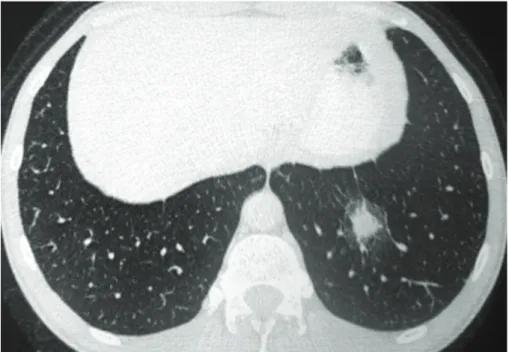
comorbidities nor a contact with environments with mold. The patient was negative for HIV by ELISA. Laboratory tests revealed leukocytosis, elevated C-reactive protein levels (33 mg/L), and hypertransaminasemia (three-fold higher than the upper limit of normal). CT scanning revealed a pulmonary nodule with a halo sign in his left lower lobe (Figure 1) and left para-hilar lymph nodes. Histological examination of a pulmonary biopsy sample obtained during bronchoscopy showed nonspecific inflammatory alterations. Cultures for mycobacteria and fungi were negative. Serology by counterimmunoelectrophoresis was positive for histoplasmosis.
Treatment was initiated with itraconazole (400 mg/day). After 30 days, a CT scan showed that the size of the nodule was reduced by half.
Figure 1 - A CT scan at the level of the lower lobes showing a nodule surrounded by ground-glass attenuation (halo sign) in the left lower lobe.
524 Pereira GH, Almeida LY, Okubo RS, Marchiori E.
J Bras Pneumol. 2013;39(4):523-524 J Bras Pneumol. 2013;39(4):523-524
524
The patient showed progressive improvement of clinical symptoms and a reduction of the para-aortic adenopathy. Treatment was maintained for one year, during which the patient remained asymptomatic, the nodule completely disappeared, and the lymphadenopathy resolved. However, histoplasmosis by counterimmunoelectrophoresis remained positive.
We reported the case of an immunocompetent patient who presented with a halo sign and lymphadenopathy caused by histoplasmosis, showing a good clinical response after one year of treatment with itraconazole (400 mg/day). Histoplasma capsulatum is a major endemic human fungal pathogen that can cause a broad variety of clinical presentations, ranging from mild, focal pulmonary disease to life-threatening systemic infections.(1) In one study, open lung biopsies of
155 patients with undiagnosed lung lesions showed that infectious etiologies were responsible for 19% of the lesions, the most common diagnosis in this group being histoplasmosis (41%).(2)
The halo sign is characterized by a ground-glass opacity surrounding a nodule, mass, or round area of consolidation.(3) This sign is frequently an
early indicator of invasive pulmonary aspergillosis in immunocompromised patients,(4) but it might
also indicate other invasive fungal diseases. Our patient required treatment with high doses of itraconazole for one year. Azoles, such as itraconazole and voriconazole, and amphotericin B have significant activity against H. capsulatum. However, approximately 10% of the patients hospitalized due to histoplasmosis die,(5) despite the lack of antifungal resistance
among recovered isolates.(6)
In conclusion, a halo sign on CT might indicate active histoplasmosis, which may require long-term treatment, even in immunocompetent patients.
Graziella Hanna Pereira
Infectious Disease Specialist,
Hospital
Nipo-Brasileiro de São Paulo
, São Paulo,Brazil
Liliane Yuri Almeida
Pulmonologist,
Hospital Nipo-Brasileiro
de São Paulo
, São Paulo, BrazilRosa Shizuda Okubo
Radiologist,
Hospital Nipo-Brasileiro de
São Paulo
, São Paulo, BrazilEdson Marchiori
Radiologist, Federal University of Rio de Janeiro, Rio de Janeiro, Brazil
References
1. Chong S, Lee KS, Yi CA, Chung MJ, Kim TS, Han J. Pulmonary fungal infection: imaging findings in immunocompetent and immunocompromised patients. Eur J Radiol. 2006;59(3):371-83. http://dx.doi.org/10.1016/j. ejrad.2006.04.017 PMid:16725293
2. Georgiadou SP, Sampsonas FL, Rice D, Granger JM, Swisher S, Kontoyiannis DP. Open-lung biopsy in patients with undiagnosed lung lesions referred at a tertiary cancer center is safe and reveals noncancerous, noninfectious entities as the most common diagnoses. Eur J Clin Microbiol Infect Dis. 2013;32(1):101-5. http://dx.doi. org/10.1007/s10096-012-1720-9 PMid:22895891 3. Silva CI, Marchiori E, Souza Júnior AS, Müller NL; Comissão
de Imagem da Sociedade Brasileira de Pneumologia e Tisiologia. Illustrated Brazilian consensus of terms and fundamental patterns in chest CT scans. J Bras Pneumol. 2010;36(1):99-123. http://dx.doi.org/10.1590/S1806-37132010000100016 PMid:20209314
4. Greene RE, Schlamm HT, Oestmann JW, Stark P, Durand C, Lortholary O, et al. Imaging findings in acute invasive pulmonary aspergillosis: clinical significance of the halo sign. Clin Infect Dis. 2007;44(3):373-9. http://dx.doi. org/10.1086/509917 PMid:17205443
5. Nosanchuk JD, Zancopé-Oliveira RM, Hamilton AJ, Guimarães AJ. Antibody therapy for histoplasmosis. Front Microbiol. 2012;3:21. http://dx.doi.org/10.3389/ fmicb.2012.00021 PMid:22347215 PMCid:3270318 6. Brilhante RS, Fechine MA, Mesquita JR, Cordeiro RA, Rocha
MF, Monteiro AJ, et al. Histoplasmosis in HIV-positive patients in Ceará, Brazil: clinical-laboratory aspects and in vitro antifungal susceptibility of Histoplasma capsulatum isolates. Trans R Soc Trop Med Hyg. 2012;106(8):484-8. http://dx.doi.org/10.1016/j. trstmh.2012.05.003 PMid:22703696