Introduction
Paracoccidioidomycosis is a systemic fungal infection whose clinical manifestations are predominantly pulmonary and whose etiologic agent is the dimorphic fungus Paracoccidioides brasiliensis. Its geographic distribution is limited to Latin America, from Mexico to Argentina. Although it has sporadically been isolated from nature, its natural reservoir is unknown.(1) A small proportion of individuals develop clinical manifestations of disease. The incidence of paracoccidioidomycosis in Uruguay is unknown. Studies and personal communications have reported that 1 or 2 cases are diagnosed
per year, and it is likely that the condition is underdiagnosed. It usually affects males and rural workers, especially farm workers and woodcutters.(2) Smoking has been reported as a risk factor for the development of the disease.(3) In culture media at 37°C, P. brasiliensis appears as spherical yeast surrounded by a refractive, double-contoured wall, formation of intracytoplasmic lipid inclusions being typically observed. It reproduces by multiple budding, in which blastoconidia are connected to a mother cell in an arrangement resembling a “ship’s steering wheel”, which gives it a particular morphological characteristic. At
Concomitant pulmonary paracoccidioidomycosis
and pulmonary histoplasmosis: a rare case*
Un caso excepcional de paracoccidioidomicosis e histoplasmosis pulmonares de presentación concomitante
Veronica Torres Esteche, Zaida Arteta, Gabriela Torres, Andrea Vaucher, Elbio Gezuele, Raquel Balleste
Abstract
The incidence of pulmonary fungal infections is very low in Uruguay, and such infections typically affect immunocompromised patients. We report the case of an immunocompetent patient presenting with a two-month history of cough, dyspnea, and fever. The patient resided in a rural area. Imaging tests revealed extensive pneumonitis and pulmonary fibrosis. On the basis of direct mycological examination, culture, and serological testing, we made a diagnosis of concomitant histoplasmosis and paracoccidioidomycosis. The patient presented arterial hypotension that was diagnostic of adrenocortical insufficiency. Although the pulmonary fibrosis and pneumonia were irreversible, the clinical condition of the patient improved after antifungal treatment. This was an exceptional case of two pulmonary fungal infections occurring simultaneously in the same patient.
Keywords: Paracoccidioidomycosis; Histoplasmosis; Lung diseases, fungal.
Resumen
La incidencia de las micosis pulmonares en Uruguay es muy baja, y estas usualmente aparecen en pacientes inmunocomprometidos. Se discute el caso de un paciente inmunocompetente proveniente de área rural, que presenta tos, disnea y fiebre de dos meses de evolución. La imagenología mostró una neumonitis extensa y fibrosis pulmonar. Los test micológicos directos, cultivo y serológicos muestran histoplasmosis y paraccocidioidomicosis en forma concomitante. El paciente presentó hipotensión arterial diagnosticándose una insuficiencia suprarrenal. A pesar de que la extensa fibrosis pulmonar y la neumonitis no fueron reversibles, el paciente mejoró clínicamente con el tratamiento antifúngico. Se trata de un caso excepcional de dos micosis pulmonares en un mismo paciente.
Descriptores: Paracoccidioidomicosis; Histoplasmosis; Enfermedades pulmonares fúngicas.
* Study carried out at the University of the Republic of Uruguay School of Medicine, Montevideo, Uruguay. Correspondence to: Veronica Torres Esteche. 2815/201 Zudañez Jaime, CP 11300, Montevideo, Uruguay. Tel. 598 94 624978. E-mail: torres.esteche@gmail.com
Financial support: None.
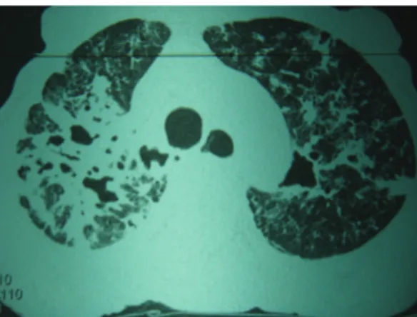
bilateral fibrotic lung involvement, cavities containing buds, and mediastinal adenopathy with necrosis (Figure 1). An axial CT scan of the abdomen showed enlarged adrenal glands. In view of his prolonged respiratory symptoms, fever, poor overall health status, and bilateral fibrotic lung involvement, with multiple cavities and nodules, we hypothesized the following: pulmonary tuberculosis or chronic deep fungal infection, the latter being due to extensive fibrosis and being active because of the fever, ground-glass lesions, and cavity contents. The findings of mediastinal adenopathy with necrosis and adrenal enlargement with necrosis were consistent with our hypotheses. Because of the clinical presentation, imaging test results, and patient history, we established a presumptive diagnosis of paracoccidioidomycosis or histoplasmosis, given that both deep fungal infections are endemic in Uruguay. The differential diagnosis included systemic vasculitis, Churg-Strauss syndrome (because the type of skin involvement was not typical of the deep fungal infections hypothesized), and Wegener’s granulomatosis (because of the presence of cavitary lung involvement). The laboratory test results were as follows: hemoglobin, 10.2 g/dL; platelet count, 563,000/mm3; white blood cells, 9,400/mm3; eosinophils, 3%; ESR, 130 mm/h; HIV testing, negative; urea level, normal; antinuclear antibody testing, negative; rheumatoid factor testing, negative; anti-DNA antibody testing, negative; antineutrophil cytoplasmic antibody testing, negative; BAL fluid, normal; direct bacteriological examination for nonspecific pathogens, negative; culture for nonspecific pathogens, negative; testing for temperatures of less than 26°C, it develops as
a mold. The major antigenic component of P. brasiliensis is a 43-kDa glycoprotein, which binds to the wall and participates in the adhesion, invasion, and pathogenesis of the fungus.(1,4) The fungus is acquired through inhalation of spores of the mycelial phase, which reach the lungs and readily convert to the yeast form, multiplying in the parenchyma and inducing a host response characterized by the formation of a primary complex, followed by the formation of granulomas. In this primary infection, systemic dissemination occurs via the hemolymphatic system. These fungi can remain dormant for years or progress to disease, depending on the host immune response.(3)
In terms of the gateway and pathogenic mechanism, histoplasmosis is similar to paracoccidioidomycosis. In Uruguay, histoplasmosis is a common diagnosis in HIV-AIDS patients, being less common in immunocompetent patients, as was the case with our patient. Histoplasma capsulatum var. capsulatum is a dimorphic fungus that is ubiquitous in nature and is of relevance in the Río de la Plata. The pulmonary forms predominate in immunocompetent patients, whereas the disseminated forms predominate in immunocompromised patients.
H. capsulatum var. capsulatum grows on Sabouraud medium at 28°C, appearing as filaments that typically have branched septate hyaline mycelia, verrucose macroconidia, and abundant microconidia. These characteristics allow the fungus to be specifically identified by mycological examination. In tissue sections stained with Giemsa, the fungus appears as small yeast with an eccentric nucleus and an inhomogeneous staining pattern.
Case report
We report the case of a 56-year-old male smoker who was a rural worker and a hunter. The patient presented with a three-month history of fever, cough, expectoration, and dyspnea. He was given amoxicillin and showed partial improvement. One month later, the symptoms worsened, and this had a negative impact on his overall health status. Physical examination revealed the following: erythema multiforme on the face, trunk, and limbs; respiratory failure; and diffuse dry crackles. A chest X-ray and an HRCT scan of the chest showed extensive
H. capsulatum (Figure 3). Double diffusion serology detected antibodies against P. brasiliensis and H. capsulatum.
The patient had asthenia and dizziness with orthostatic hypotension, which gave rise to the hypothesis of adrenocortical insufficiency. That hypothesis was confirmed by the adrenocorticotropic hormone curve and the increased baseline adrenocorticotropic hormone level. The patient received treatment with amphotericin B and showed improvement after one month. Subsequently, he received itraconazole for six months. At this writing, the patient was stable but had residual fibrosis.
Discussion
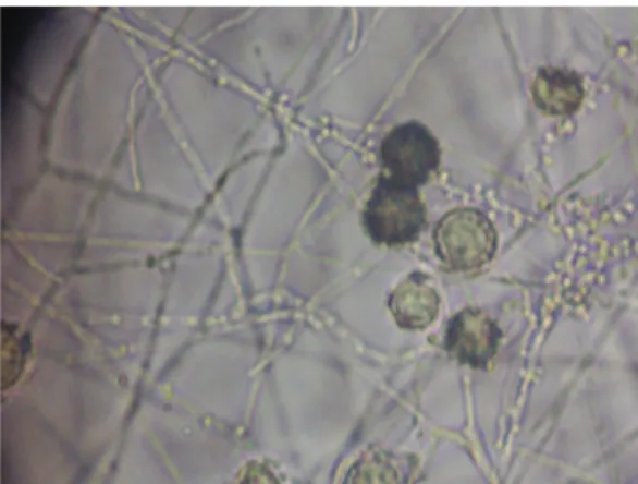
In the case reported here, the diagnosis was difficult because endemic deep fungal infections are uncommon in Uruguay. The clinical presentations of paracoccidioidomycosis and histoplasmosis depend on many factors that are related to the host, the host response, the parasite growth, and the environment. Respiratory involvement is common. It is of note that the case reported here involved an immunocompetent patient, who had severe and extensive lung involvement with a subacute clinical course, and that the diagnosis was confirmed by direct mycological examination, culture, and serological testing. Direct mycological examination revealed abundant multi-budding yeasts (10 to 30 microns in diameter) with refractive walls and vacuolar contents, findings that were consistent with the morphological characteristics of P. brasiliensis (Figure 2). Direct mycological examination with Gomori staining revealed yeasts that had the same characteristics as those described above. Sabouraud cultures (n = 6) maintained at 28°C for 4 weeks showed growth of P. brasiliensis and H. capsulatum, and the different culture tubes were identified on the basis of the micromorphology of the culture. Figure 3 shows the micromorphological pattern of the H. capsulatum colonies isolated. After the detection of both species of fungi on culture and given the fact that this is extremely rare, the stained slides were reexamined. The Giemsa-stained slides revealed the presence of very few yeasts characteristic of H. capsulatum, which had gone unnoticed in the first examination.
Although the diagnosis was confirmed by the isolation of both species of fungi, we screened for circulating antibodies by performing a simple Mycobacterium tuberculosis, negative; mycological
examination, negative; bronchial brushing, negative; serial blood cultures for bacteria, negative; PPD testing, negative; serial sputum smear microscopy, negative; direct examination, negative; and culture, negative.
Given the difficulty in making a diagnosis and the fact that his condition persisted despite the empirical treatment for nonspecific pathogens, we decided to transfer the patient to a referral center, where he underwent a second fiberoptic bronchoscopy with BAL for mycological examination. Direct mycological examination of the BAL fluid revealed abundant multi-budding yeasts with the morphological characteristics of P. brasiliensis (Figure 2). Direct mycological examination with Giemsa and Gomori stainings revealed P. brasiliensis and H. capsulatum yeasts. Sabouraud cultures maintained at 28°C for 4 weeks showed growth of P. brasiliensis and
Figure 2 - Direct examination of BAL fluid:
Paracoccidioides brasiliensis.
in up to 15% of cases of paracoccidioidomycosis, only 9% of such cases are symptomatic,(12) as occurred in the present case.
References
1. Conti I, Rilla F. Hipótesis sobre el nicho ecológico de Paracoccidioides brasiliensis. Rev Med Uru. 1989;5(2/3):97-103.
2. dos Santos WA, da Silva BM, Passos ED, Zandonade E, Falqueto A. Association between smoking and paracoccidioidomycosis: a case-control study in the State of Espírito Santo, Brazil [Article in Portuguese]. Cad Saude Publica. 2003;19(1):245-53.
3. Laguna M, Conti Díaz IA, Navarrete Pedocchi H. Paracoccidioidomicosis. A propósito del primer caso uruguayo en un paciente de sexo femenino. Rev Med Uru. 1998;14(2):171-5.
4. Fonseca LC, Mignone C. Paracoccidioidomicose do intestino delgado. Aspectos anátomo-clínicos e radiológicos de 125 casos. Rev Hosp Clin Fac Med Sao Paulo. 1976;31:199-207. 5. Salfelder K, Doehnert G, Doehnert HR.
Paracoccidioidomycosis. Anatomic study with complete autopsies. Virchows Arch A Pathol Pathol Anat. 1969;348(1):51-76.http://dx.doi.org/10.1007/BF00542835 6. Souza AS Jr, Gasparetto EL, Davaus T, Escuissato DL,
Marchiori E. High-resolution CT findings of 77 patients with untreated pulmonary paracoccidioidomycosis. AJR Am J Roentgenol. 2006;187(5):1248-52.PMid:17056912. http://dx.doi.org/10.2214/AJR.05.1065
7. do Valle AC, Guimarães RR, Lopes DJ, Capone D. Thoracic radiologic aspects in paracoccidioidomycosis [Article in Portuguese]. Rev Inst Med Trop Sao Paulo. 1992;34(2):107-15. PMid:1340023.
8. Muniz MA, Marchiori E, Magnago M, Moreira LB, Almeida JG. Paracoccidioidomicose pulmonar: aspectos na tomografia computadorizada de alta resolução. Radiol Bras. 2002;35(3):147-54. http://dx.doi.org/10.1590/ S0100-39842002000300005
9. Costa MA, Carvalhalo TN, Araujo Jr CR, Borba AO, Veloso GA, Texeira KS. Manifestações extrapulmonares de Paracoccidioidomicose. Radiol Bras. 2005;38(1):45-52. http://dx.doi.org/10.1590/S0100-39842005000100010 10. Santos JW, Michel GT, Lazzarotto M, Figaro JK, Spilmann
D, Homrich GK. Chronic cavitary pulmonary histoplasmosis. J Bras Pneumol. 2009;35(11):1161-4. PMid:20011854. http://dx.doi.org/10.1590/S1806-37132009001100016 11. Unis G, Roesch EW, Severo LC. Acute pulmonary
histoplasmosis in the state of Rio Grande do Sul, Brazil. J Bras Pneumol. 2005;31(1):52-9. http://dx.doi.org/10.1590/ S1806-37132005000100010
12. Agudelo CA, Muñoz C, Ramírez A, Gutierrez J, Velez S, Perez JC, et al. Identification of Paracoccidioides brasiliensis in adrenal glands biopsies of two patients with paracoccidioidomycosis and adrenal insufficiency. Rev Inst Med Trop Sao Paulo. 2009;51(1):45-8. http:// dx.doi.org/10.1590/S0036-46652009000100008
double diffusion test, which detected antibodies against P. brasiliensis and H. capsulatum.
We used specific P. brasiliensis and H. capsulatum antigens and obtained one band of identity for P. brasiliensis and two bands of identity for H. capsulatum; those findings suggested that specific circulating antibodies were present in sufficient quantity to determine positivity for both species of fungi.(5)
About the authors
Veronica Torres Esteche
Adjunct Professor of Clinical Medicine. University of the Republic of Uruguay School of Medicine, Montevideo, Uruguay.
Zaida Arteta
Adjunct Professor of Parasitology and Mycology. University of the Republic of Uruguay School of Medicine, Montevideo, Uruguay.
Gabriela Torres
Resident in Clinical Medicine. Department of Internal Medicine, Maciel Hospital, Montevideo, Uruguay.
Andrea Vaucher
Assistant in Clinical Medicine. University of the Republic of Uruguay School of Medicine, Montevideo, Uruguay.
Elbio Gezuele
Associate Professor. Department of Parasitology and Mycology, University of the Republic of Uruguay School of Medicine, Montevideo, Uruguay.
Raquel Balleste