Fibroblasts participate in the wound repair process through proliferation and migration as well as the synthesis of factors growth and extracellular matrix molecules. However, cell aging and the individual himself can lead to reduction of cell functions and consequently, the ability of tissue repair. This study evaluated the activity of gingival fibroblasts from young (Y) and elderly (Y) patients and their responsiveness to tumor necrosis factor alpha (TNF-α). Gingival fibroblasts were isolated from six patients (3Y; and 3E) and seeded in complete culture medium (DMEM). For cell viability analysis, total protein production and collagen synthesis, fibroblasts were cultured in 96-well plates for 24, 48 or 72 h (n=36). Cell responses to TNF-α, was evaluated by application of this cytokine to cultured cells (100 ng/mL) for 24 h, followed by evaluation of reactive oxygen species (ROS), nitric oxide (NO) and CCL5 production (n=36). Data were analyzed by Kruskal-Wallis and the Mann-Whitney U tests (α = 0.05). Viability of E fibroblasts was higher than Y fibroblasts for 24 and 48 h, but these cells showed gradual reduction of viability over the course of time. For Y cells, reduced collagen synthesis was observed at 48 h. No difference was observed in ROS production for both cells after TNF-α exposure. However, both cultures showed increased production of NO and CCL5 in the presence of TNF-α. Functional differences and distinct responsiveness to TNF-α were observed according to patient’s age.
Functional Differences In Gingival
F i b r o b l a s t s O b t a i n e d f r o m
Young and Elderly Individuals
Taisa Nogueira Pansani1, Fernanda Gonçalves Basso2, Diana Gabriela Soares1, Josimeri Hebling2, Carlos Alberto de Souza Costa3
1Department of Dental Materials
and Prosthodontics, UNESP - Univ Estadual Paulista, Araraquara School of Dentistry, Araraquara, SP, Brazil
2Department of Orthodontics and
Pediatric Dentistry, UNESP - Univ Estadual Paulista, Araraquara School of Dentistry, Araraquara, SP, Brazil
3Department of Physiology and
Pathology, UNESP - Univ Estadual Paulista, Araraquara School of Dentistry, Araraquara, SP, Brazil
Correspondence: Carlos Alberto de Souza Costa, Rua Humaitá, 1680, Centro, 14801-903 Araraquara, SP, Brazil. Tel: +55-16-3301-6477. E-mail: [email protected]
Key Words: cell aging, wound healing, tumor necrosis factor alpha
Introduction
Previous studies have shown that aging can cause morphological and functional cellular changes, such as reduction of proliferative capacity, number of organelles, growth factor expression and collagen synthesis, and that these changes can delay the repair process (1,2). Fibroblasts actively participate in the tissue repair process by proliferating, migrating and filling the wound, beyond the synthesis of growth factors and extracellular matrix molecules (2). An effective tissue repair, especially in cases of surgical implants installation leads to an aesthetic and favors the longevity of the rehabilitation treatment (3). Therefore, diminished metabolism and proliferation of these cells could delay wound healing (4). Studies verified that gingival fibroblasts present an increased repair capacity compared with skin fibroblasts, due to the enhanced migration capability and rapid re-population of the wound. These cells also exhibit higher synthesis of growth factors and the deposition of a more organized and effective surrounding matrix (3,5), supporting the rapid repair of the oral mucosa even around the dental implants.
Tissue repair process consists of essentially four phases, which may overlap: hemostasis, inflammation, proliferation and tissue remodeling (1,5). The inflammatory phase, a direct consequence of the injury, plays an important role at the beginning of the healing process, aimed at
morphological and functional recovery of the tissue (6). Additionally, this phase is essential for the elimination of bacteria and microbial contaminants that can delay the repair process. It has been demonstrated that bacteria and endotoxins can increase and prolong the production of pro-inflammatory cytokines such as interleukin 1 (IL-1β) and tumor necrosis factor alpha (TNF-α), thus extending the inflammatory phase (7). This, moreover, may also be related to the delay of tissue repair, due to a decreased proliferative capacity of the cells involved in this process (8) which affect negatively the aesthetic and function of osseointegrated oral implants. However, few studies have reported comparatively functional differences between gingival fibroblasts from young and elderly individuals and their responsiveness to inflammatory process (9,10).
T
. N
. P
ansani et al.
have analyzed the influence of the aging process on the functions of cells effectively involved in oral mucosal repair (2) that might also influence during the rehabilitation treatment using dental implants. Thus, there is only limited knowledge about the influence of aging on the oral mucosal repair capacity of elderly patients and the inflammatory responses of oral mucosal cells in these patients. The aim of this research was to evaluate the viability and the metabolism of gingival fibroblasts obtained from young and elderly individuals and the responses of these cells when exposed to TNF-α.
Material and Methods
The project was initially approved by the Research Ethics Committee of the Araraquara School of Dentistry/UNESP, SP, Brazil (CAAE #13514813.6.0000.5416). All participants signed the Instrument of Consent.
Six primary cultures were obtained from gingival fragments collected during a tooth extraction procedure or implant placement in three young adults (from 18 to 24 years of age) and three elderly individuals (over 65 years of age). Exclusion criteria, the following factors were considered: presence of systemic diseases, medication use, smoking and periodontal disease, and this information was obtained through anamnesis and clinical assessment. For cell isolation, fragments were incubated in culture medium (DMEM, Dulbecco’s Modified Eagle’s Medium; Gibco, Carlsbad, CA, USA) containing type I collagenase (Worthington Biochemical Corp.; Lakewood, NJ, USA) and were maintained in an incubator at 37 °C in a 5% CO2 atmosphere (Isotemp; Fisher Scientific, Pittsburgh, PA, USA) for 24 h. After this period, the tissue fragments were transferred to a 15 mL Falcon tube and centrifuged for 2 min (4000 x g Centrifuge T; Fanem, Guarulhos, SP, Brazil). The cell pellet was re-suspended in DMEM (1 mL) containing 10% of fetal bovine serum (FBS - Gibco) and transferred to a cell culture flask of 75 cm2 to obtain the amount necessary for the experiment.
Cell Culture
The cells (5-8 passages) in the same passage were cultured in 96-well plates (TPP- Techno Plastic Products, Trasadingen, Switzerland) (9x10³ cells/cm²) in complete DMEM (DMEM with 10% of fetal bovine serum). For each repetition, samples from 6 individuals were selected (3 young and 3 elderly) and 6 samples were seeded per patient (18 samples in Y group and 18 samples in E group), and this study was development in duplicate, totalized n=36 per group (young and elderly). After 24 h, the culture medium was replaced by a fresh serum-free DMEM. For comparison of the functional activity of fibroblasts obtained from young and elderly individuals, a viability test (MTT Assay),
total protein production (TP) and collagen synthesis (Sirius Red Assay) were performed after experimental incubation periods of 24, 48 and 72 h. For comparative evaluation, the responses of gingival fibroblasts obtained from young and elderly individuals, whether exposed or not to the inflammatory mediator, 100 ng/mL of TNF-α in serum-free DMEM, were maintained in contact with the cells for 24 h, based in a study that compared the dose-response by this inflammatory mediator (unpublished data). After this period, the production of reactive oxygen species (ROS) and nitric oxide (NO) and the synthesis of the chemokine CCL5 were evaluated.
Cell Viability Evaluation (MTT Assay)
Cell viability was determined by means of the methyl thiazolyl tetrazolium (MTT) colorimetric assay (12) This method determines the respiratory activity of the cell culture by cleavage of MTT salt [3-(4,5-dimethyltriazol-2YL) -2,5-diphenyl bromide tetrazolium (Sigma-Aldrich, St. Louis, MO, USA), by the succinate dehydrogenase enzyme system, resulting in soluble crystals (formazan crystals).
On the cells cultivated in 96-well plates, 90 μL of culture medium (DMEM) without FBS and 10 μL of MTT solution (5 mg/mL in PBS) were added to the samples. The cells remained in contact with the MTT solution in a CO2 incubator at 37 °C for 4 h, when the formation of formazan crystals was observed. After this period, the MTT solution was aspirated and replaced with 100 μL of acidified isopropanol solution (0.04 N of HCl), for solubilization of the crystals, generating a homogeneous solution which was subjected to absorbance analysis in a spectrophotometer (Synergy H1; BioTek, Winooski, VT, USA) at a wavelength of 570 nm. Absorbance expressed in numerical values was transformed into percentage according to the cell groups of the young individuals, over a 24 h period, and subjected to statistical analysis (11).
Total Protein Production
Differences in young and elderly fibroblasts Collagen Synthesis (Sirius Red Assay)
The concentration of collagen synthetized by the cultured gingival fibroblasts was determined by the Sirius Red assay. This method is based on the selective binding of the Direct Red dye (Sigma-Aldrich), in a saturated solution of picric acid, to the collagen fiber types I to IV, identifying and quantifying the total soluble collagen present in the sample.
For this assay to be performed, the culture medium was collected and stored at -20 °C until the test was performed. To the 200 μL of culture medium collected and 200 μL of the Direct Red solution (0.5 M in picric acid) were added, followed by incubation under agitation for 60 min (400 rpm, room temperature) in a shaker (Thermomixer-Confort; Eppendorf, Hamburg, Germany). Next, this solution was centrifuged in a cooled microcentrifuge (Eppendorf 5415R) at 12,000 rcf for 10 min. The supernatant was discarded, and 300 μL of HCL 0.1M were added, followed by centrifugation at 12,000 rcf for 10 min. The supernatant was discarded one more time, and 250 μL of NaOH 0.5 M were added for homogenization of the samples under vigorous agitation. Aliquots of 100 μL, in duplicate, of the solution were transferred to 96-well plates, and absorbance analysis was performed at a wavelength of 555 nm in a spectrophotometer (Synergy H1; BioTek). The concentration of total collagen was evaluated relative to the percentage of the group of young individuals in the 24 h period.
Production of Reactive Oxygen Species (ROS) Increased production of reactive oxygen species is directly related to oxidative stress, which can result in molecule and organelle peroxidation and lead to irreversible damage. To determine ROS production by gingival fibroblasts obtained from young and elderly patients, a fluorescent probe (H2DCFDA; Invitrogen) was used. This probe is permeable to the cell membrane and allowed for the detection of these intracellular ROS, capable of binding these molecules permanently. The probe was added to the culture medium previous to the application of TNF-α and was maintained for 24 h in association with treatment, to allow for the immediate identification of ROS, as these molecules are highly reactive and unstable.After 24 h, the culture medium was aspirated, and 500 μL of PBS were added to each sample. The amount of ROS was determined by the fluorescence intensity of the probe in each sample when each compartment was scanned (excitation, 488 nm; emission, 540 nm) by a fluorimeter (Synergy H1; BioTek).
Nitric Oxide Production (NO)
Nitric oxide is also an important mediator in cellular inflammatory and tissue responses and, in low concentrations, is also capable of promoting cell
proliferation. In high concentrations, NO can cause severe tissue damage (12).The quantification of NO produced by cells in culture was determined by detection of nitrite accumulation in the cell culture supernatant by the diazotisation reaction with Griess reagent (Sigma-Aldrich).
A 50 μL aliquot of the culture medium maintained in contact with the cells for the proposed treatment was transferred to another 96-well plate, and Griess reagent was added in the same proportion. After 10 min incubation at ambient temperature in the absence of light, the concentration of NO was determined by the absorbance of the solution determined at 540 nm in a spectrophotometer (Synergy H1; BioTek).
CCL5 Chemokine Expression
The CCL5 chemokine is highly expressed during the inflammatory process and is associated with the migration of other inflammatory cells (13).Evaluation of the expression of this chemokine was performed by ELISA immunoassay (enzyme-linked immunosorbent assay), based on an antigen-antibody reaction, which was performed according to manufacturer’s recommendations (R&D Systems, Minneapolis, MN, USA). Thus, the culture medium in contact with the cells was collected and stored at -20 °C until being tested.
For the ELISA assay, 96-well plates were treated with specific primary antibodies (2 μg/mL) and incubated overnight. Washes were then performed (2.5% wash solution), followed by incubation with blocking solution (1% BSA) for 1 h at room temperature. After being washed, 100 μL of each sample and standard curve concentrations were added, and the plates were incubated for 2 h at room temperature, followed by further washing. Next, the secondary antibody was added (400 ng/mL) and incubated for an additional 2 h. Samples were washed, and streptavidin (1:400) was added for 20 min in the dark. New washes were performed, and the substrate solution was added and maintained for 20 min. Finally, Stop solution was added to stop the reaction, and the optical density of the obtained samples was measured by spectrophotometry (Synergy H1; BioTek) at a 450 nm wavelength. The CCL5 concentrations were determined according to the standard curve.
Cell Morphology (Phase Contrast Microscopy) Images of fibroblastic cells from young and elderly individuals were obtained using a phase contrast microscope in 4× magnification (TS 100 Nikon, Tokyo, Japan) equipped with digital image acquisition system. The periods of evaluation were 24, 48 and 72 h of cell culture.
Statistical Analysis
T
. N. P
ansani et al.
the non-parametric Kruskal-Wallis test, complemented by the Mann-Whitney U test, was applied, at a significance level of 5%.
Results
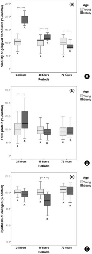
For cells from young gingival tissue (Y), no statistically significant difference was observed for the different time intervals evaluated. However, for elderly individuals cells (E), a gradual reduction in cell viability was observed over the course of the incubation periods (p<0.05). When the groups of Y and E individuals were compared, for each study period, there was increased cell viability in the E individuals group at 24 and 48 h, whereas the reverse was observed within 72 h (p<0.05) (Fig. 1A).
Gingival fibroblasts from the E individuals group showed higher total protein production at 24 h (p<0.05). When the groups of E and Y individuals’ cells were compared within each period, the total protein production was higher only in the 24 h period for E cells (Fig. 1B).
No statistically significant difference in the collagen synthesis by the Y group cells was observed, independent of the period (p>0.05). For E individuals’ cells, reduced synthesis was observed at 48 h, also presenting lower collagen production for the same period (p<0.05) (Fig. 1C).
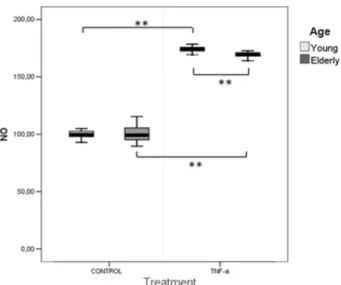
The ROS production by Y individuals’ fibroblasts did not differ statistically significantly from that observed for E individuals’ cells when exposed to TNF-α (Fig. 2). However, NO production was increased when the cells were in contact with TNF-α, independently of the ages of patients (p<0.05) (Fig. 3).
For the CCL5 synthesis expression, there was a statistically significant difference between the cells obtained from Y and E individuals’ groups exposed to TNF-α (Fig. 4).
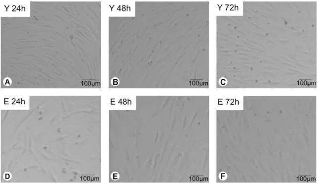
Images of Y and E fibroblasts under phase contrast microscope (TS 100, Nikon, Tokyo, Japan) at 24, 48 and 72 h of culture showed that those cells exhibited similar morphology. However, for E group it seems that a lower number of fibroblasts was present on the substrate when compared to Y group (Fig. 5).
Discussion
Aging is a gradual and continuous process involving numerous cellular and tissue changes on morphological, structural and functional levels (14).Previous studies showed that during the aging process there is a decrease in the proliferative capacity of cells, which also present lower protein metabolism and gene flow (14). However, the influence of the individual’s aging on oral mucosal cell metabolism, such as gingival fibroblasts, has not been completely elucidated. Unlike most studies found in the literature, (2) in which cellular senescence was promoted
Differences in young and elderly fibroblasts by increasing in vitro cell subculture, it was evaluated in
the present investigation the influence of aging on gingival fibroblasts by isolating primary cultures of oral mucosa fibroblasts obtained from young and elderly individuals. In this study, the authors also assessed the possible discrepancy in the response of these oral mucosa cells exposed to tumor necrosis factor (TNF-α) inflammatory cytokine. Therefore, since different behavior of young and elderly cells obtained from health individuals has been poorly presented in the literature, mainly under the data of this study certainly contribute with the research field.
In the present study, it was observed that viability, total protein synthesis and collagen production by Y gingival fibroblasts were not significantly affected by the time in culture (24, 48 and 72 h), whilst for cells obtained from E individuals, there was a time-dependent reduction in cell viability (Fig. 1). However, greater viability occurred when E individuals’ cells were compared to the Y individuals’ fibroblasts at 24 and 48 h. These data, obtained in initial periods, can be related to the premature adhesion of senescent cells, (2) which may allow E individuals’ cells to start the mitosis process more quickly. Some studies have demonstrated changes in collagen production by cells from E individuals. Cultured late-passages fibroblasts also showed significant increased production of collagenases when compared to homologous early passages, (15) which can induce a degradation of soluble collagen synthetized by the cells, as observed in this study in E individuals’ cells at 48 h.
The repair process of oral mucosal injury is also dependent on other factors, such as inflammatory mediators (16).The presence of inflammation in tissues with high concentrations of chemokines and inflammatory mediators induces the generation of reactive oxygen species (ROS) by various cells (17,18).At low concentrations, ROS can accelerate the repair process; however, at higher concentrations or prolonged exposure, ROS can cause tissue damage by lipid peroxidation in different organelles, such as the cell membranes (17).TNF-α is a pro-inflammatory cytokine, secreted from macrophages and other cells that participate in the inflammatory phase during the repair process. Low concentration of this cytokine in tissues is capable of stimulating repair by Figure 2. Reactive Oxygen Species (% young control) by gingival
fibroblasts compared at different ages (young and elderly) with or without TNF-α. Values indicate median (25th-75th percentile), n = 36. Mann-Whitney (p<0.05). No statistically significant difference in ROS production by Y and E groups.
Figure 3. Nitric oxide (% young control) by gingival fibroblasts compared at different ages (young and elderly) with or without TNF-α. Values indicate median (25th-75th percentile), n = 36. Mann-Whitney (p<0.05). **p<0.001. NO production was increased with TNF-α stimuli for Y and E groups.
T
. N. P
ansani et al.
Figure 5. Cell imaging in phase contrast microscopy of fibroblastic cells. Y 24 h: Fibroblasts from young individuals at 24 h of culture; Y 48 h: Fibroblasts from young individuals at 48 h of culture; Y 72 h: Fibroblasts from young individuals at 72 h of culture; E 24 h: Fibroblasts from elderly individuals at 24 h of culture; E 48 h: Fibroblasts from elderly individuals at 48 h of culture; E 72 h: Fibroblasts from elderly individuals at 72 h of culture.
activating macrophages and stimulating the production of growth factors. Conversely, at high concentrations or when it remains for extended periods in damaged tissue, this cytokine can hinder the local repair by increasing the synthesis of metalloproteinases and inhibiting the synthesis of extracellular matrix moleculesand if inflammation of the peri-implant tissues remains, there could be gingival resection and even implant loss (19). In the present study, the authors selected TNF-α to simulate, in a limited way and in in vitro conditions, the activity of fibroblasts in inflamed tissue. Thus, it was possible to demonstrate that the production of ROS by cells exposed to TNF-α contact did not increase in both patient groups. TNF-induced ROS production via p38, JNK, and NF-kB by positive feedback loop and the innate response was TNF-mediated by ROS regulation (20). Additionally, the activation of NF-kB pathway, can stimulate proinflammatory cytokine production by ROS (21). However, significant differences in NO production by gingival fibroblasts from Y and E patients were observed. This inflammatory mediator shows several physiological functions, including bactericidal properties. However, as for ROS, NO in high concentrations can promote cytotoxic effects and tissue damage particularly in inflamed tissues, such as in case of periodontitis and peri-implantitis (22).
The data obtained in this study were corroborated by Chung et al (18), who also demonstrated increased synthesis
of NO related to inflammatory responses against cell aging. These authors reported that the largest increase in activation of the NFκB pathway also promoted a rise in some inflammatory cytokine synthesis, such as TNF-α, interleukin 1 beta (IL-1β), interleukin 6 (IL-6) and cyclooxygenase-2 (COX-2), leading to inflammatory diseases in the elderly.
Differences in young and elderly fibroblasts statistically significant difference was observed for Y and
E fibroblast groups, it was shown the increasing expression of CCL5 trends in E patients’ cells, which can also lead to a delay in the repair process for these patients (24).
According to the methodology used in this in vitro study and considering the limitations of the laboratorial results presented, it can be stated that the fibroblasts functions discrepancies observed in cells obtained from young and elderly individuals, associated with the data previously described in the literature (9,10,15), indicate a relationship between senescence and reduced repair capacity and even changes in the regulation of inflammatory cytokines (25) especially in periodontal tissues. Moreover, during the aging process the cells undergo phenotypic changes and respond differently, mainly towards molecules of the inflammatory process (18).
According to this study, patients’ age is a factor that influences the activity of gingival fibroblasts. Thus, cell viability, total protein production and collagen synthesis by gingival fibroblasts were adversely affected by the increasing age of the individuals, with reduced cellular functions for cells obtained from elderly patients. However, patient age did not influence the responsiveness of these cells against the stimulation with TNF-α, showing maintenance of the inflammatory response capability against pathogens.
Resumo
Fibroblastos participam no processo de reparação de ferida através da proliferação e migração, bem como a síntese de fatores de crescimento e moléculas da matriz extracelular. No entanto, o envelhecimento celular e o próprio indivíduo podem levar à redução de funções celulares e, consequentemente, a capacidade de reparação de tecidos. Este estudo avaliou a atividade dos fibroblastos gengivais de pacientes jovens (J) e idosos (I) e sua capacidade de resposta frente ao fator de necrose tumoral alfa (TNF-α). Fibroblastos gengivais foram isolados de seis pacientes (3J e 3I) e semeados em meio de cultura completo (DMEM). Para a análise de viabilidade celular, a produção de proteína total e a síntese de colágeno, fibroblastos foram cultivados em placas de 96 poços, durante 24, 48 ou 72 h (n = 36). Respostas celulares frente ao TNF-α, foram avaliadas por aplicação desta citocina (100 ng/mL) nas células cultivadas durante 24 h, seguida por avaliação de espécies reativas de oxigênio (EROs), produção de óxido nítrico (NO) e produção CCL5 (n= 36). Os dados foram analisados por testes de Kruskal-Wallis e Mann-Whitney U (α = 0,05). A viabilidade de fibroblastos I foi mais elevada do que os fibroblastos J para 24 e 48 h, mas estas células mostraram uma redução gradual de viabilidade ao longo do tempo. Para as células de J, foi observada redução da síntese de colágeno em 48 h. Não foi observada diferença na produção de EROs para ambas as células após exposição ao TNF-α. No entanto, ambas as culturas apresentaram aumento da produção de NO e CCL5 na presença de TNF-α. Diferenças funcionais e alteração na capacidade de resposta ao TNF-α foram observadas de acordo com a idade do paciente.
Acknowledgements
The authors thank he São Paulo Research Support Foundation (FAPESP – grant 2015/19364-8) and the National Council for Scientific and Technological Development (CNPq – Grants 443153/2014-0, 303599/2014-6, 400984/2015-303599/2014-6, and 307696/2014-6) for financial support.
References
1. Gosain A, DiPietro LA. Aging and wound healing. World J Surg 2004;28:321-326.
2. Hwang ES, Gyesoon Y, Kang HT. A comparative analysis of the cell biology of senescence and aging. Cell Mol Life Sci 2009;66:2503-2524. 3. An N, Rausch-fan X, Wieland M, Matejka M, Andrukhov O, Schedle
A. Initial attachment, subsequent cell proliferation/viability and gene expression of epithelial cells related to attachment and wound healing in response to different titanium surfaces. Dent Mater 2012;28:1207-1214. 4. Smith KC. Laser (and LED) therapy is phototherapy. Photomed Laser Surg
2005;23:78-80.
5. Enoch S, Peake MA, Wall I, Davies L, Farrier J, Giles P, et al.. ‘Young’ oral fibroblasts are geno/phenotypically distinct. J Dent Res 2010;89:1407-1413.
6. Li J, Chen J, Kirsner R. Pathophysiology of acute wound healing. Clin Dermatol 2007;25:9-18.
7. Guo S, DiPietro LA. Factors affecting wound healing. J Dent Res 2010;89:219-229.
8. Menke NB, Ward KR, Witten TM, Bonchev DG, Diegelmann RF. Impaired wound healing. Clin Dermatol 2007;25:19-25.
9. Swift ME, Burns AL, Gray KL, DiPietro LA. Age-related alterations in the inflammatory response to dermal injury. J Invest Dermatol 2001;117:1027-1035.
10. Cáceres M, Oyarzun A, Smith PC. Defective wound-healing in aging gingival tissue. J Dent Res 2014;93:691-697.
11. Basso FG, Pansani TN, Oliveira CF, Turrioni AP, Soares DG, Hebling J, et al.. Cytotoxic effects of zoledronic acid on human epithelial cells and gingival fibroblasts. Braz Dent J 2013;24:551-558.
12. Kendall HK, Marshall RI, Bartold PM. Nitric oxide and tissue destruction. Oral Dis 2001;7:2-10.
13. Deshmane SL, Kremlev S, Amini S, Sawaya BE. Monocyte chemoattractant protein-1 (MCP-1): an overview. J Interferon Cytokine Res 2009;29:313-326.
14. Magalhães JP. From cells to ageing: a review of models and mechanisms of cellular senescence and their impact on human ageing. Exp Cell Res 2004;300:1-10.
15. Sottile J, Mann DM, Diemer V, Millis AJT. Regulation of collagenase and collagenase mRNA production in early- and late-passage human diploid fibroblasts. J Cell Physiol 1989;138:281-290.
16. Glim JE, Egmond M, Niessen FB, Everts V, Beelen RHJ. Detrimental dermal wound healing: What can we learn from the oral mucosa? Wound Rep Reg 2014;21:648-660.
17. Sculley DV, Langley-Evans SC. Salivary antioxidants and periodontal disease status. Proc Nutr Soc 2002;61:137-143.
18. Chung HY, Lee EK, Choi YJ, Kim JM, Kim DH, Zou Y, et al.. Molecular inflammation as an underlying mechanism of the aging process and age-related diseases. J Dent Res 2011;90:830-840.
19. Iglhaut G, Schwarz F, Winter RR, Mihatovic I Stimmelmayr M, Schliephake H. Epithelial attachment and downgrowth on dental implant abutments. J Esthet Restor Dent 2014;26:324-31.
20. Blaser H, Dostert C, Mak TW, Brener D. TNF and ROS crosstalk in inflammation. Trends Cell Biol 2016;26:249-261.
21. Morgan MJ, Liu ZG. Crosstalk of reactive oxygen species and NF-kappa B signaling. Cell Res 2011;21:103-115.
22. Gutiérrez-Venegas G, Kawasaki-Cárdenas P, Arroyo-Cruz SR, Maldonado-Frías S. Luteolin inhibits lipopolysaccharide actions on human gingival fibroblasts. Eur J Pharmacol 2006;541:95-105.
23. Silva TA, Garlet GP, Fukada SY, Silva JS, Cunha FQ. Chemokines in oral inflammatory disease: apical periodontitis and periodontal disease. J Dent Res 2007;86:306-319.
24. Terheyden H, Stadlinger B, Snaz M, Garbe AI, Meyle J. Inflammatory reaction - communication of cells. Clin Oral Implants Res 2013;25:399-407.
25. Benatti BB, Silvério KG, Casati MZ, Sallum EA, Nociti FH Jr. Inflammatory and bone-related genes are modulated by aging in human periodontal ligament cells. Cytokine 2009;46:176-181.