Mucograft® is a resorbing porcine matrix composed of type I and type III collagen, used for soft tissue augmentation in guided tissue bony regeneration procedures. This in vitro study aimed to evaluate the biological behavior of Mucograft® in human gingival fibroblasts, as well as the ability of the matrix to induce production of extracellular matrix. Six resorbing Mucograft® matrices (MCG) were cut into 3 x 2 mm rectangles and 5 x 5 mm squares and were placed in 96- and 24-well plates, respectively. The control group (CTRL) consisted of cells plated on polystyrene without the MCG. After one, two, three and seven days, cell proliferation and viability were assessed using the Trypan exclusion method and MTT test, respectively. Type III collagen (COL 3A1) and vimentin (VIM) expression were also evaluated at 10 and 14 days, using Western blotting. Statistical analysis, using ANOVA with post hoc Bonferroni test, revealed that human gingival fibroblasts from MCG showed similar results (p>0.05) for proliferation and viability as the cells cultured on CTRL. After 14 days, a significant decrease in COL 3A1 expression (p<0.05) was observed when cultured with the MCG. VIM expression showed no significant difference at any time period (p>0.05). Although no increase in extracellular matrix secretion was observed in this in vitro study, Mucograft® presented cellular compatibility, being an option for a scaffold whenever it is required.
Evaluation of the Biological Behavior
of Mucograft
®
in Human Gingival
F i b r o b l a s t s : A n
In Vitro
S t u d y
Rafaela S. R. e Lima1, Daiane C. Peruzzo1 Marcelo H. Napimoga2, Eduardo Saba-Chujfi1, Silvio Antonio dos Santos-Pereira1, Elizabeth F. Martinez3
1Department of Periodontology,
SLMANDIC - São Leopoldo Mandic Institute and Research Center, Campinas, SP, Brazil
2Department of Immunology,
SLMANDIC - São Leopoldo Mandic Institute and Research Center, Campinas, SP, Brazil
3Department of Pathology and Cell
Biology, SLMANDIC - São Leopoldo Mandic Institute and Research Center, Campinas, SP, Brazil
Correspondence: Elizabeth Ferreira Martinez, Rua José Rocha Junqueira, 13, 13045-610 Campinas. SP, Brasil. Tel: +55-19-3211-3600. e-mail: [email protected]
Key Words: gingival tissue graft, Mucograft® collagen matrix, cell culture, guided tissue regeneration.
Introduction
Periodontal soft tissue grafts are primarily used for root coverage, to thicken a gingival site or improve the crestal volume in pre-prosthetic surgery. Soft tissue grafts are also advised to create a favorable environment for peri-implant mucosa (1). Soft tissue grafts may be harvested from the palate, retromolar pads or edentulous sites. Disadvantages of harvesting the graft from the retromolar pad and edentulous sites are the minimal amount of tissue availability, as well as recuperation of thinner grafts only. Therefore, the preferred site for harvesting soft tissue grafts is the palate (1), which requires a second surgical site, increasing morbidity in terms of post-operative discomfort and procedure time (2).
Biomaterials are being used as a replacement for palatal tissue harvest, with the aim of reducing morbidity. Biomaterials may be considered as such when it allows for adequate tissue integration, without inducing an immune response, chronic inflammation or sensitivity that may interfere with healing and, hence, harm the patient. The advantages of biomaterials are their unlimited availability, decreased surgical time, reduced discomfort due to lack of a donor site and fewer post-operative complications (3). Among the biomaterials used for periodontal tissue regeneration, collagen matrices have received significant attention. Mucograft® is a resorbing porcine matrix
composed of type I and type III collagen, which is used for soft tissue augmentation in both guided tissue and bone regeneration procedures. It is composed by a porcine collagen bilayer structure (4,5). The compact layer, which consists of compact collagen fibers with occlusive cellular properties, allows tissue adherence as a prerequisite for favorable wound healing. This layer not only protects against bacterial infiltration during open healing conditions, it also contains adequate elastic properties to accommodate suturing. The second layer consists of a thick, porous, spongy collagen structure, which is placed next to the host tissues to facilitate organization of the blood clot and promote neoangiogenesis and tissue integration (5,6). Although clinical and histological studies with Mucograft® have demonstrated the induction of a mild tissue reaction, excellent tissue integration was observed (5). A recent study comparing Mucograft® with BioGuide®, which is another biomaterial, demonstrated that the former facilitated cell proliferation and promoted early tissue reaction in vitro and in vivo, respectively (7). This matrix has been extensively studied in clinical setting (4,8-10), with several studies showing promising esthetic results (4,9-12). Additional studies that report on cell proliferation and viability, and the potential to induce connective tissue synthesis are, however, lacking.
603
Biological behavior of Mucograft® in fibroblasts
biological behavior of Mucograft® on human gingival fibroblasts, as well as its ability to induce production of extracellular matrix.
Material and Methods
Specimen Preparation
In order to investigate the action of Mucograft® (MCG) on human gingival fibroblast viability, as well as its capacity to induce cellular proliferation and protein expression, six resorbing Mucograft® matrices (Geistlich Biomaterials, Wolhusen, Switzerland) measuring 3 x 2 mm and 5 x 5 mm were placed in 96- and 24-well plates for the proliferation and viability tests, and Western blotting, respectively. The control group (CTRL) consisted of cells plated on polystyrene without the Mucograft® collagen matrix.
Cell Cultures
Gingival fibroblasts were obtained from explants of healthy attached human gingiva from three different donors, obtained from periodontal surgery for crown lengthening (13,14). This study was approved by the São Leopoldo Mandic Institute and Research Center Institutional Review Board (IRB - #2012/0308).
The cells were cultured in Dulbecco’s Modified Eagle Medium (DMEM) (Sigma, St Louis, MO, USA) supplemented with 1% antimycotic-antibiotic solution (10,000 units of penicillin, 10 mg of streptomycin and 25 μg of amphotericin B per mL, in 0.9% sodium chloride; Sigma), containing 10% donor calf serum (DCS; GIBCO, Buffalo, NY, USA), plated in 60 mm diameter plastic culture dishes and incubated under standard cell culture conditions (37 °C, 100% humidity, 95% air, and 5% CO2). Once the cells reached subconfluence, they were detached using 0.05% trypsin and subcultured at a density of 110 cells/mm2. The cells were used at subculture levels 3 or 4 for all experimental assays.
Cell Proliferation and Cell Viability Assays
Cells were grown on 24 and 96 wells plates (Costar®, Corning, NY, USA) at an initial concentration of 1.9x10⁴ cells/mL and 0.42x10⁴ cells/mL per well, respectively for the proliferation and viability tests. After 1, 2, 3 and 7 days, the cells were detached using 0.05% trypsin and counted in a Neubauer chamber to calculate proliferation indices. In a different set of plates under the same conditions, 10 µL of MTT solution (5 mg/mL in PBS) and 90 µL of base medium were added to each well. Cells were incubated for 3 h at 37 °C, 5% CO2, 95% air and complete humidity. After 3 h, the MTT solution was removed and replaced with 100 µL of dimethyl sulfoxide (DMSO). The plate was further incubated for 15 min at room temperature (RT) and the optical density (OD) of the wells was determined at a wavelength of 590 nm in a SpectraMax Plus microplate reader (Molecular
Devices). The experiments were repeated twice under the same conditions to ensure accuracy.
Western Blotting
Cells were grown on the membranes for 10 and 14 days, homogenized and centrifuged at 15,000 g for 15 min at 4 °C. The protein concentration was measured by a BCA assay (Pierce, Rockford, IL, USA). Protein extracts were separated on 15% sodium dodecylsulfate–polyacrylamide gels, transferred onto polyvinylidene difluoride membranes (Hybond; Amersham Biosciences, Piscataway, NJ, USA), exposed for 1 h to the primary antibodies anti-vimentin (VIM, mouse, 1:1000, Dako Corp., Carpenteria, CA, USA) and anti-type III collagen (COL3A1, mouse, 1:1000, Abcam, Cambridge, UK), and diluted in TBST and 5% low fat milk. The primary antibody GAPDH was used as an endogenous control (1:5000; Santa Cruz Biotechnology, Santa Cruz, CA, USA). After incubation with the secondary monoclonal antibody of either mouse or rabbit origin (1:2500), the reaction was developed using Bio-Rad Laboratories (Hercules, CA, USA) Western blotting chemiluminescent detection reagents (Opti-4CN) onto x-ray films (GE Healthcare, Fairfield, CT, USA). Measurements of optical density were performed using the NIH Image J 1.37 (National Institutes of Health, Bethesda, MD, USA) for scanned membranes.
Statistical Analysis
Data were first examined for normality using the Shapiro-Wilk test. Two-way analysis of variance with
post hoc Bonferroni test was then applied to all assays, at a significance level of 0.05. The results were expressed as mean ± standard deviation.
Results
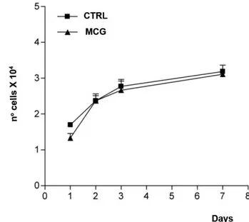
Mucograft® biological behavior was evaluated using cell proliferation counts and the Trypan vital exclusion method. In the control group (CTRL), human gingival fibroblasts, which were cultured in DMEM only, presented similar results to the cells cultured on the Mucograft® resorbing matrices at 1, 2, 3 and 7 days, demonstrated by the lack of a significant difference between the groups (p=0.7582) (Fig. 1).
The MTT assay for cell viability revealed no significant difference between CTRL and MCG (p=0.7003), as shown in Figure 2.
R.S.R. Lima et al.
finding demonstrated that the matrix was not capable of inducing COL 3A1 synthesis in 10 days, but a considerable reduction of collagen expression occurred after 14 days when compared to the control group.
A numeric decrease in Vimentin (VIM) expression was observed when the cells were cultured on the matrix when compared to CTRL, although it was not statistically significant (p>0.05).
Discussion
Biomaterials have been developed as tissue substitutes,
using the concept of guided tissue regeneration (GTR) (15,16). An adequate resorbing material for oral procedures should allow cell adhesion, proliferation and migration in order to prevent exposure to oral microorganisms (17,18). Collagen is one of the most researched resorbing materials, being the main component of the periodontal connective tissue matrix, with significant hemostatic properties, thus aiding in early stabilization of the surgical wound.
Mucograft® is a collagen matrix of porcine origin used as a substitute in cases of loss of the connective tissue structure (4,8-11,19). It has been used to replace the connective tissue graft from the palate, as well as for recession coverage and regeneration of keratinized mucosa around teeth and implants (4,12,20). Additionally, Mucograft® has shown promising results for use as a graft for socket seal in ridge preservation procedures (21). Its mechanism of action is the creation of a three-dimensional scaffold that allows the ingrowth and repopulation of fibroblasts, blood vessels and epithelium from surrounding tissues, eventually transformed into keratinized tissue. Despite its extensive use in clinical procedures, few studies have demonstrated in vitro the biological behavior of the Mucograft® collagen matrix. The present study aimed to investigate whether human gingival fibroblasts would increase their proliferation potential and extracellular matrix protein expression in the presence of Mucograft®, The results revealed no significant difference in cell growth or viability on the Mucograft® surface when compared to the control group.
A pre-clinical trial in mice used two prototype collagen matrices, namely 1 (CM1) and 2 (CM2), to study their tissue integration, biodegradation and new blood vessel formation (20). These matrices were composed of native porcine
Figure 1. Evaluation of cell proliferation via the Trypan exclusion method for human gingival fibroblasts at 1, 2, 3 and 7 days. The curve was based on biological triplicates with values expressed as the mean (±SD). Two-way ANOVA and Bonferroni test (p=0.7582).
Figure 2. Cell viability test (MTT) in human gingival fibroblasts at 1, 2, 3 and 7 days. The curve was based on biological triplicates with values expressed as the mean (±SD). Two-way ANOVA and Bonferroni test (p=0.7003).
605
Biological behavior of Mucograft® in fibroblasts
collagen I and III, which differed by the degree of additional chemical cross-linking, with CM1 and CM2 presenting a denser and looser network structure, respectively. Results from the histological analysis demonstrated that the level of cross-linking had a significant influence on the amount of new blood vessel and connective tissue formation, as well as on degradation of the collagen network. The less dense CM2 did indeed offer an improved angiogenic pattern and enhanced connective tissue formation compared to the denser network of CM1. More recently, Willershausen et al. (7) performed a detailed surface and morphological ultrastructure analysis of Mucograft® and compared it with BioGuide® (BG). Cellular growth patterns and proliferation rates of human fibroblasts on Mucograft® and BG were analyzed in vitro. The early tissue reaction of CD-1 mouse on these materials was also analyzed by histological and histomorphometrical techniques. The results demonstrated that both matrices facilitated in vitro cell proliferation. In vivo, these two materials induced a comparable early tissue reaction. The present in vitro study did not demonstrate an increase in cellular proliferation and viability. Mucograft works simply as a scaffold, since it did not induce significant changes in the expression of collagen III and Vimentin.
Different barrier membranes, growth and differentiation factors and soft tissue substitutes have been used to promote healing and soft tissue regeneration. When used for the treatment of localized gingival recessions, barrier membranes have provided improved histological outcomes in terms of reduced epithelial attachment and greater amounts of new cementum, connective tissue attachment and bone. However, these improved histological outcomes had limited clinical significance and not predictably present in all studies (15). Some studies have investigated the clinical and histological outcome of Mucograft® for procedures surrounding teeth and dental implants (4,8,10,12). The matrix showed acceptable tissue integration, even in open healing conditions. Application of the collagen matrix significantly reduced the time spent in the surgical chair when compared with autologous grafting (2). Areas of regeneration have shown a similar appearance to that of the surrounding natural soft tissues, both in terms of texture and color, which makes its use preferable in esthetic areas that are difficult to match with palatal transplants (10,12,22). Additionally, the use of collagen matrix removes the need for painful tissue harvesting procedures and significantly reduces postoperative pain (4,19). However, despite the advantages presented by Mucograft®, the gold standard for esthetic soft tissue management for teeth and dental implants is still the subepithelial connective graft (9,22).
It is difficult to establish a suitable comparison between
in vivo and in vitro studies, since laboratory conditions
allow good control of the variables for the latter, while the former comprises greater structural, cellular and tissue complexity, making isolated analysis of the regeneration or stimulation potential of the matrices more difficult. Therefore, it may be concluded that although no increase in extracellular matrix secretion was observed, Mucograft® presented cellular compatibility, therefore being a viable option as biomaterial when a scaffold is required.
Resumo
A Mucograft® é uma matriz reabsorvível, de origem suína, composta
de colágenos do tipo I e III, utilizada para aumento de tecido mole em regeneração óssea guiada. Este estudo in vitro teve como objetivo avaliar o comportamento biológico da Mucograft®, em fibroblastos gengivais
humanos, bem como a indução da síntese de matriz extracelular. Seis matrizes reabsorvíveis de Mucograft® (MCG) foram cortadas em retângulos
e quadrados medindo 3 x 2 mm e 5 x 5 mm e alocadas em placas de 96 e 24 poços, respectivamente. O grupo controle (CTRL) consistiu no plaqueamento celular em poliestireno, sem MCG. Após um, dois, três e sete dias, a proliferação e a viabilidade celular foram avaliadas utilizando o corante vital azul de Trypan e o teste MTT, respectivamente. Além disso, a expressão de colágeno tipo III (COL 3A1) e vimentina (VIM) foi avaliada após 10 e 14 dias, por meio de Western-blotting. Após análise estatística (Anova e pós teste de Bonferroni), pode-se observar queos fibroblastos gengivais humanos, cultivados sobre MCG, apresentaram proliferação e viabilidade semelhantes em comparação às células que foram cultivadas apenas no poliestireno (CTRL). Após 14 dias, notou-se uma diminuição significativa da expressão de COL 3A1 (p<0,05) quando as células foram cultivadas sobre a MCG. A expressão da VIM não mostrou diferença significativa em nenhum dos períodos estudados (p>0,05). No presente estudo in vitro pode-se concluir que apesar de não ter sido observado aumento da síntese de matriz extracelular, a Mucograft® apresentou
compatibilidade celular, sendo uma opção de biomaterial em casos que o arcabouço é necessário.
Acknowledgements
The authors wish to thank Pollyanna Tombini Montaldi and Jerusa Bossonaro Pinheiro for their excellent technical expertise.
References
1. Thoma DS, Benić GI, Zwahlen M, Hämmerle CH, Jung RE. A systematic
review assessing soft tissue augmentation techniques. Clin Oral Implants Res 2009;20(Suppl 4):146-165.
2. Barros RMR, Novaes Junior AB, Grisi MFM, Souza SLS, Taba JR, Palioto DB. A 6-month comparative clinical study of a conventional and a new surgical approach for a root coverage with acellular dermal matrix. J Periodontol 2004;75:1350-1356.
3. Urist MR. Bone morphogenetics protein induced bone formation in experimental animals and patients with large bone defects. In: CIBA Foundation Symposium. Cell and molecular biology of vertebrate hard tissues. London: John Willey & Sons; 1988.
4. Sanz M, Lorenzo R, Aranda JJ, Martin C, Orsini M. Clinical evaluation of
a new collagen matrix (Mucograft® prototype) to enhance the width
of keratinized tissue in patients with fixed prosthetic restorations: a randomized prospective clinical trial. J Clin Periodontol 2009;36:868-876.
5. Ghanaati S, Schlee M, Webber JM, Willershausen I, Barbeck M, Balic E, et al.. Evaluation of the tissue reaction to a new bilayered collagen matrix in vivo and its translation to the clinic. Biomed Mater 2011;6:015010.
R.S.R. Lima et al.
7. Willershausen I, Barbeck M, Boehm N, Sader R, Willershausen B, Kirkpatrick CJ, et al.. Non-cross-linked collagen type I/III materials enhance cell proliferation: in vitro and in vivo evidence. J Appl Oral Sci 2014;22:29-37.
8. Herford AS, Akin L, Cicciu M, Maiorana C, Boyne PJ. Use of a porcine collagen matrix as an alternative to autogenous tissue for grafting oral soft tissue defects. J Oral Maxillofac Surg 2010;68:1463-1470. 9. McGuire MK, Scheyer ET. Xenogeneic collagen matrix with coronally
advanced flap compared to connective tissue with coronally advanced flap for the treatment of dehiscence-type recession defects. J Periodontol 2010;81:1108-1117.
10. Nevins M, Nevins ML, Kim SW, Schupbach P, Kim DM. The use of mucograft collagen matrix to augment the zone of keratinized tissue around teeth: a pilot study. Int J Periodontics Restorative Dent 2011;31:367-373.
11. Cardaropoli D, Tamagnone L, Roffredo A, Gaveglio L. Treatment of gingival recession defects using coronally advanced flap with a porcine collagen matrix compared to coronally advanced flap with connective tissue graft: a randomized controlled clinical trial. J Periodontol 2012;83:321-328.
12. Schmitt CM, Moest T, Lutz R, Wehrhan F, Neukam FW, Schlegel KA. Long-term outcomes after vestibuloplasty with a porcine collagen matrix (Mucograft®) versus the free gingival graft: a comparative prospective clinical trial. Clin Oral Implants Res 2015;27.Doi:10.1111/ clr.12575. [Epub ahead of print].
13. Martinez EF, Araújo VC. In vitro immunoexpression of extracellular
matrix proteins in dental pulpal and gingival human fibroblasts. Int Endod J 2004;37:749-755.
14. Martinez EF, Araújo VC, Sousa SO, Arana-Chavez VE. TGF-beta1 enhances the expression of alpha-smooth muscle actin in cultured human pulpal fibroblasts: immunochemical and ultrastructural analyses. J Endod 2007;33:1313-1318
15. Vignoletti F, Nunez J, Sanz M. Soft tissue wound healing at teeth, dental implants and the edentulous ridge when using barrier membranes, growth and differentiation factors and soft tissue substitutes. J Clin Periodontol 2014;41Suppl15:S23-S35.
16. Zucchelli G, Mounssif I. Periodontal plastic surgery. Periodontol 2000 2015;68:333-368.
17. Kellomäki M, Niiranen H, Puumanen K, Ashammakhi N, Waris T, Törmälä P. Bioabsorbable scaffolds for guided bone regeneration and generation. Biomaterials 2000;21:2495-2505.
18. Genon CR. Comparative clinical study of guided tissue regeneration with a bioabsorbable bilayer collagen membrane and subeptelial connective tissue graft. J Periodontol 2001;72:1258-1264.
19. Fu JH, Su CY, Wang HL. Esthetic soft tissue management for teeth and implants. J Evid Based Dent Pract 2012;12:129-142.
20. Thoma DS, Sancho-Puchades M, Ettlin DA, Hämmerle CH, Jung RE. Impact of a collagen matrix on early healing, aesthetics and patient morbidity in oral mucosal wounds - a randomized study in humans. J Clin Periodontol 2012;39:157-165.
21. Thoma DS, Hämmerle CH, Cochran DL, Jones AA, Görlach C, Uebersax L, et al.. Soft tissue volume augmentation by the use of collagen-based matrices in the dog mandible - a histological analysis. J Clin Periodontol 2011;38:1063-1070.
22. Chambrone L, Tatakis DN. Periodontal soft tissue root coverage procedures: a systematic review from the AAP Regeneration Workshop. J Periodontol. 2015;86(2 Suppl):S8-S51.