RBCCV 44205-1579 DOI: 10.5935/1678-9741.20140064
New contribution to the study of ventricular
remodeling and valve rings in dilated
cardiomyopathy: anatomical and histological
evaluation
Nova contribuição ao estudo do remodelamento ventricular e dos anéis valvares na cardiomiopatia
dilatada: avaliação anátomo-histopatológica
Moise Dalva
1, MD, PhD; Aristides Tadeu Correia
2; Natalia de Freitas Jatene
1; Paulo Hilário
Nascimento Saldiva
3, MD, PhD; Fabio Biscegli Jatene
1, MD, PhD
1Instituto do Coração da Hospital das Clínicas daFaculdade de Medicina da Universidade de São Paulo, InCor-HCFMUSP, São Paulo, SP, Brazil. 2Surgical Anatomical Museum of Hospital das Clínicas da Faculdade de Medicina da Universidade de São Paulo, InCor-HCFMUSP, São Paulo, SP, Brazil.
3Pathology Department of the Hospital das Clínicas da Faculdade de Medici-na da Universidade de São Paulo, InCor-HCFMUSP, São Paulo, SP, Brazil.
This study was carried out at Hospital das Clínicas of the Faculdade de Medicina da Universidade de São Paulo, InCor-HCFMUSP, São Paulo, SP, Brazil.
No inancial support.
Correspondence address: Moise Dalva
Instituto do Coração do Hospital das Clínicas da Faculdade de Medicina da Universidade de São Paulo InCor-HCFMUSP
Rua Dr. Enéas de Carvalho Aguiar, 44 – Cerqueira César – São Paulo, SP, Brazil - Zip Code: 05403-000
E-mail: [email protected]
Article received on December 29th, 2013 Article accepted on March 16th, 2014
Abstract
Introduction: Idiopathic dilated cardiomyopathy causes great impact but many aspects of its pathophysiology remain unknown.
Objective: To evaluate anatomical and histological aspects of hearts with idiopathic dilated cardiomyopathy and compare them to a control group, evaluating the behavior of the perimeters of the atrioventricular rings and ventricles and to compare the
per-centage of collagen and elastic ibers of the atrioventricular rings. Methods:Thirteen hearts with cardiomyopathy and 13 normal hearts were analysed. They were dissected keeping the ventricular mass and atrioventricular rings, with lamination of segments 20%, 50% and 80% of the distance between the atrio-ventricular groove and the atrio-ventricular apex. The sections were subjected to photo scanning, with measurement of perimeters. The atrioventricular rings were dissected and measured digitally to evaluate their perimeters, later being sent to the pathology
laboratory, and stained by hematoxylin-eosin, picrosirius and oxidized resorcin fuccin.
Results: Regarding to ventricles, dilation occurs in all seg-ments in the pathological group, and the right atrioventricular ring measurement was higher in idiopathic dilated cardiomy-opathy group, with no difference in the left side. With respect
to collagen, both sides had lower percentage of ibers in the pathological group. With respect to the elastic ibers, there was
no difference between the groups.
Conclusion: There is a change in ventricular geometry in cardiomyopathy group. The left atrioventricular ring does not dilate, in spite of the fact that in both ventricles there is lowering of collagen.
despite this relevant fact, the rings expansion mechanism is not completely understood[2-4].
The presence of valvular insuficiency may contribute to increased morbidity in heart valve disease patients[5] or
those presenting a DCM of various etiologies[6]. There is a
tendency to consider the valve tissues as inert, because of its simple histological structure and sparse cell population, however, this fact seems less reasonable in light of the huge mechanical load imposed on these structures throughout life and the consequent need for maintenance of its tissue integrity at the cost of balance between collagen production and degradation[7].
Although the presence of myocytes and coronary circula-tion are capital for the funccircula-tioning of the heart as a pump, the components of the extracellular matrix (ECM), particularly the collagen ibers of types I and III are recognized as funda -mental to the maintenance of the cardiac cycle[8]. Among its
many functions, the most important are to provide structural framework for myocytes and vessels as well as provide the body resistance and resilience properties, providing systolic and diastolic tone, helping the heart to maintain its confor-mation[8-12].
Abbreviations, acronyms and symbols
RVR Right ventricular ring LVR Left ventricular ring
CAPPesq Ethics Committee for Research Project Analysis DCM Dilated cardiomyopathy
idDCM Idiopathic dilated cardiomyopathy iscDCM Ischemic dilated cardiomyopathy
DistAV-AP Distance of atrioventricular groove to the left ventricular apex
SD Standard deviation USP University of São Paulo
HCFMUSP Clinics Hospital of the Faculty of Medicine, University of São Paulo
HE hematoxylin-eosin CHF congestive heart failure INCOR Heart Institute
ISFC International Society and Federation of Cardiology MAGPs Microibrils associated with glycoproteins MMPs Metalloproteinases
RFO Weigert Resorcin-fuchsin with previous oxidation by oxone
SPSS Statiscal package for the social sciences SVOC Coroner’s Service of São Paulo USA United States of America WHO World Health Organization
INTRODUCTION
Congestive heart failure (CHF) is the entity causing major impact in terms of morbidity and mortality, and its main cause is dilated cardiomyopathy (DCM), which in its various etiologies, constitute a serious public health problem, with an estimated prevalence of 4-8 cases per 100,000 people per year and an estimated incidence of 36.5 per 100,000 people[1].
The key pathophysiological aspect of such entities is the severe systolic dysfunction caused by the loss of eficiency of the heart to act as hydraulic pump. Although myocellular component is present in an important way, other mechanisms such as the remodeling and activation of the renin-angioten-sin-aldosterone system are factors contributing to the perpet-uation of the presentation[1].
Ventricular remodeling characterized by both right and left morphogeometrical changes, provides vicious cycle of functional deterioration, since the heart loses its original ana-tomical conformation, which is critical to its eficiency. In this context, the valve insuficiency caused primarily by expansion of the atrioventricular rings is of paramount importance[1], but
Resumo
Introdução: A cardiomiopatia dilatada idiopática (CMDId)
é causadora de grande impacto, porém aspectos de sua isiopa -tologia são desconhecidos.
Objetivo: Avaliar aspectos anatomo-histológicos de corações com CMDId comparando-os a corações normais, com medidas perimetrais dos anéis atrioventriculares direito (AVD) e esquerdo (AVE) e dos ventrículos direito (VD) e esquerdo (VE) e a
porcen-tagem de ibras colágenas e elásticas dos anéis.
Métodos: Foram avaliados 13 corações de cadáveres porta-dores de CMDId e 13 corações normais, que foram dissecados mantendo-se os anéis atrioventriculares e a massa ventricular, com laminação em segmentos correspondentes a 20%, 50% e 80% da distância entre o sulco atrioventricular e o ápice ventricular.
Os cortes foram submetidos à digitalização fotográica, sendo
comparadas as medidas. Os anéis foram dissecados, medidos e enviados ao laboratório de anatomia patológica, sendo realiza-das colorações por meio de hematoxilina-eosina, picrossírius e resorcina fuccina oxidada.
Resultados: Com relação aos ventrículos, no grupo CMDId ocorre dilatação nos segmentos apical, equatorial e basal. A me-dida do AVD foi maior no grupo CMDId, não havendo diferença
no AVE entre os grupos. Com relação ao percentual de ibras
colágenas, há diminuição no grupo CMDId em relação ao grupo
normal. Com relação às ibras elásticas, não houve diferença
entre os grupos.
Conclusão: Ocorre alteração da geometria ventricular com dilatação no grupo CMDId. Na CMDId observou-se aumento no perímetro do AVD. Não se observou aumento do perímetro do AVE. Houve diminuição percentual na área total de colágeno tanto no AVD quanto no AVE em corações com CMDId.
The breakdown of these ibers may occasionally per -sist even after removal of the underlying disease in many situations. In patients with disease of the mitral valve with secondary tricuspid insuficiency, there can be no normal -ization of tricuspid regurgitant low even with the correction of mitral valve disease[13]. This fact raises controversy in the
literature about the real necessity of repair of the tricuspid valve ring when it is secondarily dilated[13], and to what
ex-tent the non-standardization of relux may be associated with irreversible histological changes in atrioventricular rings. This fact is of crucial importance, given the fact that about half of patients with mitral valve disease requiring surgery presents signiicant tricuspid insuficiency[14]. The authors
who advocate not performing repair insist on the fact that the correction of mitral lesion leads to normalization of the afterload of the right ventricle by reducing the pressure of the pulmonary vascular bed[15]. In contrast, those who advocate the
realization of repair of the tricuspid ring support the fact that the ring expansion cannot be naturally reversible in advanced cases, despite the total correction of the mitral valve[16,17]. This
fact could possibly be due to microscopic structural changes in the atrioventricular rings with occurrence of collagenolysis and replacement of collagen ibers for tissue of other nature, compromising its integrity.
The anatomical concept that the ibrous skeleton of the heart does not dilate has been refuted[2,3,18], and its
enlarge-ment has been proved in cases of severe heart failure due to dilated cardiomyopathy of ischemic (iscDCM) or nonchagasic idiopathic (idDCM) etiologies[2,3]; however, it does not exist
in the literature comparative histological study of right and left atrioventricular rings in cases of idDCM in light of this new concept. At the same time, available knowledge about the role played by ECM in terms of control and regulation of this process is still scarce so that there is broad ield of research being done in this area.
Objective
The aims of this study are:
1-Evaluating and comparing the perimeters of the right and left ventricles in different segments and right and left atrio-ventricular rings in normal hearts and patients with IdDCM.
2- Comparing the percentage by area of collagen and elastic ibers of the right and left atrioventricular rings between the normal hearts and patients with idDCM.
METHODS
The design of this study was initially submitted to and approved by the Institutional Research Ethics Committee.
Material
Specimens of normal and dilated hearts were studied. Normal hearts came from the Coroner’s Service of São Paulo
(SVOC-USP), and dilated hearts from the Anatomic Pathol-ogy Laboratory of the Heart Institute of the Clinics Hospital, University of São Paulo (InCor-HC USP).
A total of 26 specimens were grouped as follows: Group 1 - (idDCM) Composed of 13 hearts from individ-uals with idDCM
Group 2 - (NORMAL) Composed of 13 hearts from individuals without cardiomyopathy and considered normal
After collection, samples were ixed in formalin, followed by removal of large vessels and the atria, leaving only the atrioventricular rings and ventricular mass.
After the steps of preparation and assessment of parts, cross-sections of ventricular mass were performed, starting from the atrioventricular groove towards the apex of the heart (DistAV-AP). The cross-sections were performed at a level corresponding to 80% (baseline), 50% (equatorial) and 20% (apical) from this distance, following being photographed with a digital camera (Sony, model Cyber Shot DSC W 200) which was set at a table through a bucky 15 cm distally from the parts. The images were transferred to a computer where measurements were performed with the software Image Tool, (Department of Dental Diagnostic Science of the University of Texas Health Science Center, San Antonio, USA). All parts were photographed next to a rule which served as a reference for measurements.
After the completion of the photographs, the right and left atrioventricular rings were completely dissected, but were not separated, keeping them together by the central ibrous body. The rings were placed in boxes and sent to the pathology department, following being waxed and laminated, and per-formed histological sections of 5 micrometers thick and used the following staining methods:
• Hematoxylin-eosin (HE) - Standard coloring in pathology services, and is used for identiication of technical artifacts and histopathological changes that eventually could compromise analysis by other methods.
• Picrosirius - coloring used to study collagen ibers. • Weigert’s resorcinol-fuchsin with previous oxidation by oxone (RFO) - coloring used to study elastic ibers
Quantitative morphometric analysis was performed by means of digital image analysis using system consisting of optical microscope Leyca DMR (Leyca Microsystems Wetzlar Gmb H, Germany) connected to a computer by a video camera.
After obtaining the macroscopic data, comparisons of the average ventricular circumference of each segment were performed (apical and basal Equatorial) between idDCM and normal groups and the right and left ventricles as well as comparisons of means of ventricular perimeter between each segment (Apical, Equatorial and Basal) within each group (idDCM and NORMAL). The perimeters of the right and left atrioventricular rings were also compared.
Regarding the microscopic data, the average of the per-centages comparisons were performed by area of collagen and elastic ibers of the right and left atrioventricular rings between each group ( idDCM and NORMAL).
Regarding the statistics, descriptive analyzes were per-formed, presenting means along with the related standard deviations (± SD) and minimum and maximum values. The assumptions of normal distribution in each group and the homogeneity of variances between groups were assessed, respectively, with the Shapiro-Wilk test and the Levene test. The inferential analysis for ventricular perimeter was performed using analysis of variance (ANOVA) for repeated measures to compare the means of each segment between the groups (intergroup factor). The t test was used to evaluate the average perimeters of the right and left atrioventricular rings and the mean percentage of elastic and collagen ibers of the rings. The descriptive and inferential
statistical analyzes were performed using SPSS version 13 (SPSS 13.0 for Windows).
RESULTS
One of the objectives was to assess the perimeters of the right and left ventricles (apical, equatorial and basal segments) for each group ( idDCM and NORMAL), but the point for section of the apical segment (20%) did not include the right ventricular cavity of hearts from idDCM the NORMAL groups and in most cases. Thus, in the right ventricles of the hearts from idDCM and NORMAL groups were analyzed only the perimeters of the equatorial and basal segments.
Descriptive data for variables of right ventricle perimeters (equatorial and basal segments) and left (apical, equatorial and basal segments) for each group are presented in Tables 1 and 2 and the descriptive results for the perimeters of variables of right and left atrioventricular rings for each group are presented in Tables 3 and 4.
The descriptive results for the variables percentages by area of collagen ibers of the right and left atrioventricular rings for each group are shown in Tables 5 and 6, and the descriptive results for the variables percentages by area of elastic ibers of the right and left atrioventricular rings for each group are presented in Tables 7 and 8.
Table 1. Descriptive measures of equatorial and basal perimeter variables in idDCM and NORMAL (mm) right ventricle groups.
Groups
idDCM
NORMAL
Segment Equatorial
Basal
Equatorial
Basal
N 13
13
13
13
Mean 170.812
223.339
112.66
173.38
Standard deviation 44.60938
29.03743
20.58866
24.82283
Minimum 84.36
190.58
74.78
123.59
Maximum 242.99
287.34
142.72
216.54
idDCM: Idiopathic dilated cardiomyopathy
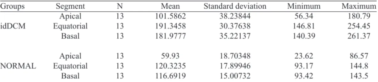
Table 2. Descriptive measures of variables apical, equatorial and basal perimeter in idDCM and NORMAL (mm) left ventricle groups.
Groups
idDCM
NORMAL
Segment Apical Equatorial
Basal
Apical Equatorial
Basal
N 13 13 13
13 13 13
Mean 101.5862 191.3458 181.9777
59.93 120.3235 116.6919
Standard deviation 38.23844 30.37638 35.22137
18.70348 17.89946 15.00732
Minimum 56.34 146.81 140.39
23.62 93.17 93.42
Maximum 180.79 254.45 261.37
86.57 144.8 143.5
In the mean comparison analysis of right ventricular pe-rimeters of equatorial and basal segments between NORMAL and idDCM groups, statistically signiicant differences were found (P<0.05) in all studied segments.
In the mean comparison analysis of the right atrioventric-ular perimeters of the rings between idDCM and NORMAL groups statistically signiicant difference was found (P<0.05).
In the mean comparison analysis of left ventricular apical perimeters of equatorial and basal segments between idDCM and NORMAL groups, statistically signiicant differences were found (P<0.05) in all segments.
The results of the Post-Hoc test with Bonferroni correction for multiple comparisons showed statistically signiicant dif -ferences for all comparisons, except with respect to the com-Table 3. Descriptive measures of variables RVR ring perimeter in idDCM groups and Normal (mm).
Groups idDCM NORMAL
N 13 13
Mean 120.1915 104.0046
Standard deviation 15.33305 13.88195
Minimum 94.85 75.77
Maximum 141.78 128.89
RVR=Right ventricular ring; idDCM: Idiopathic dilated cardiomyopathy
Table 4. Descriptive measures of variables LVR perimeter in idDCM and Normal (mm) groups.
Groups idDCM NORMAL
N 12 13
Mean 108.3233
97.2723
Standard deviation 13.76889 16.40091
Minimum 87.35 69.02
Maximum 128.01 118.09
RVR=Right ventricular ring; idDCM: Idiopathic dilated cardiomyopathy
Table 5. Descriptive measures of variables collagen ibers of RVR in idDCM and NORMAL groups (percentage). Groups
idDCM NORMAL
N 13 13
Mean 19.2332 38.5756
Standard deviation 14.19502 21.51783
Míinimum 1.51 13.43
Maximum 60.73 88.89
RVR=Right ventricular ring; idDCM: Idiopathic dilated cardiomyopathy
Table 6. Descriptive measures of variables collagen ibers of LVR in idDCM and NORMAL groups (percentage). Groups
idDCM NORMAL
N 13 13
Mean 22.0962 38.4603
Standard deviation 12.85746 14.75941
Minimum 1.44 14.85
Maximum 59.55 59.55
LVR=Left ventricular ring; idDCM: Idiopathic dilated cardiomyopathy
Table 7. Descriptive measures of variables elastic ibers of RVR in idDCM and NORMAL groups (percentage). Groups
idDCM NORMAL
N 13 13
Mean 19.5032 17.5873
Standard deviation 11.33865 13.42513
Minimum 8.12 0.29
Maximum 45.4 43.46
RVR=Right ventricular ring; idDCM: Idiopathic dilated cardiomyopathy
Table 8. Descriptive measures of variables of elastic ibers of LVR in idDCM and NORMAL groups (percentage). Groups
idDCM NORMAL
N 13 13
Mean 21.0929 18.1184
Standard deviation 11.16968 13.63213
Minimum 7.13 1.26
Maximum 43.78 50.78
parison between equatorial and basal ventricular perimeters. In the mean comparison of the perimeters analysis of left atrioventricular rings between the idDCM and NORMAL groups, there was no signiicant statistical difference (P> 0.05).
In the mean comparison analysis of the collagen ibers of the right and left atrioventricular rings between the idDCM and NORMAL groups, there was a statistically signiicant difference (P<0.05).
In the mean comparison analysis of the elastic ibers of the right and left atrioventricular rings between the idDCM groups and NORMAL, there was no statistically signiicant difference (P>0.05).
DISCUSSION
The medical understanding of the CHF has undergone substantial change since the irst records of this entity, which can be traced in writings attributed to Hippocrates, and we can identify its historical evolution, which progressed in symmetry with the advancement of scientiic knowledge[19].
From a functional standpoint, the loss of pumping function occurs due to energy dissipation, a fact derived from mech-anisms as increased heart weight, ventricular dilation, and thrombi in the heart chambers and dilation of atrioventricular rings[1].
The ECM had its role reviewed in the genesis of idDCM. Initially, its components were taken as part of passive support in which the myocytes are intertwined, but recent studies point to the fact that these components play an active role in all phases of the normal cardiac cycle, for giving the heart fundamental properties such as resistance, resilience and elasticity, with consequent amendment of these features in pathological cardiac cycle, whose main characteristic is ven-tricular remodeling, which can be observed in macroscopic and microscopic level[8,20].
The normal myocardial collagen comprises predominantly Type I (corresponding to about 80% of the total collagen mass) and III, which form a three dimensional network structure which includes valves, chordae tendineae and perivascular interstitial collagenous components, which is organized in bundles. They are called epimisium (which covers each muscle iber individually), perimysium (covering myocytes groups) and endomysium (found between each myocyte)[8].
Regarding the behavior of the collagen ibers in cardio -myopathies, studies show conlicting results, and there may be increased[21] or decrease[20] of the collagenous component
as well as breakdown of normal structure. Weber et al.[8], in
histological study analyzing three hearts of patients who died due to idDCM, reported that there was a decrease of type I collagen (tougher) and increased collagen type III (less resistant) compared to the normal pattern and loss of normal functional architecture of collagenous ibers. They found that the increase in the less resistant collagen is likely
responsible for the remodeling mechanism, with decreased contractile eficiency.
This discrepancy results seem to be related in part to the methodology used and partly due to the fact that collagen may take different forms in the case of normal myocardial collagen or ibrosis.
Although myocardial ECM changes have already been investigated in cases of idDCM, two factors remain unknown, namely any modiication of the histological composition of the ECM of right and left atrioventricular rings and the behavior of the collagen ibers in terms of balance among its produc -tion, degradation and organization. These facts motivated the present study.
From a macroscopic point of view, it was noted that there dilatation in both ventricles of the idDCM group, albeit with distinct morphology, since the expansion of the RVR accompanies the expansion of the equatorial and basal ventricular segments, contrary to what happens in LVR which presents no signiicant expansion compared to the control group, although there was dilation of equatorial and basal segments at left. In relation to LVR, these indings conirm the results of Juliani[22] and Hueb et al.[2,3], who
claim to not be the degree of left ventricular dilation that determines the degree of dilation of the mitral ring, since they occur independently. This statement has always been a matter of controversy in the literature. In a study that ex-amined the measure of LVR in 102 hearts, 78 of which had left ventricular dilation, Bulkley & Roberts[23] conclude that
the isolated expansion of the left ventricular rarely causes failure in the left atrioventricular valve. They mention that the contrary afirmative has long been regarded as true, as postulated by great names of cardiology, as Flint and Osler in books dated end of the nineteenth century.
The association of left atrioventricular valve insuficiency increases the morbidity and mortality of patients with CHF caused by idDCM[1]. Although often seen as secondary only
to ventricular remodeling and it is therefore classiied as “functional”, recent studies indicate that possibly there are in-trinsic components to the valve structure as a whole that acting differently can be responsible for the observed failure[4,6]. The
valve lealets, although considered to be only inert, because apparently they are not committed in cases of idDCM, contrary to what happens in other types of valvular regurgitation, pres-ent their own characteristics that must be taken into account, such as afferent and efferent innervation, intrinsic contractile properties, and spatial orientation of collagenous ibers that allows optimal distribution of mechanical stress, so that the remodeling of the lealets possibly plays an intrinsic role in the genesis of valve failure, as demonstrated by Timek et al.[4]
causes failure. The fact that there was dilation statistically signiicant of the RVR may optionally be associated to a lack of a full collagenous ring around the right atrioventricular oriice, unlike what occurs on the left side, where this hole is effectively surrounded by strong collagenous ring, which may reduce the propensity for ring dilation.
According Juliani[22], there was cross left ventricular
dilation in idDCM, which is mainly caused by changes in the baseline and equatorial segments. The fact that dilation of LVR can be a signiicant component of the ventricular remodeling process because anatomically the ring is part of the ventricle containing it, and because of its non-expansion may not occur changes in ventricular anatomic coniguration, with tapering of its superior portion. This fact occurs only at left because at right occurs dilation of the basal, equato-rial and RVR segments, a fact that gives rise to a different ventricular conformation and morphology of the wider top. These indings seem to corroborate the results of Hueb[3],
who observed the fact that impairment of RVR valve ac-companies the enlargement of the right ring, which does not occur on the left.
The proportionality between the different types of collagen seems to represent major role in maintaining normal geomet-ric conformation of the heart[8]. Although the amount of total
collagen in the myocardium may increase, due mainly to the replacement of such ibers by scar tissue, with a consequent functional breakdown, there appears to be evidence points to the fact that the ECM of myocardium composition may vary according to the anatomic location. Gunja-Smith et al.[24] in
study comparing 8 hearts with idDCM extracted from heart transplant recipients, with 12 normal hearts, stated emphati-cally that “it deals with a simpliication to assume that every heart has a similar composition.”
In this study, we assessed the ECM regarding its collage-nous and elastic components exclusively in the region of the atrioventricular rings. As far as we know, there is no similar study in literature, a reason that can possibly explain the incon-sistencies found with respect to the total amount of collagen, because in this study we noted, under microscopic inference analysis, that the percentage amount of total collagen ibers was signiicantly lower in idDCM group relative to the control group in both atrioventricular rings.
Study limitations
With regard to the selection of the sample, although we had calculated the power to explain any change found in macroscopic terms such as above 80%, it was not possi-ble to calculate it in microscopic terms, since there are no studies in the literature about the speciic change in EMC of atrioventricular rings in idDCM that would serve as the basis to a similar calculation. The possibility of a pilot study was not contemplated by the impossibility of obtaining more
participants, since the only available were effectively used in the study. This limitation may eventually be overcome from studies in animal models, since there are already developed models for this purpose.
Microscopic analysis was limited by issues relating to staining methods used, especially in relation to the study of elastic ibers. Staining with the use of resorcin-fuchsin oxidized allows evaluating the totality of the elastic ibers present, but it does not adequately provide for differentiation between three different types of such ibers (oxytalan, elastic and mature). Studies with use of alternative colors, such as Verhoeff method for exclusive coloring mature elastic ibers and Weigert resorcin-fuchsin, for recognizing the mature and elastic ibers may help elucidate the elastic behavior of the system qualitatively and not only quantitative.
Regarding the collagen ibers, the methodology used allowed the quantiication of the total ibers present, how -ever, it did not allow the differentiation of different types of ibers present, so that it could not be assessed the quan -tity of the collagen studied that was composed of healing material and ibrosis.
In the present study, there was the aim to investigate the variation of the total quantity of collagen and elastic ibers in different anatomic regions of the heart, which could lead to the conirmation of the hypothesis that the ECM structure varies according to the anatomical region, presenting a standard of increase of some structures and decrease in others.
Final considerations
The anatomical variation of the atrioventricular ring in terms of presence of ECM has been demonstrated. Angelini et al.[25] analyzed after autopsy the left atrioventricular
junc-tion in 13 subjects, 7 of them free of heart disease and 6 pa-tients with mitral valve prolapse and concluded that except for the intertrigonal distance, where lies the mitral-aortic continuity, there is great ECM array arrangement, with the presence of variable size ibrous portions permeating areas where atrial myocardium and the ventricular myocardium are located, and further found that the amount of collagen in the ring ranged thick and easily identiiable portions and also thin portions.
This same hypothesis was raised by Juliani[22], who in
CONCLUSION
The results showed that:
1) There was an increase of ventricular perimeters in id-DCM group compared to the normal group at right and left in different segments evaluated. The perimeter of the RVR was higher in idDCM group compared to the NORMAL group, with no signiicant difference in relation to LVR between the two groups.
2) With regard to the percentage by area of collagen ibers, the right and left atrioventricular rings showed lower percent-age of ibers in idDCM group compared to the control group. Regarding the percentage by area of elastic ibers, there was no difference between groups.
Authors’ roles & responsibilities
MD Analysis and/or interpretation of data, statistical analysis, inal approval of the manuscript, conception and design of the study, implementation of operations and/or experiments, manuscript writing or critical review of its contents ATC Analysis and/or interpretation of data, statistical analysis,
inal approval of the manuscript, conception and design of the study, implementation of operations and/or experiments, manuscript writing or critical review of its contents NFJ Implementation of operations and/or experiments
PANS Conception and design, manuscript writing or critical review of its contents
FBJ Analysis and/or interpretation of data, inal approval of the manuscript, study design, manuscript writing or critical review of its contents
REFERENCES
1. Hare JM. The dilated, restrictive and iniltrative cardiomyopathies.
In. Libby P, Bonow RO, Mann DL, Zipes DP, eds. Braunwald’s heart disease: a textbook of cardiovascular medicine. 8th ed. Philadelphia: Saunders Elsevier; 2008. p.1739-62.
2. Hueb AC, Jatene FB, Moreira LFP, Pomerantzeff PMA, Mioto BM, Chabelmann RC, et al. Estudo comparativo do anel valvar mitral e do ventrículo esquerdo na cardiomiopatia dilatada. Rev Bras Cir Cardiovasc. 2001;16(4):354-63.
3. Hueb AC, Jatene FB, Moreira LF, Pomerantzeff PM, Kallás E, Oliveira SA. Ventricular remodeling and mitral valve
modiications in dilated cardiomyopathy: new insights from
anatomic study. J Thorac Cardiovasc Surg. 2002;124(6):1216-24.
4. Antoniali F, Braile DM, Potério GMB, Costa CE, Lopes MM, Ribeiro GCA, et al. Proporção entre os segmentos do anel da valva tricúspide normal: um parâmetro para realização de anuloplastia valvar. Braz J Cardiovasc Surg. 2006;21(3): 262-71.
5. Matsuyama K, Matsumoto M, Sugita T, Nishizawa J, Tokuda Y, Matsuo T. Predictors of residual tricuspid regurgitation after mitral valve surgery. Ann Thorac Surg. 2003;75(6):1826-8.
6. Breda JR, Palma JHA, Teles CA, Branco JNR, Catani R, Buffolo
E. Miocardiopatia terminal com insuiciência mitral secundária:
tratamento com implante de prótese e remodelamento interno do ventrículo esquerdo. Braz J Cardiovasc Surg. 2006;21(3):283-8.
7. Henney AM, Parker DJ, Davies MJ. Collagen byosinthesis in normal and abnormal human heart valves. Cardiovasc Res. 1982;16(11):624-30.
8. Weber KT. Cardiac interstitium in health and disease: the ibrillar
collagen network. J Am Coll Cardiol. 1989;13(7):1637-52.
9. Montes GS, Junqueira LC. The use of the Picrosirius-polarization method for the study of the biopathology of collagen. Mem Inst Oswaldo Cruz. 1991;86(Suppl 3):1-11.
10. Melo ECM, Lemos M, Ximenes Filho JA, Sennes LU, Saldiva PHN, Tsuji DH. Distribution of collagen in the lamina propria of the human vocal fold. Laryngoscope. 2003;113(12):2187-91.
11. Buhler RB, Sennes LU, Mauad T, Melo EC, Silva LF, Saldiva
PH. Collagen iber and versican distribution within the lamina
propria of fetal vocal folds. Laryngoscope. 2008;118(2):371-4.
12. Sakae FA, Imamura R, Sennes LU, Mauad T, Saldiva PH, Tsuji
DH. Disarrangement of collagen ibers in Reinke’s edema.
Laryngoscope. 2008;118(8):1500-3.
13. Sagie A, Schwammenthal E, Padial LR, Vasquez de Prada JA, Weyman AE, Levine RA. Determinants of functional tricuspid regurgitation in incomplete tricuspid valve closure: Doppler
color low study of 109 patients. J Am Coll Cardiol. 1994;24(2):
446-53.
14. Dreyfus GD, Corbi PJ, Chan KM, Bahrami T. Secondary tricuspid regurgitation or dilatation: which should be the criteria for surgical repair? Ann Thorac Surg. 2005;79(1):127-32.
15. Braunwald NS, Ross J Jr, Morrow AG. Conservative management of tricuspid regurgitation in patients undergoing mitral valve replacement. Circulation. 1967;35(4 Supp):I63-9.
16. Cohen SR, Sell JE, McIntosh CL, Clark RE. Tricuspid regurgitation in patients with acquired, chronic, pure mitral regurgitation. II. Nonoperative management, tricuspid valve annuloplasty, and tricuspid valve replacement. J Thorac Cardiovasc Surg. 1987;94(4):488-97.
successful mitral valve replacement for rheumatic mitral valve disease. Br Heart J. 1991;66(4):295-301.
18. McCarthy PM. Does the intertrigonal distance dilate? Never say never. J Thorac Cardiovasc Surg. 2002;124(6):1078-9.
19. Katz AM. The ’’modern’’ view of heart failure: how did we get here? Circ Heart Fail. 2008;1(1):63-71.
20. Spinale FG. Myocardial matrix remodeling and the matrix
metalloproteinases: inluence on cardiac form and function.
Physiol Rev. 2007;87(4):1285-342.
21. Nunes VL, Ramires FSA, Pimentel WS, Fernandes F, Ianni BM, Mady C. O papel do acúmulo de colágeno no interstício miocárdico na sobrevida dos pacientes com cardiomiopatia dilatada idiopática e chagásica. Arq Bras Cardiol. 2006;87(6):757-62.
22. Juliani PS. Avaliação morfogeométrica do ventrículo esquerdo
e do anel valvar mitral na cardiomiopatia dilatada isquêmica
ou idiopática: estudo comparativo computadorizado [Tese de doutorado]. São Paulo: Faculdade de Medicina, Universidade São Paulo; 2008.
23. Bulkley BH, Roberts WC. Dilatation of mitral annulus. A rare cause of mitral regurgitation. Am J Med. 1975;59(4):457-63.
24. Gunja-Smith Z, Morales AR, Romanelli R, Woessner JF Jr. Remodeling of human myocardial collagen in idiopathic dilated cardiomyopathy. Role of metalloproteinases and pyridinoline cross-links. Am J Pathol. 1996;48(5):1639-48.
25. Angelini A, Ho SY, Anderson RH, Davies MJ, Becker AE. A histological study of the atrioventricular junction in hearts with
normal and prolapsed lealets of the mitral valve. Br Heart J.