Rat Subcutaneous Tissue Response to Macrogranular
Porous Anorganic Bovine Bone Graft
Willian Fernando ZAMBUZZI1 Rodrigo Cardoso de OLIVEIRA1
Felipe Ladeira PEREIRA2 Tânia Mary CESTARI2
Rumio TAGA2 José Mauro GRANJEIRO3
1Department of Biochemistry, Institute of Biology, State University of Campinas, Campinas, SP, Brazil 2Department of Biological Sciences, School of Dentistry of Bauru, University of São Paulo, Bauru, SP, Brazil 3Department of Cellular and Molecular Biology, Institute of Biology, Fluminense Federal University, Niterói, RJ, Brazil
The ideal bone graft must present biocompatibility, osteoconductive and osteoinductive properties, resistance and plasticity. Xenogenic grafts of bovine cancellous bone origin are particularly interesting due to their biologically designed porous structure that enhance both cellular and vascular invasion. The purpose of this study was to evaluate the tissue response induced by bovine macrogranular porous anorganic bone implanted in rat subcutaneous tissue. Forty rats were assigned to 2 groups, as follows: the control group received empty collagen capsules and the test group received subcutaneous implants of the test material. Samples were collected after 10, 20, 30 and 60 days and processed histologically. Histological analysis showed at 10 days a granulomatous inflammatory infiltrate, rich in multinucleated giant cells and free of lymphocytes or plasma cells, similarly to mineralized allograft implanted in rat subcutaneous. In later periods, there was a significant decrease in the inflammatory infiltrate and an increase in fibrosis around graft particles. In conclusion, the test material induced a foreign body-type granuloma with subsequent fibrosis around the graft particles implanted in rat subcutaneous and did not elicit any immune response, thus being considered biocompatible.
Key Words: xenogenic graft, bovine cancellous bone, biocompatibility, subcutaneous tissue.
Correspondence: Prof. Dr. José Mauro Granjeiro, Departamento de Biologia Celular e Molecular do Instituto de Biologia, Universidade Federal Fluminense, Outeiro de São João Baptista, S/N, Campus do Valonguinho, Centro, 24020-150 Niterói, RJ, Brasil. Tel: +55-21-2629-2324. Fax: +55-21-3701-1617. e-mail: [email protected]
INTRODUCTION
Autogenous bone has been widely accepted and used as a pattern for the treatment of bone loss (1). Nevertheless, the limitations of autogenous grafts are availability, morbidity of the donor site and unsatisfying biomechanical properties (2). Consequently, there has been a continuous search for other alternatives such as alografts (3), xenografts (4) and aloplastic materials (5). Several companies have developed bone substitute ma-terials of bovine origin, such as, Schering (Luboc™), Sulzer (Ne-osteo™), Geislich (BioOss™) and Baumer (Gen-Ox™). It is possible to obtain biomaterials from bovine long bones, such as deproteinized anorganic cortical or cancellous bone (6) and demineralized
cor-tical or cancellous matrix (7), in granules or blocks, and also demineralized resorbable membrane (4).
proteins similarly to allografts (9).
This study evaluated the tissue response induced by bovine macrogranular porous anorganic bone im-planted in rat subcutaneous tissue.
MATERIAL AND METHODS
The tested material was macrogranular porous anorganic bone (1-2 mm granules) of bovine cancellous bone origin, deproteinized at 100o C (Gen-Ox™; Baumer
S.A., Mogi Mirim, SP, Brazil). The material was placed into clear collagen capsules (0.1 g per capsule) for implantation in the animals.
Forty male Wistar rats (weight 160 g) obtained from the Vivarium of the School of Dentistry of Bauru were randomly assigned to 2 groups (n=20), as follows: control group: subcutaneous implantation of empty collagen capsules; test group: implantation of collagen capsules containing 0.1 g of graft material.
The animals were anesthetized with an intramus-cular dose of Dopalen® (Agribrands do Brasil Ltda,
Paulinia, SP, Brazil; 0.5mL/kg body weight) and Anasedan® (Agribrands do Brasil) at a 1:1 (v:v) ratio.
After shaving and antisepsis of the dorsal region, a linear incision was made in the skin, the subcutaneous tissue was dissected and the capsule implanted. Suture of the surgical wound and antisepsis were done thereafter.
The rats received a normal diet and water ad libitum throughout the study. After 10, 20, 30 and 60 days, 5 animals of each group were sacrificed per period by anesthetic overdose. Samples were immediately removed, fixed in 10% phosphate buffered formalin for 24 h, dehydrated in ethanol, clarified in xylol and embedded in Histosec® (paraffin + synthetic resin;
Merck, Darmstadt, Germany). Alternate 5-μm-thick sections were cut and stained by hematoxylin and eosin (HE). The study was conducted following the guidelines of the Brazilian College of Animal Experimentation.
Inflammatory infiltrate and fibrosis intensity around the graft particles was analyzed semi-quantitatively under light microscopy, attributing scores according to the level of inflammatory reactions (inflammatory infiltrate) and reparative processes (fibrosis, angioblastic and fibroblastic proliferation).
Scores were given as follows: absent (0), mild (1), moderate (2) and intense (3). Data of both groups were analyzed by Kruskal-Wallis non-parametric test at 5% significance level.
RESULTS
The results are illustrated in Figure 1 (graphic presentation) and Figure 2 (histologic pannel).
10 Days
In the control group (Fig. 2A) it was possible to observe a granulation tissue rich in fibroblasts and blood capillaries and also the presence of large mac-rophages with hyaline material in the cytoplasm, resem-bling fragments of phagocyted collagen capsules. In the test group, a moderate to intense inflammatory infiltrate characterized by the presence of a large number of macrophages and inflammatory multinucle-ated giant cells (IMGC) (Fig. 2B) was seen around the graft particles in addition to a discrete presence of polymorphonuclear leukocytes and lymphocytes. Mod-erate fibroblastic and angioblastic proliferation and mild fibrosis were detected around the grafts particles (Fig. 1). The more peripherally situated particles were encapsulated while the more central ones were involved by a larger number of inflammatory cells.
20 Days
A moderate inflammatory infiltrate as well as
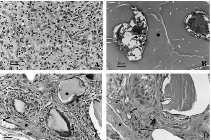
Figure 2. SEM micrographs of histological sections at different experimental periods. (A) Control group at 10 days: granulation tissue rich in mononuclear cells. (B) Test group at 10 days: implant particles (•) involved by inflammatory infiltrate showing polymorphonuclear leukocytes and IMGC () inside the particles’ pores. (C) Test group at 30 days: IMGC () in close contact with implant particles (•); note the presence of blood vessels (arrow) and fibrosis (). (D) Test group 60 days: fibrosis () and blood vessels (arrow) around particles were observed. HE staining.
angioblastic and fibroblastic proliferation were ob-served in the control group. Small foci of macrophages containing phagocyted material were seen, similarly as in the 10-day period. There was a decrease in the intensity of the inflammatory infiltrate (moderate to mild) and an increase in the fibrosis around the graft particles in the test group. A capsule was formed in the outer limit exhibiting similar characteristics to the original connective tissue, with fat cells and blood vessels. The number of polymorphonuclear leukocytes and lymphocytes was reduced significantly and the number of macrophages and IMGC remained similar to the previous period.
30 Days
There was an increase in collagen fiber density
and angiogenesis compared to previous control periods. In the test group (Fig. 2C), there was invasion of graft particle pores by connective tissue and endothelial cells. Moderate fibrosis and mild inflammatory infiltrate was also detected. IMGC were the most abundant cell type in contact with the surface of the particles despite the reduction in the number of macrophages and the in-crease in the number of blood vessels.
60 Days
be emphasized, however, that the presence of these cells occurs in an attempt of the organism to resorb the material and does not necessarily imply lack of biocompatibility. There are a couple of hypothesis that try to explain the nature of these IMGC. Glowacki and Cox (14) identified these cells as osteoclasts, including a brush border, while other authors disagree (15). According to Baggi and Miller (16), two types of giant cells can be observed around mineralized particles: one, with IMGC typical morphological characteristics, and the other, in a lesser extent, with osteoclastic morphology, although without its unique brush border. Despite the presence of IMGC in contact with implant particles, brush border was detected here, which is in agreement with a previous study (15) that demonstrated IMGC recruitment in response to implantation of particles, but without osteoclastic morphological aspects.
The porosity of the material certainly contributes for an increase in surface area, favoring the recruitment and posterior contact of a large number of inflammatory cells with the material. However, at 10 days post implantation, few cells were detected in the central area of the reactive tissue, probably due to the total volume and compaction of the graft particles. It should be noted that the interconnected biological pores might increase bone growth (17), as they provide a tunnel through which cells, blood vessels and growth factors may migrate. Aloplastic materials that mimic such characteristic have been developed as synthetic carriers for growth factors (18) and bioengineering (19).
Anorganic bovine bone matrix type is a natural, porous, xenogenic hydroxyapatite, approved by the Food and Drug Administration for the use as a filling material in bone defects (20). Thermal deproteinization allows elimination of protein contents while keeps the natural hydroxyapatite structure and surface. Its biologically structured surface allows cellular adhesion, proliferation and migration (8), thus enhancing terminal differentiation and maturation of new bone cells. Xe-nografts may be associated to bioactive molecules increasing their osteoconductive property or providing an osteoinductive potential for the treatment of bone defects (20).
Within the limitation of this study, it may be concluded that the xenograft of macrogranular anor-ganic porous bovine bone was well tolerated in rat subcutaneous tissue and did not elicit any immune response.
amount than that of the previous period, was also observed and exhibited characteristics of slow-turn-over granuloma (foreign body-type reaction).
DISCUSSION
In the search for development of biocompatible materials to be used as bone substitutes, bovine bone has been continuously investigated as a reliable source, showing promising results both in vivo (4-8) and in clinical studies (10). Thousands of surgeries using bone grafts are made every year for the treatment of large bone defects, and autogenous bone remains the main bone graft used. In dentistry, a large number of cases demands this surgical procedure and, sometimes, the graft may be obtained from a donor area intraorally (11). The shortcomings and risks of autogenous bone grafts (2) have stimulated the search for alternative bone substitutes that combine properties such as biocompatibility and osteoconduction. In this context, materials of xenogenic origin have been investigated as biomaterials since the 1960s (12).
Adequate mechanical, chemical and thermal pro-cessing used for the production of xenografts allows obtaining biocompatible bone substitute materials. The findings of a previous study (13) with porous anorganic bone grafting in experimental bone defects showed significant bone formation, attributing osteoconductive properties to this material. Another study (8) found an interaction of osteoblasts on the surface of these materials, enhancing cell adhesion and proliferation.
In the present study, the evaluation of the tissue response to a biomaterial produced from bovine cancel-lous bone by a Brazilian laboratory – particulate porous anorganic bone – implanted in rat subcutaneous tissue showed during the experimental periods, chronic in-flammatory infiltrate rich in macrophages and multi-nucleated giant cells, and few polymorphonuclear leukocytes, plasma cells and eosinophils.
At 10 days post-implantation, the biologically designed pores of the graft particles were already filled with reparative tissue. Reduction of the inflammatory infiltrate and increase of the fibrosis around the graft particles were observed throughout the experiment.
RESUMO
O enxerto ósseo ideal deve possuir características como biocompatibilidade, capacidade osteocondutora, osteoindutora, resistência e plasticidade. Dentre os implantes xenogênicos de origem bovina, os produzidos com o osso esponjoso revestem-se de particular interesse devido a sua arquitetura constituída de poros biologicamente desenhados que favorecem a invasão celular e vascular até o centro do defeito. O objetivo deste estudo foi avaliar a resposta tecidual ao material de osso inorgânico medular bovino macrogranular. Quarenta ratos foram divididos em 2 grupos (n=20): o grupo controle recebeu cápsulas de colágeno vazias, e o grupo experimental recebeu implante subcutâneo do material teste. As amostras foram coletadas após 10, 20, 30 e 60 dias de implantação e processadas histotecnicamente. A análise histológica mostrou aos 10 dias pós-cirúrgicos que o infiltrado inflamatório era do tipo granulomatoso rico em células gigantes multinucleadas, mas livre de linfócitos ou plasmócitos, quadro similar ao observado para aloenxertos mineralizados implantados em subcutâneo de ratos. Com o avançar do tempo experimental houve significante diminuição do infiltrado inflamatório inicial concomitantemente ao aumento no grau de fibrosamento ao redor das partículas implantadas. Concluiu-se que o material de enxerto testado em tecido conjuntivo subcutâneo de ratos induziu um granuloma tipo corpo estranho e fibrose ao redor das partículas implantadas, resposta semelhante do mesmo tecido aos aloenxertos mineralizados, e não desencadeou nenhuma resposta imune, sendo portanto biocompatível.
ACKNOWLEDGEMENTS
The authors wish to thank Ovídio dos Santos Sobrinho, Thelma Lopes da Silva, Daniele Santi Ceolin, Luiz Carlos da Silva and Erasmo Gonçalves da Silva for their technical assistance and to FAPESP (Process N. 02/03928-0) for financial support.
REFERENCES
1 . Bell RB, Blakey GH, White RP, Hillebrand DG, Molina A. Staged reconstruction of the severely atrophic mandible with autogenous bone graft and endosteal implants. J Oral Maxillofac Surg 2002;60:1135-1141.
2 . Joshi A, Kostakis GC. An investigation of post-operative morbidity following iliac crest graft harvesting. Br Dent J 2004;196:167-171
3 . Chen TM, Wang HJ, Salyer KE. Cranioplasty using allogeneic perforated demineralized bone matrix with autogenous bone paste. Ann Plast Surg 2002;49:272-279.
4 . Oliveira RC, Silva RM, Cestari TM, Buzalaf MAR, Taga EM, Taga R, Granjeiro JM. Tissue response to a membrane of demineralized bovine cortical bone implanted in the subcuta-neous tissue of rats. Braz Dent J 2004;15:3-8.
5 . De Groot K. Bioceramics consisting of calcium phosphate salts. Biomaterials 1980;1:147-150.
6 . Oliveira RC, Sicca MC, Silva TL, Cestari TM, Oliveira DT,
Buzalaf MAR, Taga R, Taga EM, Granjeiro JM. Effect of deproteinization temperature on the preparation of microgranular bovine cortical bone. Microscopic and bio-chemical analysis in rat subcutaneous tissue. J Appl Oral Sci (formerly Rev Fac Odontol Bauru) 1999;7:85-93.
7 . Sanada JT, Rodrigues JGR, Canova GC, Cestari TM, Taga EM, Taga R, Buzalaf MAR, Granjeiro JM. Histologic, radiographic and imunoglobuline profile analysis after implantation blocks of demineralized bovine cancellous bone graft in muscle of rats. J Appl Oral Sci 2003;11:209-215.
8 . Stephan E, Jiang D, Lynch S, Bush P, Dziak R. Anorganic bovine bone supports osteoblastic cell attachment and prolif-eration. J Periodontol 1999;70:364-369.
9 . Marins LV, Cestari TM, Sottovia AD, Granjeiro JM, Taga R. Radiographic and histological study of perennial bone defect repair in rat calvaria after treatment with blocks of porous bovine organic graft material. J Appl Oral Sci 2004;12:62-69. 10. Yukna CN, Yukna RA. Multi-Center evaluation of bioabsorbable collagen membrane for guided tissue regenera-tion in human class II furcaregenera-tions. J Periodontol 1996;67:650-657.
11. Dortbudak O, Haas R, Bernhart T, Mailath-Pokorny GJ. Inlay autograft of intra-membranous bone for lateral alveolar ridge augmentation: a new surgical technique Oral Rehabil 2002;29:835-841.
12. Melcher AH, Dent HD. The use of heterogenous anorganic bone as an implant material in oral procedures. Oral Surg Oral Med Oral Pathol 1962;15:996-1000.
13. Hürzeler M, Quiñones C, Kirsh A. Maxillary sinus audmentation using different grafting materials and dental implants in monkeys. Clin Oral Implants Res 1997;8:476-486.
14. Glowacki J. Osteocastic feature of cells that resorb bone implants in rats. Calcif Tissue Int 1986;39:327-331. 15. Kelly JD, Schneider GB. Morphological and Histochemical
comparison of cells elcitedly ectopic bone implants and tibial osteoclasts. Amer J Anat 1991;192:45-49.
16. Bagi CM, Miller SC. Osteoclast features of cells that resorb demineralized and mineral-containing bone implants in rats. Scanning Microsc 1989;3:963-970.
17. Mushipe MT, Revell P, Shelton JC. Cancellous bone repair using bovine trabecular bone matrix particles. Biomaterials 2002;23:365-370.
18. Gallardo J, Galliano PG, Porto Lopez JM. Preparation and in vitro evaluation of porous silica gels. Biomaterials 2002;23:4277-4284.
19. Taboas JM, Maddox RD, Krebsbach PH, Hollister SJ. Indirect solid free form fabrication of local and global porous, biomimetic and composite 3D polymer-ceramic scaffolds. Biomaterials 2003;24:181-184.
20. Yukna RA, Krauser JT, Callan DP, Evans GH, Cruz R, Martin M. Multi-center clinical evaluation of combination anorganic bovine-derived HA matrix (ABM)/Cell binding peptide (P-15) as bone replacement graft material in human periodontal osseous defects. 6-months results. J Periodont 2000;69:655-663.