Authors
Rodrigo Dias de Meira1 Cinthia Esbrile Moraes Carbonara1,2
Kélcia Rosana da Silva Quadros1,2 Carolina Urbini dos Santos1 Patrícia Schincariol1 Gustavo de Souza Pêssoa3 Marco Aurélio Zezzi Arruda3 Vanda Jorgetti4
Rodrigo Bueno de Oliveira1,2
1 Universidade Estadual de Campinas, Faculdade de Ciências Médicas, Departamento de Medicina Interna, Campinas, SP, Brasil.
2 Universidade Estadual de Campinas, Departamento de Medicina Interna (Nefrologia) - Faculdade de Ciências Médicas, Laboratório para o Estudo do Distúrbio Mineral e Ósseo em Nefrologia (LEMON), Campinas, SP, Brasil.
3 Universidade Estadual de Campinas, Instituto de Química, Grupo de Espectrometria, Preparo de Amostras e Mecanização, Departamento de Química Analítica, Campinas, SP, Brasil.
4 Universidade de São Paulo, Departamento de Medicina Interna, São Paulo, SP, Brasil.
Submitted on: 08/01/2017. Approved on: 09/24/2017.
Correspondence to: Rodrigo Bueno de Oliveira. E-mail: rodrigobueno.hc@gmail.com
The enigma of aluminum deposition in bone tissue from a
patient with chronic kidney disease: a case report
O enigma da deposição de alumínio no tecido ósseo de um paciente
com doença renal crônica: relato de caso
Cerca de quatro décadas atrás, a relação entre demência relacionada à diálise e alu-mínio (Al) começou a ser estabelecida. A restrição de medicamentos contendo Al e melhorias na qualidade da água utilizada na diálise resultaram no desaparecimento clínico da intoxicação por Al. Contudo, no Brasil continua a ser identificada uma eleva-da prevalência de deposição de Al no tecido ósseo de pacientes em diálise. O presente re-lato de caso de um paciente em hemodiálise (HD) há um ano com deposição significativa de Al no tecido ósseo nos leva a especular se esse problema não tem sido subestimado. Realizamos uma ampla investigação para identificar possíveis fontes de exposição ao Al, com uma revisão cuidadosa do histórico de medicação e dos controles de qualidade da água. A concentração de Al foi medida por diferentes métodos, incluindo espectro-metria de massa, nos concentrados polie-letrolíticos para hemodiálise e soluções de diálise peritoneal, na tentativa de elucidar as possíveis fontes de contaminação. O ob-jetivo do presente relato de caso é alertar a comunidade médica sobre uma possível elevada prevalência de deposição de Al no tecido ósseo e discutir as possíveis fontes de contaminação nos pacientes com doença re-nal crônica (DRC).
R
ESUMOPalavras-chave: Doença Renal Crônica; Diálise; Doenças Ósseas Metabólicas; Alumínio.
About four decades ago, the relationship between dialysis-dementia and aluminum (Al) began to be established. The restric-tion of drugs containing Al and improve-ments on water quality used for dialysis resulted in the clinical disappearance of Al intoxication. However, high prevalence of Al deposition in bone tissue from Brazil-ian dialysis patients is still being detected. Through the case report of a patient on hemodialysis (HD) for one year, present-ing significant Al deposition in bone tis-sue, we speculated if this problem is not being underestimated. We used extensive investigation to identify potential sources of Al exposure with a careful review of medication history and water quality con-trols. Al concentration was measured by different methods, including mass spec-trometry, in poly-electrolyte concentrate solutions and solution for peritoneal di-alysis, in an attempt to elucidate the pos-sible sources of contamination. The ob-jective of this case report is to alert the medical community about a potential high prevalence of Al deposition in bone tissue and to discuss the possible sources of contamination in patients with chronic kidney disease (CKD).
A
BSTRACTKeywords: Kidney Failure, Chronic; Di-alysis; Bone Diseases, Metabolic; Alumi-num.
I
NTRODUCTIONAluminum (Al) is the most abundant me-tal on earth and human beings are often
exposed to it.1 The accumulation and
to-xicity of this metal was noted in hemo-dialysis (HD) patients in the 1970’s, and osteomalacia, anemia, and dementia were
associated with exposure to water, dialy-sate preparations, or drugs containing
Al.2-4 Since improvements on water
treat-ment were established and the use of non--Al-containing phosphate (P) binders be-came standard practice, the prevalence of Al intoxication with clinical signs almost
that Al-related bone diseases would also have disa-ppeared. This potential misconception was supported by clinical and serum Al levels evaluations only, inste-ad of the gold standard method: bone biopsy stained by solochrome azurine.
Brazil is one of the countries with the largest num-ber of dialysis patients in the world and has about 700 dialysis units. Most units use reverse osmosis for water treatment, and quality requirements are simi-lar to the European and American guidelines, being
controlled under Federal legislation.6-7 Four
laborato-ries in Brazil are specialized on renal osteodystrophy and perform bone histomorphometric analysis and histological studies for Al detection. These centers have an accumulated experience of more than 5,000 bone biopsies from chronic kidney disease (CKD) pa-tients. Recently, the Brazilian Registry of Bone Biopsy (REBRABO) was created as a research platform on
this field.8 Data analysis has detected a high
preva-lence of Al deposition in bone samples from Brazilian
CKD patients over the decades.9,10 Therefore, we
claim attention to potential under-diagnosis of Al de-position in bone tissue in other countries as well.
We present the case of a patient who had been on HD for just one year and was diagnosed with Al deposition in bone tissue. An extensive investigation was carried out to identify potential sources of Al exposure.
C
ASE REPORTA 36-year-old man with CKD of undetermined etio-logy started peritoneal dialysis (PD). After 3 years, he switched to HD due to an episode of fungal peritoni-tis. He remained clinically stable during the first year of HD and never presented any signs or symptoms related to mineral and bone metabolism disorders, such as bone pain, pruritus, muscular weakness, pa-thological fracture, signs of vascular calcification or neurological symptoms. His physical examination was normal. Overtime he developed asymptomatic hyperparathyroidism, presenting serum intact para-thyroid (iPTH) levels of 467 pg/mL, P of 3.8 mg/dL, calcium (Ca) of 9.5 mg/dL, alkaline phosphatase (AP) of 92 IU/L, and Al of 13 mcg/L [methodology: graphi-te furnace-atomic absorption spectrometry (GFAAS);
reference range: < 30 mcg/L].7
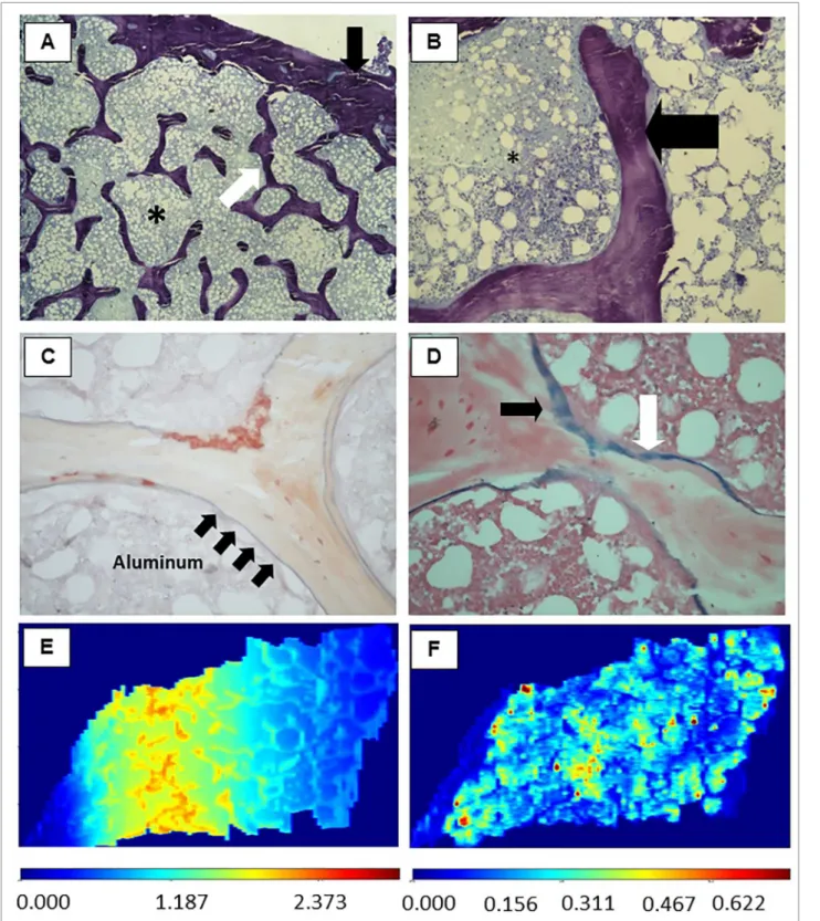
At this moment, the patient was included in a clini-cal study, and a transiliac bone biopsy was performed. The sample obtained consisted of two cortical and trabecular bone samples revealing the diagnosis of osteitis fibrosa. Unexpectedly, the coloration of solo-chrome azurine was positive for Al, covering 50% of
the bone surface.11-14 Pearls’ staining was positive for
iron in a similar extent (Figure 1A to 1D). Treatment with desferoxamine at 5 mg/kg once a week for 6 months was initiated, with follow-up exams revealing serum levels of Ca 10.2 mg/dL, P 2.2 mg/dL, iPTH 263 pg/mL, AP 47 IU/mL, and Al 4.7 mcg/L. At the end of the treatment, the patient was still asymptom-atic and without signs of Al intoxication or bone dis-ease. One year after being submitted to bone biopsy the patient underwent renal transplantation.
The unexpected diagnosis of Al deposition has led to the investigation of sources of exposure, such as medi-cations, water for HD, polyelectrolyte concentrates, and PD solution bags. Review of medical records has shown the patient had never used antacids, Al-based P binders, or any medications that could deliberately contain Al. In the last 3 years, he had never presented alterations in annual serum Al levels (GFAAS,
refer-ence range: < 30 mcg/L).7 Al detection analyses in HD
water treated by reverse osmosis provided negative re-sults (two samples, separated by one year) (methodol-ogy: inductively-coupled plasma optical emission
spec-trometry; reference range < 10 μg/L).7
We tested bone tissue samples, water used in the dialysis unit, polyelectrolyte concentrate solutions, and PD solution bags using inductively-coupled plasma mass spectrometry (ICP-MS) with laser abla-tion (LA) techniques. The chemical elements present in the sample were ionized by high plasma
tempera-ture. Only ions Fe+ and Al+ were selected, generating
a signal proportional to their quantities in the sam-ples. The technique is based on the use of a laser for ablating the sample, and the vapor generated in the process is transported by an inert gas (argon) to the inductively coupled plasma torch. LA-ICP-MS lecture can be converted to an imaging mode containing the
distribution of metal in the tissue.15-16 This
Samples Al concentration (µg/L)
PCHD (acid) trademark A 46.7 ± 0.8
PCHD (acid) trademark B (sample 1) 50.2 ± 0.9
PCHD (acid) trademark B (sample 2) 50.3 ± 1.5
SCB trademark A (sample 1) 46.9 ± 0.5
SCB trademark A (sample 2) 47.6 ± 0.5
Peritoneal dialysis solution trademark C 50.4 ± 0.7
Reverse osmosis outlet water (sample 1) 51.5 ± 0.6
Reverse osmosis outlet water (sample 2) 51.5 ± 0.7
Pre-treatment inflow water (sample 1) 51.5 ± 1.0
Pre-treatment inflow water (sample 2) 49.2 ± 0.5
Dialysate at the input of the HD machine 49.7 ± 0.5
HD: hemodialysis; PCHD: polyelectrolyte concentrate for hemodialysis; SCB: sodium bicarbonate concentrate; Al: aluminum.
TABLE 1 QUANTIFICATIONOF ALUMINUMBY ICP-MS INDIFFERENTWATERSAMPLESANDSOLUTIONSUSEDINDIALYSIS
UNIT. THECONCENTRATIONOF ALINALLSAMPLESWASVERYCLOSETOTHEVALUEOFTHENORMALIZATION CONCENTRATIONADDEDTOEACHSAMPLE
Wave–UP213). The images were treated with the
soft-ware LA-iMageS.16 Using a slide obtained from the
same fragment of bone tissue, the presence of Al and Fe deposits was confirmed, with clear discrimination between them (Figure 1 E-F).
Samples of water (N = 4), polyelectrolyte concen-trate solutions (N = 5; two different trademarks), and PD solution bags (N = 1), were normalized with the addition of a standard concentration of 50 μg/L of Al. The accuracy of the method was evaluated using the certified reference material of trace elements in natu-ral waters (SRM 1640A), obtaining a value of 52.9 ± 1.2 μg/L, compared with the certified value of 52.6 ± 1.8 μg/L. The results show that all analyzed samples by means of the ICP-MS method were negative for Al (Table 1).
D
ISCUSSIONAl intoxication in dialysis patients with classic signs and symptoms of Al-encephalopathy and osteomala-cia has ceased to be considered a clinical problem for
several years and is rather considered a rare event.2,3
However, deposition of Al in bone tissue, especially in the mineralization front (“bone intoxication by Al”) has a high prevalence in Brazil; a multicenter study found 2,507 bone biopsies from patients with clini-cal, radiologiclini-cal, or laboratory indications of bone disease. A prevalence of Al intoxication was 61.3% between 1985-1990, 38.7% between 1991-1996, and
42.5% between 1997-2001.9 A survey in 2008 from
data of the REBRABO study revealed a prevalence of
Al intoxication of 42% in 149 samples.8,10
Therefore, we believe that Al intoxication is still an important problem in Brazil and perhaps in other coun-tries. We hypothesize that its clinical manifestation is currently attenuated, with potential repercussions on anemia and bone disease. Al causes a decrease in heme synthesis and interferes with iron metabolism leading to microcytic anemia. Rao et al. studied 18 HD patients un-der erythropoietin (EPO) treatment and observed a trend for poor EPO-response in those with high deposition of
Al in osteoid surfaces.17 The accumulation of this metal
in bone tissue causes osteomalacia and adynamic bone disease. These effects are mediated through interferences on parathyroid hormone synthesis and release. Studies have reported Al deposition in parathyroid glands and
disturbances of calcium-sensing-receptor activity.18,19
However, discriminating the consequences or symptoms of Al toxicity can be difficult because they are usually nonspecific and are present in sev-eral diseases that affect patients with CKD. Related symptoms are proximal muscle weakness, bone pain, spontaneous fractures, acute alteration in mental sta-tus, and premature osteoporosis. It should be noted that serum Al levels are not reliable markers of organ deposition and bone biopsy is the definitive approach for the diagnosis of Al-related bone disease.
Two other possibilities for Al contamination are from medicine and food. Medications for patients un-dergoing dialysis may contain Al, especially in intra-venous form, such as dipyrone, erythropoietin, and
iron sulfate.20 The impact of this contamination is
- 0.1%) are absorbed from food sources. Factors that may influence absorption and its bioavailability are compounds that bind to Al in the intestinal lumen,
gastric acidity, and hardness of water consumed.21
Patients with celiac disease may have increased in-testinal permeability to Al, and can thus develop
Al-related bone disease.22 None of these conditions was
observed in our patient.
Unfortunately, we did not evaluate Al content in the ingested water and intravenous drugs used by the patient. We believe that the main source of Al expo-sure for CKD patients is the water used for dialysis, although we could not prove this. The ICP-MS could be a differential and complementary technique for a frequent evaluation of fluids and drugs used in the treatment of these patients, aiming to avoid expo-sure to Al. Additionally, its complementary technique (LA-ICP-MS) can discriminate safely which metal is deposited in the tissue. In this case report a limited amount of samples was analyzed, while the patient had contact with 360 L or more of water per week for years. We cannot affirm that polyelectrolyte concen-trates and PD solution bags were not sources of con-tamination, since only a few samples were analyzed.
C
ONCLUSIONAl intoxication may be largely under-diagnosed, perhaps in several regions of the world. There is an ur-gent need for clinical studies with bone biopsy in this field in order to confirm our hypothesis. Considering that doses of Al in fluids have limited diagnostic value and bone biopsy is an invasive procedure and restric-ted to a few centers, both ICP-MS and LA-ICP-MS are promising techniques that can be used to unders-tand the phenomenon of Al intoxication in patients on dialysis, helping in the identification of contami-nation sources. Systemic Al intoxication is an unusual event nowadays, but deposition of Al in bone tissue can be a frequent event, which can cause important clinical outcomes, such as fractures and death.
A
CKNOWLEDGMENTThe authors thank Espaço da Escrita - Coordenadoria Geral da Universidade - UNICAMP - for the language services provided, and Wagner Vasques Dominguez for the technical assistance.
R
EFERENCES2. Dunea G. Dialysis dementia: an epidemic that came and went. ASAIO J 2001;47:192-4.
3. Mahurkar SD, Salta R, Smith EC, Dhar SK, Meyers L Jr, Dunea G. Dialysis dementia. Lancet 1973;1:1412-5.
4. Sandhu G, Djebali D, Bansal A, Chan G, Smith SD. Serum con-centrations of aluminum in hemodialysis patients. Am J Kidney Dis 2011;57:523-5.
5. Malluche HH. Aluminum and bone disease in chronic renal failure. Nephrol Dial Transplant 2002;17:21-4.
6. Oliveira MB, Romão JE Jr, Zatz R. End-stage renal disease in Brazil: epidemiology, prevention, and treatment. Kidney Int Su-ppl 2005:S82-6.
7. Comission of the European Community (CEC). Resolution 86/ C184/04 of the Council concerning the protection of dialysis patients by minimizing the exposure to aluminum. Off J Eur Communities 1986;C184.
8. de Oliveira RB, Barreto FC, Custódio MR, Gueiros JE, Neves CL, Karohl C, et al. Brazilian Registry of Bone Biopsy (REBRA-BO): design, data elements and methodology. Braz J Nephrol 2014;36:352-9.
9. Araújo SM, Ambrosoni P, Lobão RR, Caorsi H, Moysés RM, Barreto FC, et al. The renal osteodystrophy pattern in Brazil and Uruguay: an overview. Kidney Int Suppl 2003; 85:S54-6. 10. Carbonara CEM, dos Reis LM, Sampaio EDA, Canziani MEF,
Moysés RMA, de Carvalho AB, et al. Relação entre o tipo de osteodistrofia renal e manifestações clínicas em pacientes com DMO – DRC. In: 28th Congresso Brasileiro de Nefrologia; 2016 Sep 14-17; Maceió, AL, Brazil.
11. Taylor A, Walker AW. Measurement of aluminium in clinical samples. Ann Clin Biochem 1992;29:377-89.
12. Buchanan MR, Ihle BU, Dunn CM. Haemodialysis related os-teomalacia: a staining method to demonstrate aluminium. J Clin Pathol 1981;34:1352-4.
13. Ellis HA, Pang MM, Mawhinney WH, Skillen AW. Demons-tration of aluminium in iliac bone: correlation between alu-minon and solochrome azurine staining techniques with data on flameless absorption spectrophotometry. J Clin Pathol 1988;41:1171-5.
14. Fernández-Martín JL, Menéndez P, Acuña G, Canteros A, Gó-mez C, Cannata JB. Staining of bone aluminium: comparison between aluminion and solochrome azurine and their correla-tion with bone aluminium contant. Nephrol Dial Transplant 1996;11:80-5.
15. Muñoz JJ, Drigo SA, Barros-Filho MC, Marchi FA, Scapula-tempo-Neto C, Pessoa GS, et al. Down-Regulation of SLC8A1 as a Putative Apoptosis Evasion Mechanism by Modulation of Calcium Levels in Penile Carcinoma. J Urol 2015;194:245-51. 16. López-Fernández H, de S Pessôa G, Arruda MA,
Capelo-Mar-tínez JL, Fdez-Riverola F, Glez-Peña D, et al. LA-iMageS: a sof-tware for elemental distribution bioimaging using LA–ICP–MS data. J Cheminform 2016;8:65-75.
17. Rao DS, Shih MS, Mohini R. Effect of serum parathyroid hor-mone and bone marrow fibrosis on the response to erythro-poietin in uremia. N Engl J Med 1993;328:171-5.
18. Díaz-Corte C, Fernández-Martín JL, Barreto S, Gómez C, Fernández-Coto T, Braga S, et al. Effect of aluminium load on parathyroid hormone synthesis. Nephrol Dial Transplant 2001;16:742-5.
19. Felsenfeld AJ, Machado L, Bover J, Trinidad P, Rodriguez M. Effect of aluminium on the development of hyperparathyroi-dism and bone disease in the azotaemic rat. Nephrol Dial Transplant 1993;8:325-34.
20. Bohrer D, Bertagnolli DC, de Oliveira SM, do Nascimento PC, de Carvalho LM, Pomblum SG. Drugs as a hidden source of aluminium for chronic renal patients. Nephrol Dial Transplant 2007;22:605-11.
21. Drüeke TB. Intestinal absorption of aluminium in renal failure. Nephrol Dial Transplant 2002;17:13-6.