www.bjorl.org.br
Brazilian Journal of
OtOrhinOlaryngOlOgy
1808-8694/$ - see front matter © 2014 associação Brasileira de Otorrinolaringologia e Cirurgia Cérvico-Facial. Published by Elsevier Editora ltda. all rights reserved.
DOi: 10.5935/1808-8694.20140015
REVIEW ARTICLE
Oral manifestations resulting from chemotherapy in children with
acute lymphoblastic leukemia
Everton Freitas de Morais
a,*, Jadson Alexandre da Silva Lira
a,
Rômulo Augusto de Paiva Macedo
a, Klaus Steyllon dos Santos
a,
Cassandra Teixeira Valle Elias
b, Maria de Lourdes Silva de Arruda Morais
a,ca Universidade Potiguar, Natal, RN, Brazil
b Liga Norte-Riograndense Contra o Câncer, Natal, RN, Brazil
c Universidade Estadual do Rio Grande do Norte (UERN), Natal, RN, Brazil
Received 30 July 2013; accepted 22 September 2013
KEYWORDS
Child; Drug therapy; Leukemia;
Oral manifestations
PALAVRAS-CHAVE
Criança; Quimioterapia; Leucemia;
Manifestações bucais
Abstract
Introduction: Acute lymphocytic leukemia is a type of cancer most common in children and it is characterized by excessive and disordered immature leukocytes in the bone marrow.
Aim: Identify most frequent oral manifestations in children with acute lymphocytic leukemia under chemotherapy treatment.
Methodology: The research was conducted on the eletronic database PubMed/Medline, Science Direct, Scielo and Scopus. It has been sought papers with full presentation, wrote in Portuguese, English and Spanish, published between January 1992 and April 2013.
Results: From studies primarily selected, only eight met the criteria of inclusion. All studies per-formed intraoral examinations to diagnose oral lesions. According to results, the most frequent lesions were mucositis, candidiasis, periodontitis and gingivitis. The oral health condition from acute lymphocytic leukemia carriers varied according oral hygiene of the patient.
Conclusion: The results of studies identiied such a great part of patients with ALL presented
some lesion in oral cavity during or after chemotherapy treatment. The dentist surgeon needs to recognize oral manifestations and intervene in the oral health of patients with ALL, contributing and helping with treatment.
© 2014 Associação Brasileira de Otorrinolaringologia e Cirurgia Cérvico-Facial. Published by Elsevier Editora Ltda. All rights reserved.
Manifestações orais decorrentes da quimioterapia em crianças portadoras de leucemia linfocítica aguda
Resumo
Introdução: A leucemia linfocítica aguda é um dos tipos de câncer mais comuns em crianças e é caracterizada pela produção excessiva e desordenada de leucócitos imaturos na medula óssea. Objetivo: Identiicar as manifestações orais mais frequentes em crianças portadoras de leuce -mia linfocítica aguda sob o tratamento quimioterápico.
Metodologia: A pesquisa foi realizada nas bases de dados eletrônicas PubMed/Medline, Science
Please cite this article as: Morais EF, Lira JAS, Macedo RAP, Santos KS, Elias CTV, Arruda-Morais MLS. Oral manifestations resulting from chemotherapy in children with acute lymphoblastic leukemia. Braz J Otorhinolaryngol. 2014;80:78-85.
* Corresponding author.
Introduction
Leukemia is a disease characterized by progressive and ex-cessive production of leukocytes in the bone marrow, whose immature forms start circulating in blood.1,2 The
dissemi-nated proliferation of blasts leads to a substitution of nor-mal bone marrow elements, resulting in the accumulation of immature cells in the blood. The etiology of leukemia is still uncertain, but many studies point to causal factors such as viral infection and radiation and chemical exposure. There are three different subtypes of lymphocytes, and thus
there are different types of leukemia, which are classiied
according to the cell involved, as well as the duration and characteristics of the disease.1-4
Acute lymphoblastic leukemia (ALL) represents approx-imately 80% of leukemias, and occurs mostly in children.1
ALL results in uncontrolled and excessive production of lym-phoid blasts, hindering the normal production of red and white cells, as well as platelets. The chances of survival have increased with advances in anticancer treatment mo-dalities.2,3
The irst signs of leukemia can regularly occur in the oral
cavity, especially in the acute phase of cancer, as common le-sions at this stage of the disease that can be observed and recognized primarily by the dentist.4,5 The most common
man-ifestations of leukemia in the oral cavity are gingival bleeding, hyperplasia, opportunistic infections, and bone alterations.
During the antineoplastic treatment, the lesions become even more severe, since chemotherapy acts on poorly dif-ferentiated or high-metabolism cells, affecting not only blast cells, but also normal body cells.3,4
The dentist needs to be aware of lesions caused by leu-kemia and the anticancer treatment in order to improve the patient’s oral health. Thus, this study aimed to per-form a systematic review of the literature on oral compli-cations secondary to chemotherapy performed in children with ALL.
Methods
A systematic search of articles in English, Portuguese, and Spanish published between January of 1992 and April of 2013 was performed in the PubMed/MEDLINE, Science Di-rect, Scopus, and SciELO databases. Studies whose the tar-get population was children aged 2 to 18 years with ALL and that evaluated the complications of anticancer treatment in the oral cavity were selected.
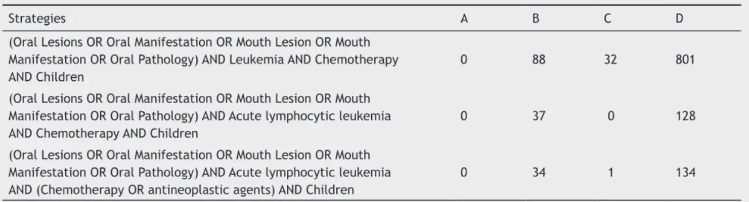
The search used the following terms: oral manifesta-tions, Leukemia, Acute Lymphoblastic Leukemia, ALL, che-motherapy, and children, as well as the corresponding words in Portuguese and Spanish. The Boolean operators AND, OR, NOT were used in the databases, when possible. The search strategies are shown in Table 1.
Direct, Scielo e Scopus. Procurou-se por artigos apresentados na íntegra, escritos em português, inglês e espanhol, publicados entre janeiro de 1992 e abril de 2013.
Resultados: Dos estudos selecionados primariamente, apenas oito atenderam aos critérios de inclusão. A população avaliada foi um grupo de crianças portadoras de leucemia linfócitica aguda. Todos os estudos realizaram exames intraorais para o diagnóstico das lesões bucais. De acordo com os resultados, as lesões mais frequentes foram mucosite, candidíase, periodon-tite e gengivite. A condição de saúde bucal dos portadores de leucemia linfócitica aguda variou de acordo com a higiene bucal do paciente.
Conclusão: Pacientes com LLA podem apresentar alguma lesão na cavidade oral durante ou após o início da quimioterapia. O cirurgião dentista necessita reconhecer as manifestações orais e intervir na saúde bucal do paciente com LLA, contribuindo e auxiliando no seu tratamento. © 2014 Associação Brasileira de Otorrinolaringologia e Cirurgia Cérvico-Facial. Publicado por Elsevier Editora Ltda. Todos os direitos reservados.
Table 1 Search strategies and number of articles found in the databases.
Strategies A B C D
(Oral Lesions OR Oral Manifestation OR Mouth Lesion OR Mouth Manifestation OR Oral Pathology) AND Leukemia AND Chemotherapy AND Children
0 88 32 801
(Oral Lesions OR Oral Manifestation OR Mouth Lesion OR Mouth Manifestation OR Oral Pathology) AND Acute lymphocytic leukemia AND Chemotherapy AND Children
0 37 0 128
(Oral Lesions OR Oral Manifestation OR Mouth Lesion OR Mouth Manifestation OR Oral Pathology) AND Acute lymphocytic leukemia AND (Chemotherapy OR antineoplastic agents) AND Children
0 34 1 134
After the initial search and retrieval of abstracts, four independent evaluators selected the relevant studies ac-cording to the inclusion and exclusion criteria. Longitudinal or cross-sectional controlled clinical trials that assessed oral manifestations in children with ALL were included in the sys-tematic review. Studies in languages other than those primar-ily chosen, in vitro studies, animal studies, literature reviews, case reports, and studies not associated with the target popu-lation or the oral manifestations of the disease were excluded.
The irst step in study selection was performed by an -alyzing the titles and abstracts. Subsequently, all studies whose titles or abstracts were considered relevant were ob-tained in full and thoroughly analyzed. Finally, the articles
reviewed and selected through consensus of the ive evalua -tors were included in the data systematization.
Results
Among the initially selected studies, 18 showed potential to be included in the systematic review; however, after thorough analysis of the studies, only eight met all the
in-clusion criteria. Among the selected studies, ive had been
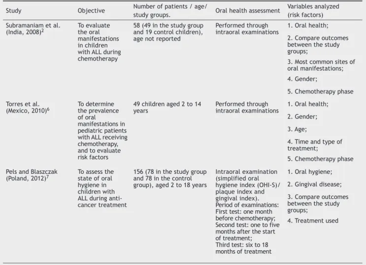
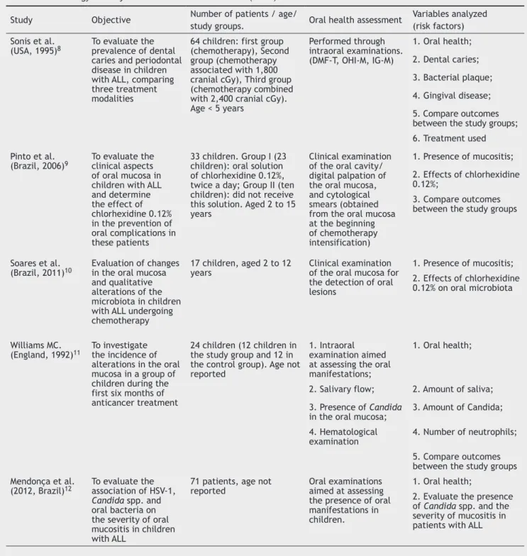
performed in developing countries and three in developed countries; the methods and results of the studies are sum-marized in Tables 2 and 3.
Regarding the proile of the studies, the sample size
ranged between 17 and 156 children, with a total sample size of 472 children aged 2 to 18 years. The selected studies were published between the years 1992 and 2012. Of the selected studies, seven were cross-sectional; only the study by Williams et al.11 was longitudinal. The main objective of
all selected studies was the evaluation of oral complications due to chemotherapy in patients with ALL.
The studies by Torres et al.6 and Subramaniam et al.2
evaluated the incidence of oral manifestations between gen-ders. In the study by Torres et al.,6 of the 49 children
stud-ied, 26 (53%) were males and 23 (47%) females. In the study by Subramaniam et al.,2 of the 58 children, 37 (63.8%) were
males and 21 (36.2%) females. The authors did not establish an association between gender and the frequency of oral manifestations. The remaining studies included in this sys-tematic review did not assess their patients according to gender. In all selected studies, the intraoral examination was used to establish the diagnosis of oral lesions during chemotherapy.
Of all evaluated studies, only Pels et al.7 reported the
performance of intraoral clinical examination before the start of chemotherapy. Among the variables analyzed by the authors were chemotherapy, type of drugs used by the children with ALL, use of chlorhexidine gluconate, oral hygiene, and place of residence.
Table 2 Methodology and objectives of the selected studies.
Study Objective Number of patients / age/
study groups. Oral health assessment
Variables analyzed (risk factors)
Subramaniam et al.
(India, 2008)2 To evaluate the oral
manifestations in children with ALL during chemotherapy
58 (49 in the study group and 19 control children), age not reported
Performed through
intraoral examinations 1. Oral health; 2. Compare outcomes between the study groups;
3. Most common sites of oral manifestations;
4. Gender;
5. Chemotherapy phase
Torres et al.
(Mexico, 2010)6 To determine the prevalence
of oral
manifestations in pediatric patients with ALL receiving chemotherapy, and to evaluate risk factors
49 children aged 2 to 14
years Performed through intraoral examinations 1. Oral health; 2. Gender;
3. Age;
4. Time and type of treatment;
5. Chemotherapy phase
Pels and Blaszczak
(Poland, 2012)7 To assess the state of oral
hygiene in children with ALL during anti-cancer treatment
156 (78 in the study group and 78 in the control group), aged 2 to 18 years
Intraoral examination (simplified oral hygiene index (OHI-S)/ plaque index and gingival index). Period of examinations: First test: one month before chemotherapy; Second test: one to five months after the start of treatment; Third test: six to 18 months of treatment
1. Oral hygiene;
2. Gingival disease;
3. Compare outcomes between the study groups;
Table 2 Methodology and objectives of the selected studies (cont.).
Study Objective Number of patients / age/
study groups. Oral health assessment
Variables analyzed (risk factors) Sonis et al.
(USA, 1995)8 To evaluate the prevalence of dental
caries and periodontal disease in children with ALL, comparing three treatment modalities
64 children: first group (chemotherapy), Second group (chemotherapy associated with 1,800 cranial cGy), Third group (chemotherapy combined with 2,400 cranial cGy). Age < 5 years
Performed through intraoral examinations. (DMF-T, OHI-M, IG-M)
1. Oral health;
2. Dental caries;
3. Bacterial plaque;
4. Gingival disease;
5. Compare outcomes between the study groups; 6. Treatment used
Pinto et al.
(Brazil, 2006)9 To evaluate the clinical aspects
of oral mucosa in children with ALL and determine the effect of chlorhexidine 0.12% in the prevention of oral complications in these patients
33 children. Group I (23 children): oral solution of chlorhexidine 0.12%, twice a day; Group II (ten children): did not receive this solution. Aged 2 to 15 years
Clinical examination of the oral cavity/ digital palpation of the oral mucosa, and cytological smears (obtained from the oral mucosa at the beginning of chemotherapy intensification)
1. Presence of mucositis;
2. Effects of chlorhexidine 0.12%;
3. Compare outcomes between the study groups
Soares et al.
(Brazil, 2011)10 Evaluation of changes in the oral mucosa
and qualitative alterations of the microbiota in children with ALL undergoing chemotherapy
17 children, aged 2 to 12 years
Clinical examination of the oral mucosa for the detection of oral lesions
1. Presence of mucositis;
2. Effects of chlorhexidine 0.12% on oral microbiota
Williams MC.
(England, 1992)11 To investigate the incidence of
alterations in the oral mucosa in a group of children during the first six months of anticancer treatment
24 children (12 children in the study group and 12 in the control group). Age not reported
1. Intraoral examination aimed at assessing the oral manifestations;
1. Oral health;
2. Salivary flow; 2. Amount of saliva;
3. Presence of Candida in the oral mucosa;
3. Amount of Candida;
4. Hematological examination
4. Number of neutrophils;
5. Compare outcomes between the study groups Mendonça et al.
(2012, Brazil)12 To evaluate the association of HSV-1,
Candida spp. and oral bacteria on the severity of oral mucositis in children with ALL
71 patients, age not reported
Oral examinations aimed at assessing the presence of oral manifestations in children.
1. Oral health;
2. Evaluate the presence of Candida spp. and the severity of mucositis in patients with ALL
Although six2,6-9,12 of the eight articles mentioned the
drugs used for chemotherapy, no clear correlation was es-tablished between oral complications and type of drug used. Pels et al.7 established an association between the phase of
chemotherapy and oral complications; oral manifestations were more common in the induction phase.
The most frequent oral lesions were mucositis,2,6,9-12
candidiasis,2,6,12 periodontitis,6-8 and gingivitis.6 The most
common sites of oral manifestation involvement were the oral mucosa and labial mucosa, according to the study by
Subramaniam et al.2 The oral manifestations and most
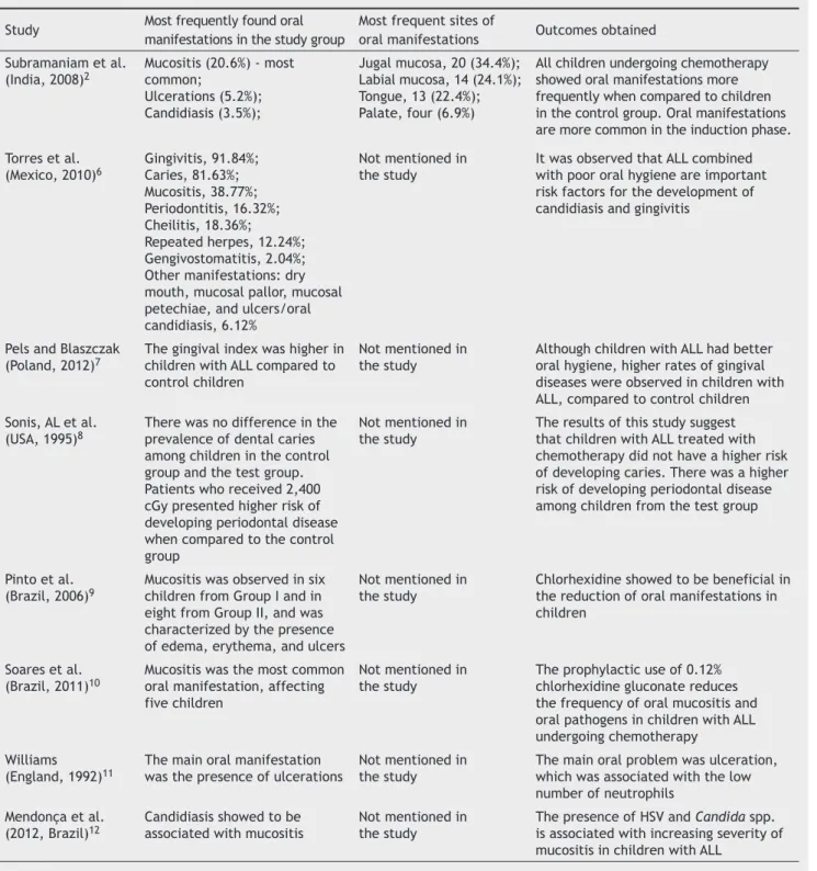
com-mon sites of the selected studies are presented in Table 3. The prevalence of oral manifestations was related to patients’ poor oral hygiene6 and antineoplastic
treat-ment;2,8 in the selected studies, no speciic drug was relat
-ed to the presence of oral manifestations. Another import-ant factor related to oral manifestations was the quality of life and social class of the patients with ALL,6 as the
Table 3 Outcomes obtained from the studies selected in the search.
Study Most frequently found oral
manifestations in the study group
Most frequent sites of
oral manifestations Outcomes obtained Subramaniam et al.
(India, 2008)2
Mucositis (20.6%) - most common;
Ulcerations (5.2%); Candidiasis (3.5%);
Jugal mucosa, 20 (34.4%); Labial mucosa, 14 (24.1%); Tongue, 13 (22.4%); Palate, four (6.9%)
All children undergoing chemotherapy showed oral manifestations more frequently when compared to children in the control group. Oral manifestations are more common in the induction phase.
Torres et al. (Mexico, 2010)6
Gingivitis, 91.84%; Caries, 81.63%; Mucositis, 38.77%; Periodontitis, 16.32%; Cheilitis, 18.36%; Repeated herpes, 12.24%; Gengivostomatitis, 2.04%; Other manifestations: dry mouth, mucosal pallor, mucosal petechiae, and ulcers/oral candidiasis, 6.12%
Not mentioned in the study
It was observed that ALL combined with poor oral hygiene are important risk factors for the development of candidiasis and gingivitis
Pels and Blaszczak (Poland, 2012)7
The gingival index was higher in children with ALL compared to control children
Not mentioned in the study
Although children with ALL had better oral hygiene, higher rates of gingival diseases were observed in children with ALL, compared to control children Sonis, AL et al.
(USA, 1995)8
There was no difference in the prevalence of dental caries among children in the control group and the test group. Patients who received 2,400 cGy presented higher risk of developing periodontal disease when compared to the control group
Not mentioned in the study
The results of this study suggest that children with ALL treated with chemotherapy did not have a higher risk of developing caries. There was a higher risk of developing periodontal disease among children from the test group
Pinto et al.
(Brazil, 2006)9 Mucositis was observed in six children from Group I and in
eight from Group II, and was characterized by the presence of edema, erythema, and ulcers
Not mentioned in the study
Chlorhexidine showed to be beneficial in the reduction of oral manifestations in children
Soares et al.
(Brazil, 2011)10 Mucositis was the most common oral manifestation, affecting
five children
Not mentioned in the study
The prophylactic use of 0.12% chlorhexidine gluconate reduces the frequency of oral mucositis and oral pathogens in children with ALL undergoing chemotherapy
Williams (England, 1992)11
The main oral manifestation was the presence of ulcerations
Not mentioned in the study
The main oral problem was ulceration, which was associated with the low number of neutrophils
Mendonça et al. (2012, Brazil)12
Candidiasis showed to be associated with mucositis
Not mentioned in the study
The presence of HSV and Candida spp. is associated with increasing severity of mucositis in children with ALL
Discussion
The selected studies in this systematic review evaluated the oral manifestations in patients with ALL. The studies by Torres et al.6 and by Subramaniam et al.2 estimated the
incidence between genders, but did not establish an associ-ation between gender and frequency of oral manifestassoci-ations; no other study showed such association.
In the study by Subramaniam et al.,2 oral manifestations
were observed in 77.8%, 36.4%, 70%, and 66.7% of children, divided according to the groups in the study. In the study by Torres et al.,6 approximately 95% of patients had some type of oral manifestation. These results corroborate the indings
from other studies.13,14
the drugs used are often secreted through the saliva, which results in drug exposure in the oral cavity.15
How-ever, among the studies selected, only six2,6-9,12
men-tioned the drug combinations used in chemotherapy, and none directly correlated the oral manifestations to a
spe-ciic type of drug.
Among the drugs most often associated with oral man-ifestations, teniposide, paclitaxel, methotrexate, idarubi-cin, epirubiidarubi-cin, doxorubiidarubi-cin, cisplatin, and cytarabine are associated with mucositis, xerostomia, gingival bleeding, and other diseases of the oral cavity.16,17 Methotrexate,
bleomycin, doxorubicin, cisplatin, vinblastine and vincris-tine are drugs that produce direct toxicity of some of their antimetabolites and other synthetic agents, such as hy-droxyurea and procarbazine hydrochloride, which lead to glandular degeneration, changes in collagen, and epithelial dysplasia.15,18 Drug combinations used during chemotherapy
in the studies are shown in Table 4.
Mucositis was the most common oral manifestation in the study by Subramaniam et al.,2 affecting 20.6% of
pa-tients with leukemia, whereas among children who were not undergoing treatment, only 3.4% had mucositis. For most researchers, mucositis is caused by chemotherapy and oc-curs due to a dynamic process, resulting in the destruction of the basal cells; the same is stated by Sonis,19 in a study
published in 1998, who also mentioned that tissue dam-age was caused by direct contact with the antineoplastic agents. In the study by Torres et al.,6 mucositis was not the
most frequent disease; nevertheless, it affected approxi-mately 38.7% of children.
In the study by Pels et al.,7 although children with ALL
had better oral hygiene, they had more oral manifestations than children from the comparison group; oral mucositis was the most common lesion among patients undergoing chemotherapy. Pinto et al.9 concluded that good oral
hy-giene associated with the use of 0.12% chlorhexidine
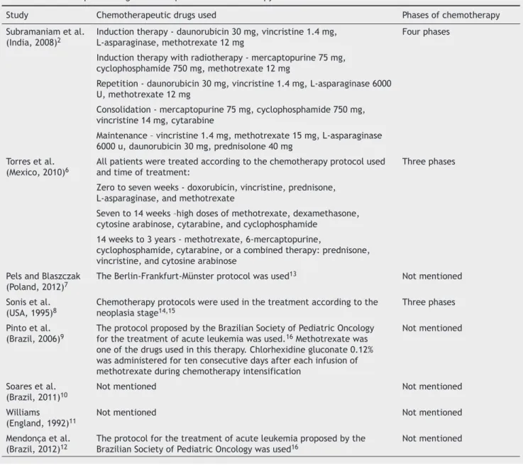
glu-Table 4 Chemotherapeutic drugs used and phases of chemotherapy.
Study Chemotherapeutic drugs used Phases of chemotherapy
Subramaniam et al.
(India, 2008)2 Induction therapy - daunorubicin 30 mg, vincristine 1.4 mg, L-asparaginase, methotrexate 12 mg Four phases
Induction therapy with radiotherapy - mercaptopurine 75 mg, cyclophosphamide 750 mg, methotrexate 12 mg
Repetition - daunorubicin 30 mg, vincristine 1.4 mg, L-asparaginase 6000 U, methotrexate 12 mg
Consolidation - mercaptopurine 75 mg, cyclophosphamide 750 mg, vincristine 14 mg, cytarabine
Maintenance – vincristine 1.4 mg, methotrexate 15 mg, L-asparaginase 6000 u, daunorubicin 30 mg, prednisolone 40 mg
Torres et al.
(Mexico, 2010)6 All patients were treated according to the chemotherapy protocol used and time of treatment: Three phases
Zero to seven weeks - doxorubicin, vincristine, prednisone, L-asparaginase, and methotrexate
Seven to 14 weeks –high doses of methotrexate, dexamethasone, cytosine arabinose, cytarabine, and cyclophosphamide
14 weeks to 3 years - methotrexate, 6-mercaptopurine,
cyclophosphamide, cytarabine, or a combined therapy: prednisone, vincristine, and cytosine arabinose
Pels and Blaszczak
(Poland, 2012)7 The Berlin-Frankfurt-Münster protocol was used
13 Not mentioned
Sonis et al. (USA, 1995)8
Chemotherapy protocols were used in the treatment according to the neoplasia stage14,15
Three phases
Pinto et al.
(Brazil, 2006)9 The protocol proposed by the Brazilian Society of Pediatric Oncology for the treatment of acute leukemia was used.16 Methotrexate was
one of the drugs used in this therapy. Chlorhexidine gluconate 0.12% was administered for ten consecutive days after each infusion of methotrexate during chemotherapy intensification
Not mentioned
Soares et al. (Brazil, 2011)10
Not mentioned Not mentioned
Williams
(England, 1992)11 Not mentioned Not mentioned
Mendonça et al. (Brazil, 2012)12
The protocol for the treatment of acute leukemia proposed by the Brazilian Society of Pediatric Oncology was used16
conate prevented oral manifestations in children with ALL, especially mucositis, due to its capacity to interfere with microorganism adhesion, when used as an antiseptic in the oral cavity.
The study by Soares et al.10 showed similar results to
the study of Pinto et al.9 children who used chlorhexidine
gluconate had fewer oral manifestations when compared to the control group. A microbiological analysis showed a re-duction in pathogenic microorganisms in the oral cavity, and mucositis, the most frequent oral manifestation, was
ob-served in ive children (29.4% of the sample). Other studies
also evaluated the use of chlorhexidine in patients with ALL, with positive results in reducing oral manifestations caused by chemotherapy.20,21
In the study by Torres et al.,6 the most common oral
manifestation was gingivitis, affecting 91.84% of the sam-ple; the high rate of gingivitis in this study was associated to poor oral hygiene, in addition to chemotherapy. The chemo-therapy phase also showed an association with the onset of gingivitis. In the study by Subramaniam et al.,2 the rate
of gingivitis was lower: less than 2% among patients. This difference may be due to the better oral hygiene of the chil-dren participating in that study,2 corroborating the indings
of Trindade et al.,14 who demonstrated that oral hygiene
should be encouraged at any stage of treatment, since it can prevent oral and systemic manifestations.
The study by Sonis et al.8 concluded that patients with ALL
treated with chemotherapy had the same probability of den-tal caries as children from the control group. For the authors, the presence of caries was associated with poor oral hygiene
of the participants. These results corroborate the indings of
Sepet et al.,22 who assessed the presence of caries among
children in the maintenance phase of chemotherapy and in a group of healthy children, observing no correlation between the presence of caries and the use of chemotherapeutic drugs. Primary herpetic gingivostomatitis was observed in 2.04% of the patients in the study by Torres et al.,6 and
recurrent herpes in 12.24% . The presence of herpes in that study could have been caused by the participants’ low im-munity, associated with exposure to the virus; this result corroborates other studies that also reported the presence of herpetic stomatitis in children with ALL.23,24
In the longitudinal study by Williams et al.,11 all patients
had ulcerations in the oral mucosa, whereas oral candidiasis was observed in 10% of tests performed during the study period. The presence of the disease was associated with the low number of neutrophils of leukemia patients in
combina-tion with low salivary low caused by chemotherapy. In the
study by Mendonça et al.,12 oral candidiasis was also
associ-ated with the presence and severity of mucositis; antifungal prophylaxis was suggested in patients with ALL with muco-sitis. This same suggestion was made in the study by Pine et al., published in 2010.25 Other oral manifestations observed
in these studies were dry mouth, gingival bleeding, oral er-ythema, toothache, and primary herpetic stomatitis.
Conclusion
The oral health status of patients with ALL varies according to a number of factors. The results of the studies demon-strated that most patients with ALL had some lesion in the
oral cavity during or after the start of chemotherapy. The dentist is responsible for recognizing these lesions and inter-vening in the oral health of these patients, thereby contrib-uting to their treatment and quality of life improvement. New studies are required to better elucidate the subject, especially regarding the prevention and control of these oral manifestations.
Conlicts of interest
The authors declare no conlicts of interest.
References
1. Costa SS, Silva AM, Macedo IAB. Conhecimento de manifesta-ções orais da Leucemia e protocolo de atendimento odontoló-gico. Rev Odontol Univ São Paulo. 2011;23:70-8.
2. Subramaniam P, Babu KL, Nagarathna J. Oral manifestations in acute lymphoblastic leukemic children under chemotherapy. J Clin Pediatr Dent. 2008;32:319-24.
3. Hamerschlak N. Leukemia: genetics and prognostic factors. J Pediatr. 2008;84:52-7.
4. Maeda YC. Manifestações bucais da leucemia e do tratamento antineoplásico [Monograph]. Piracicaba: Faculdade de Odonto-logia de Piracicaba; 2008.
5. Silva LCP, Carneiro FM, Cruz RA. Manifestações bucais das leu-cemias agudas na infância. Arq Bras Odontol. 2008;4:40-54. 6. Torres EP, Ruíz MSR, Alejo GF, Hernández SJF, Pozos GAJ. Oral
manifestations in pediatric patients receiving chemothe-rapy for acute lymphoblastic leukemia. J Clin Pediatr Dent. 2010;34:275-80.
7. Pels E, Mielnik BM. Oral hygiene in children suffering from acu-te lymphoblastic leukemia living in rural and urban regions. Ann Agric Environ Med. 2012;19:529-33.
8. Sonis AL, Waber DP, Sallan S, Tarbell NJ. The oral health of lon-g-term survivors of acute lymphoblastic leukaemia: a compari-son of three treatment modalities. Eur J Cancer B Oral Oncol. 2012;31:250-2.
9. Pinto LP, Souza LB, Gordón-Núñez MA, Soares RC, Costa EMMB, Aquino ARL, et al. Prevention of oral lesions in children with acute lymphoblastic leukemia. Int J Pediatr Otorhinolaryngol. 2006;70:1847-51.
10. Soares AF, Aquino ARL, Carvalho CHP, Nonaka CFW, Almeida D, Pinto LP. Frequency of oral mucositis and microbiologi-cal analysis in children with acute lymphoblastic leukemia treated with 0.12% chlorhexidine gluconate. Braz Dent J. 2011;22:312-6.
11. Williams MC, Martin MV. A longitudinal study of the effects on the oral mucosa of treatment for acute childhood leukaemia. Int J Paediatr Dent. 1992;2:73-9.
12. Mendonça RM, Araújo M, Levy CE, Morari J, Silva RA, Yunes JA, et al. Prospective evaluation of HSV, Candida spp., and oral bacteria on the severity of oral mucositis in pediatric acute lymphoblastic leukemia. Support Care Cancer. 2012;20:1101-7. 13. Ribas MO, Costa NP. Estudo das observações clínicas,
esto-matológicas e radiográicas das alterações dentárias e ósseas
nos pacientes com leucemia na infância. Rev Odonto Ciênc. 1995;2:151-84.
14. Trindade AKF, Biases RCCG, Guedes FG, Pereira BC, Sousa ED, Queiroga AS. Manifestações orais em pacientes pediátricos leu-cêmicos. Arq Odontol. 2009;45:22-9.
16. Fonseca SM, Almeida EPM, Massunaga VM. Administração dos quimioterápicos. In: Fonseca SM, Machado RCL, Paiva DRS, Al-meida EPM, Massunaga VM, Junior WR, et al. Manual de qui-mioterapia antineoplásica. Rio de Janeiro: Reichmann e Affon-so; 2000. p. 16-9.
17. Miller D. Quimioterapia. In: Administração de medicamentos. Rio de Janeiro: Reichmann e Affonso; 2002. p. 369-82. 18. Bensaudoun RJ, Magné N, Marcy PY, Dermard F. Chemotherapy
-and radiotherapy-induced mucositis in head and neck cancer patients; new trends in pathophysiology, prevention and treat-ment. Eur Arch Otorhinolaryngol. 2007;258:481-7.
19. Sonis ST. Mucositis as a biologic process: a new hypothesis for the development of chemotherapy-induced mucositis. Oral Oncol. 1998;34:39-43.
20. Brito CA, Araújo DS, Granja JG, Souza SM, Lima MAG, Oliveira MC. Efeito da clorexidina e do laser de baixa potência na pre-venção e no tratamento da mucosite oral. Rev Odontol UNESP. 2012;41:236-41.
21. Ferretti GA, Raybould TP, Brown AT, McDonald JS, Greenwood M, Maruyama Y, et al. Chlorhexidine prophylaxis for chemothe-rapy- and radiothechemothe-rapy-induced stomatitis: a randomized dou-ble-blind trial. Oral Surg Oral Med Oral Pathol. 1990;69:331-8. 22. Sepet E, Aytepe Z, Ozerkan AG, Yalman N, Guven Y, Anak
S, et al. Acute lymphoblastic dental health na children in maintenance therapy. J Clin Pediatr Dent. 1998;22:257-60. 23. Marques HH, Yamamoto M, Odone FV. Varicela-zoster em
imu-nodeprimidos: Estudo de 12 casos. Rev Pediat. 1981;3:335-7. 24. Childers NK, Stinnett EA, Wheeler P, Wright JT, Castleberry
RP, Dasanayake AP. Oral complications in children with cancer. Oral Surg Oral Med Oral Path. 1993;75:41-7.