http://dx.doi.org/10.1590/s2175-97902017000300215
A
r
*Correspondence: G. Zheng. Hubei University of Chinese Medicine, Wuhan 430065 - People’s Republic of China. E-mail address: [email protected]. # These authors contributed equally to this work.
Total flavonoids from Ampelopsis megalophylla suppress
proliferation of vascular smooth muscle cells in vivo and in vitro
Zhenpeng Qiu
1#, Junxuan Zhou
1#, Junjie Hu
1, Yong Wu
1, Guohua Zheng
2 *1College of Pharmacy, Hubei University of Chinese Medicine, Wuhan, People’s Republic of China, 2Laboratory of Chinese Medicine Resource and Compound Prescription (Ministry of Education), Hubei University of Chinese Medicine, Wuhan,
People’s Republic of China
Various beneits of lavonoids for ameliorating cardiovascular diseases have been demonstrated. However, the lowering efects on blood pressure caused by antiproliferative potentials of lavonoids in vascular smooth muscle cells are rare. In this study, the antihypertensive efects of total lavonoids from Ampelopsis megalophylla were investigated. The dynamic pressure values and the rate of media thickness versus
lumen diameter were measured by the tail-cuf system and H&E staining in vivo, respectively. The mRNA expressions of ACE, Ang II, eNOS, c-Myc, cyclin D1 and p27Kip1 in thoracic aorta or A7r5 cells were measured by qPCR, respectively. The protein expressions of c-Myc, Cyclin D1, p27Kip1 and β-catenin in tissues or A7r5 cells were measured by Western blot assay. Total lavonoids of A. megalophylla (TFAM) reduced the expressions of ACE and Ang II, and elevated the content of eNOS in thoracic aorta cells of SHRs. Furthermore, TFAM decreased the mRNA and protein expressions of c-Myc and cyclin D1 by
repressing the Wnt/β-catenin-mediated TCF/LEF transcriptional activation both in vivo and in vitro, which is synergetic with the up-regulation of p27Kip1 expression. Our study provided evidence for developing lavonoids from A. megalophylla as herbal supplements to prevent against cardiovascular diseases by suppressing vascular remodeling.
Keywords:Ampelopsis megalophylla. Flavonoids/efects. Cardiovascular remodeling. β-Catenin.
INTRODUCTION
Although noticeable pharmacological achievement advances on the therapeutic strategy of hypertension, the epidemiological characteristics of hypertension are less optimistic. Hypertension remains an obsessional public health challenge globally. The amount of adults with hypertension in 2025 is forecasted to elevate by approximately 60% to aggregate 1.6 billion (Kearney et al., 2005). Hence, hypertension is unambiguously considered as the main cause of cardiovascular disease (CVD) (Graham
et al., 2007). The pathological changes of hypertension are involved with the decline of cardiac-cerebral vascular function, including the proliferation of vascular smooth muscle cells, which contributes to vascular remodeling (Gibbons, Dzau, 1994). Generally, the long-term elevated blood pressure (BP) level at or above 140/90 mmHg is
deined as essential hypertension (Gu et al., 2002).Some pharmacological agents have been applied to ameliorate hypertension. Because many of these agents require lifelong treatment, numerous patients still have poorly stabilized BP
and sufer hypertensive complications.
Multiple plant-derived chemicals can signiicantly
alleviate high BP (Herrera-Arellano et al., 2007; Xiong et al., 2013). It has been indicated that dietary polyphenols may be beneficial to the prevention and treatment of hypertension (Galleano, Pechanova, Fraga, 2010). Flavonoids represent the major class of polyphenols, in addition to their antioxidant effects, which exhibit a comprehensive spectrum of pharmacological activities. Although the crucial roles of renin-angiotensin system (RAS), oxidative stress, and vascular remodeling have been elaborated in the development and the persistence of hypertension, the pathomechanism of hypertension is
apparently complicated (Beevers, Lip, O’Brien, 2001).
The emerging and largely consistent evidences demonstrate
that lavonoids can improve endothelial function and may
the anthocyanin cyanidin-3-glucoside could increase the expression of endothelial Nitric Oxide Synthase (eNOS) in
vascular endothelial cells. In fact, consumption of
lavanol-rich dark chocolate decreases BP and elevates insulin
sensitivity in healthy volunteers. Further indings support a potentially beneicial action of lavanols in chocolate on BP
and vasorelaxation in essential hypertensives (Grassi et al.,
2005). Moreover, the lavonoid quercetin also reduces the
elevated blood pressure, the cardiac and renal hypertrophy, and the dysfunctional vascular changes in spontaneously hypertensive rats (SHRs) (Duarte et al., 2001). Quercetin and its metabolites exhibit selective vasodilator effects toward the resistance vessels in the isolated rat thoracic and abdominal aorta (Perez-Vizcaino et al., 2002). Thus, using
the natural lavonoids to moderate hypertension could be an efective complementary strategy to reduce the
cardio-cerebral vascular accidents.
Dihydromyricetin (DHM) is a kind of bioactive
lavonoids and identiied as the beneit constituent in the
tender stems and leaves of Ampelopsis grossendentata
(Zhang et al., 2007a). Pharmacokinetic studies of dihydromyricetin and myricetin in rat plasma by High
Performance Liquid Chromatography with Diode Array Detector (HPLC-DAD) also have been assessed after
oral administration of Ampelopsis grossedentata (Zhang
et al., 2007b). In fact, dihydromyricetin is the main
lavonoid in other ampelopsis, including in Ampelopsis megalophylla (See supporting information). The present study demonstrates that the ameliorative potential of total
lavonoids in Ampelopsis megalophylla Diels et Gilg, a
kind of lavonoid-rich tea resources which is consumed in Western Hubei (China), have efects on the Angiotensin
II (Ang II), which is associated with the proliferation of vascular smooth muscle cells in both spontaneously hypertensive rats and A7r5 vascular smooth muscle cells.
MATERIAL AND METHODS
Plant collection and identification
Medicinal materials were collected at the Enshi, Hubei in China during July 2012. A botanist at the Herbarium of Hubei University of Traditional Chinese Medicine identified and authenticated the plants as
Ampelopsis megalophylla Diels et Gilg, and a voucher specimen 201207AM was preserved at the herbarium.
Preparation of total flavonoids
The leaves of A. megalophylla were naturally air-dried and pulverized. The powders (2.0 kg) were extracted
with ethanol (95%, v/v) under relux for 4 h by three times.
The filtrations were vacuum-concentrated to obtain a crude ethanol extract (540 g). Then the crude extract was
dissolved in water and iltrated. The water solution was
subjected to polyamide gel column chromatography eluted with ethanol (70%, v/v). Finally, the ethanol elution was
merged and concentrated. The yield of total lavonoids
was 3.4%. Phytochemical analysis of the plant extracts has previously been carried out using standard procedures as reported (Xie et al., 2014). Dihydromyricetin and
myricetin were analyzed quantitatively by HPLC-DAD,
and considered as the major components in the extracts (total flavonoids of A. megalophylla, TFAM) (See Supporting information). The extract was dispersed in sodium chloride for further intragastric administration.
Animals and in vivo experiment design
The SHR model is a kind of classical hypertensive models, which is similar to primary hypertension in humans (Bauersachs et al., 1998). SHR is applied to identify antihypertensive agents because this model provides analogous pattern of molecular pathology as in hypertensive patients. From the early stage to the terminal stage, blood pressure is elevated gradually, which is attributed to progressively increased vascular resistance and continuously activated RAS. Therefore, SHRs (purchased
from Vital River Laboratory Animal Technology Co., Ltd.,
Beijing, China) were used in present study. The SHRs received standard food and water ad libitum and at 22-24
°C in an artiicial 12 h/12 h light/dark cycle. The SHRs (20 weeks) were treated with TFAM-L, TFAM-M and TFAM-H
twice a day at 10:00 and 15:00 from 4 to 10 week of the evaluation (90, 180, 360 mg/kg, respectively, diluted in drinking water, gavage administration).
Blood pressure measurement
Systolic blood pressure (SBP) was recorded
noninvasively using tail-cuf system (Duarte et al., 2001) every 2 days. In brief, after pre-warming the SHRs at 36 °C for 15 min, SBP was measured every other week from the initiation of the evaluation. Mean blood pressures (n=5) were obtained for each animal when the pressure value is relatively stable.
Evaluation of vascular media thickness and lumen diameter of thoracic aorta
the thoracic aorta segments of SHRs were separated immediately after euthanasia and stored in Phosphate
Bufer Solution (PBS) at room temperature. After gross
inspection of the segments, the intact regions were selected for further analysis. The thoracic aorta were intersected into sequential ring segments and further stained by using
hematoxylin and eosin (H&E) for determination of the
media thickness and lumen diameter.
Analysis of serum nitric oxide content
After anesthetizing animals in the end of the experiment, the whole blood was obtained from the carotid
artery of sacriicial animals and the serum was separated
by centrifugation (3000 rpm/min). Nitrate reductase method was applied to determine the levels of nitric oxide using Nitric Oxide (NO) Assay Kit (Jiancheng, China) according to the manufacturer’s manual.
Reverse transcription and real-time quantitative PCR
Total cellular RNA was isolated by using Trizol reagent (Invitrogen, USA), and first-strand cDNA
synthesis with 1 μg of RNA was performed using MMLV
reverse transcriptase (Promega, USA), according to the manufacturer’s instructions. The PCR profile was 95 °C for 2 min, 40 cycles of 95 °C for 5 s, 55 °C for 12 s, and 55 °C for 12 s, followed by extension for 7 min at
72°C. Veriied primer sequences for amplifying mRNA
of angiotensin converting enzyme (ACE), Ang II, c-Myc,
p27Kip1 and Bcl-2 were listed in Table I. The mRNA amount
in diferent groups was normalized to relative expression of β-actin.
Western blot
Whole cell extracts and nucleoproteins were prepared as described (Wang et al., 2015). Equal amounts of samples were fractionated by sodium dodecyl sulfate polyacrylamide gel electropheresis (SDS-PAGE) of 15% tricine gels and blotted onto polyvinylidene fluoride (PVDF) membranes by Trans-Blot Turbo Blotting System (Bio-Rad, USA). The following primary antibodies were used: c-Myc (9402S, Cell Signaling Technology (CST), Beverly, MA, USA), Cyclin D1 (2922S, CST, Beverly, MA, USA), p27Kip1 (2552S, CST, Beverly, MA, USA)
and HRP-conjugated β-actin (5125S, CST, Beverly, MA, USA). For investigating the expression of β-catenin in
nucleus, the protein-loaded membranes were probed with
HRP-conjugated anti-β-catenin primary antibodies. The bands of target protein were analyzed using Image Lab
5.1 (Bio-Rad, USA). β-actin and Histone H3 (Sigma, USA) served as the internal control for quantitating protein expression in whole cell extracts (c-Myc, Cyclin D1 and p27Kip1) and the nucleus extracts (β-catenin), respectively.
Cell culture, Ang II stimulus and CCK-8 assay
A7r5 cells, which are the commercial cells derived from the aorta of fetal rat, were purchased from American Type Culture Collection (Rockville, MD, USA) and
TABLE I - Sequences of polymerase chain reaction (PCR) primer used in real-time PCR
Sequences (5’ to 3’) Length (bp)
β-actin Forward CGTTGACATCCGTAAAGACCTC 110
Reverse TAGGAGCCAGGGCAGTAATCT
c-Myc Forward CCAGCCAAGGTTGTGAGGTTAGG 176
Reverse CAGACGTAAACAGCTCCGAA
cyclin D1 Forward GAACAAACAGATCATCCGCAAACAC 231
Reverse TGCTCCTGGCAGGCCCGGAGGCAGT
eNOS Forward CTCAATGTCGTGTAATCGGTCT 98
Reverse TCCACCGTTACCAGACAACTATCC
ACE Forward TCCACCGTTACCAGACAACTATCC 119
Reverse CTGCGTATTCGTTCCACAACACCT
Ang II Forward AGCACGACTTCCTGACTTGGATAAA 245
Reverse AGACTCTGTGGGCTGCTCCTCCTC
p27 Forward CATTCAATGGAGTCAGCGAT 120
incubated as previously described (Filipeanu et al., 2001). For investigating the effects of TFAM on the Ang II-mediated vascular smooth muscle cell proliferation, Ang II (10-6 μM) was initially employed to stimulate A7r5 cells
for 24 h and subsequently the cells were administrated
with TFAM in diferent concentrations (10-50 μg/mL)
for 24 h. Cell viability was measured in 96-well plates by Cell Counting Kit-WST-8 (CCK-8) assay as our previous research (Zhou et al., 2015).
Luciferase reporter assay
The effect of Ang II on T-cell Factor (TCF)/
Lymphoid Enhancing Factor (LEF) family-dependent
gene transcription was evaluated using the TOP-flash TCF reporter plasmid containing two sets of three copies of the TCF binding site upstream of the thymidine kinase (TK) minimal promoter and luciferase open reading frame (Millipore, Billerica, MA). A7r5 cells were co-transfected
with TOP-lash or FOP-lash plasmid (1 μg), and Renilla reporter plasmid (0.1 μg) (pRL-TK; Promega). The cells were further cultured with or without Ang II (1 μM) for 12
h in serum-free medium, and then sequentially post-treated
with or without TFAM (10-30 μg/mL). Luciferase assay was determined using Luciferase Assay System (Promega, US).
Statistical analysis
Data were expressed as the means ± standard
deviation (SD) for at least three experiments (each in duplicate) and analyzed using a t-test or one-way ANOVA
for comparison. The diference was considered signiicant
if the probability was <0.05 (*) or <0.01 (**).
RESULTS
Ameliorative effects of TFAM on hemodynamic and physical parameters in spontaneously hypertensive rats
The dosages of total lavonoids in TFAM-L, TFAM-M
and TFAM-H, are 90 mg/kg, 180 mg/kg and 360 mg/kg, respectively. No significant change in body weight was observed by the TFAM treatment. The average body weights of the animals at the end of the study (7 weeks) in the SHR untreated group, TFAM treated group and positive control group were 369.17 ± 4.9, 359.06 ± 4.1 and 366.2 ± 4.13 g, respectively. Moreover, no significant differences were observed in the liver, kidney and heart weights at the end of
the experiments between diferent treatment groups.
Generally, the values of SBP and heart rate (HR) in SHRs without TFAM treatment were 182 ± 8.6 mm Hg.
To validate the antihypertensive efects of TFAM under
in vivo conditions, TFAM were orally administered to SHRs with different concentrations followed by SBP measurements every week (Figure 1A). The maximum SBP decrease (-21.1 ± 4.46 mmHg) was obtained early in the experiment at 2 weeks, and the effect gradually
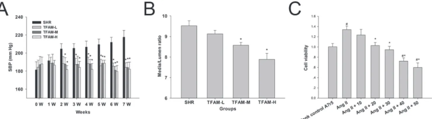
FIGURE 1 - TFAM ameliorated the hemodynamic and physical parameters in spontaneously hypertensive rats, and suppressed Ang II-induced A7r5 cell proliferation in Vitro. (A) TFAM restored the systolic blood pressure in SHRs. SBP (mmHg) values from
untreated SHRs or those treated with TFAM (TFAM-L, 90 mg/kg; TFAM-M 180 mg/kg; TFAM-H, 360 mg/kg) were recorded during light and dark cycles each week. Summary graphs to demonstrate the efects of TFAM on SBP. * indicates P < 0.01 compared
to the SHR group. (B) TFAM attenuated the rate of media thickness versus lumen diameter. The media thickness of the thoracic aorta and lumen diameter were obtained in histologic sections, respectively. Data represented as mean ± SD from n = 6 rats per treatment group in (A) and (B), the values given are the mean ± SD of three independent experiments. *P < 0.05 compared with the control SHR group. (C) CCK-8 assay was employed to evaluate the efects of TFAM on Ang II-induced A7r5 cell proliferation.
and signiicantly (p < 0.05) enhanced from 3 to 7 weeks.
Moreover, the HR in SHRs was observed with no
signiicance among groups.
TFAM reduced the rate of media thickness versus lumen diameter
The media thickness of the thoracic aorta in SHR group was highly variable, with an average thickness
of 136.96 ± 7.23 μm. Compared with SHR group, the thoracic aorta had a signiicantly lower media thickness
in TFAM treated group. We next evaluated whether the
media thickness had an efect on the functional viability of
thoracic aorta, especially on the endothelial function. To do so, the lumen diameter of thoracic aorta was investigated, and the rates of media thickness versus lumen diameter in
each group were obtained. Compared to control group, the rate of media thickness versus lumen diameter in TFAM-H
group was signiicantly reduced, suggesting that TFAM
could attenuate the remodeling process of vascular smooth muscle cells in SHRs (Figure 1B).
Serum nitric oxide level and aortic mRNA
expression of eNOS, ACE and Ang II was altered by TFAM
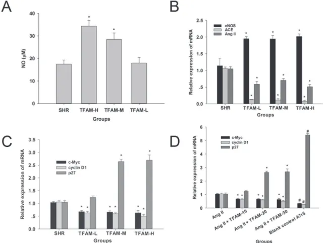
Treatment with TFAM elevated the serum level of nitric oxide (Figure 2A) and aortic mRNA expression of eNOS (Figure 2B).Aortic ACE and Ang II mRNA expression was suppressed by treatment with TFAM (P<0.05) (Figure 2B). The results indicated that the activation of RAAS could be attenuated by TFAM in SHRs.
FIGURE 2 - Efects of TFAM on the serum nitric oxide level, and expressions of RAS and proliferation related genes in vascular
smooth muscle cells. TFAM increased the release of nitric oxide in serum and altered the mRNA expressions of eNOS, ACE and Ang II. (A) The efects of TFAM on the content of nitric oxide.(B) The evaluation was estimated via qPCR analysis using mRNA from cells treated with TFAM and the process was described in Methods. (C) The efects of TFAM-L, TFAM-M, TFAM-H on the
mRNA expressions of p27Kip1, c-Myc and cyclin D1 in the thoracic aorta of SHRs. The values in (A, B, C) given are the mean ±
SD. n = 6, *P<0.05, compared with the SHR group. (D) The efects of TFAM (10-20μg/mL) on the mRNA expressions of p27Kip1,
TFAM could suppress mRNA expression of proliferative and remodeling factors in vascular smooth muscle cells
Cell cycle control is achieved by sequentially modulating multiple proteins including c-Myc, p27Kip1, and
cyclin D1, which are modulated by Wnt signaling (Taipale, Beachy, 2001), and trigger the process of cell proliferation. Up-regulated expressions of proto-oncogenes in aorta in hypertensive rat models are also reported in previous studies (Naftilan, Pratt, Dzau, 1989). In our study, TFAM
signiicantly reduced the mRNA expression of c-Myc and
Cyclin D1 in SHRs (Figure 2C). Furthermore, our results showed that the mRNA expression of p27Kip1, which could
block the cell cycle (Toyoshima, Hunter, 1994), was elevated at TFAM-H group (Figure 2C).
P r e v i o u s s t u d i e s h a v e d e m o n s t r a t e d t h a t overexpression of pro-oncogenes and aberrant proliferation of vascular smooth muscle cells (VSMCs) is Ang II
dependent (Lyall et al., 1992). In Figure 1C, the data indicated that TFAM could inhibit the Ang II-induced proliferation of A7R5 cells in a dose-dependent manner
at 10-50 μg/mL. Considering the antiproliferative efects
and cytotoxicity of TFAM in cell viability evaluation,
the concentrations, 20-30 μg/mL, were selected for
further RNA and protein quantification. To identify whether c-Myc, Cyclin D1 and p27Kip1 was involved
in the inhibitory effect of TFAM on Ang II-induced A7r5 VSMCs hypertrophy and proliferation, we also validated the expression of c-Myc, Cyclin D1 and p27Kip1
by qPCR analysis in A7r5 cells. As expected, Ang II
signiicantly enhanced c-Myc and Cyclin D1 expression,
and suppressed the mRNA expression of p27Kip1 in A7r5
cells. Moreover, c-Myc and Cyclin D1 expressions were
signiicantly inhibited by TFAM post-treatment (Figure
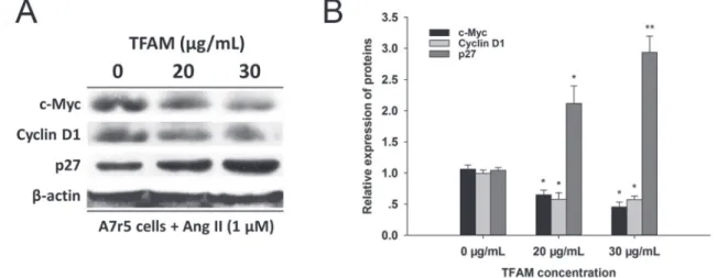
2D). The evidence was further observed in the western blot assay (Figure 3), which suggested that TFAM at 20 and 30
μg/mL could modulate the protein expressions of c-Myc,
Cyclin D1 and p27Kip1 in A7r5 cells. Thus, these data
demonstrated that the regulation of c-Myc, Cyclin D1 and p27Kip1 by Ang II was involved in TFAM inhibitory efect
on Ang II-induced VSMC hypertrophy and proliferation.
Antiproliferative effects of TFAM is involved in canonical Wnt signaling pathway
In consideration of the effects of TFAM on the expression c-Myc, we next evaluated the mediating role
of TFAM on overall β-catenin levels in thoracic aorta
cells of SHRs. However, the result was negative (Data not shown). Next, we presumed that TFAM might mediate the subcellular localization of β-catenin, and subsequently
affect theβ-catenin dependent transcriptional activity.
As shown in Figure 4A, immunoblotting assay showed
a relative decrease in nuclear β-catenin accumulation in
cells with TFAM post-treatment as compared with those in control group. This result indicated that TFAM could decrease the accumulation of β-catenin in the nucleus,
rather than the expression of β-catenin in A7r5 cells.
T h e f u n c t i o n a l c o n s e q u e n c e o f β-catenin accumulation was also estimated by using a luciferase reporter system. In Figure 4C, Ang II could induce
β-catenin-mediated gene transcriptional activity in A7r5
cells. Furthermore, the transcriptional activity induced
FIGURE 3 - TFAM suppressed protein expressions of c-Myc and Cyclin D1 and elevated the level of p27Kip1 protein. (A) Protein
expressions of c-Myc, Cyclin D1 and p27Kip1 after 24 h of administration with 20 and 30 μg/mL of TFAM. (B) Bars represent the intensity of the bands of (A) quantitated by densitometry, respectively. No treatment control group was showed as 0 μg/mL of
by Ang II was suppressed in TFAM-M and TFAM-H, indicating that TFAM could suppress canonical Wnt signaling pathway both in rat vascular smooth muscle cells.
DISCUSSION
Some epidemiological evidence indicates that consumption of antioxidant-rich diet may alleviate hypertension. An alteration to a vegetable-based diet is also efficacious to relieve hypertension (Berkow, Barnard, 2005). In this study, we explore the novel
observation that antihypertensive efects of lavonoids in
A. megalophylla are involved in afecting the remodeling,
and the expressions of tumor suppressor genes and proto-oncogenes in vascular smooth muscle cells.
Like the pathogenesis in most primary hypertensive
patients, spontaneously hypertensive rats obviously exert the characteristic of chronical cardiovascular remodeling (Duarte et al., 2001). In the present study, the artery function in SHRs was facilitated by treatment for 7 weeks
with total lavonoids in A. megalophylla. Moreover, we
explore a novel observation that antihypertensive efects of TFAM are involved in afecting the vascular remodeling,
and the expressions of tumor suppressor gene and proto-oncogene in vascular smooth muscle cells.
In essential hypertension, resistance vessels sufer
RAS-mediated continuous contraction and chronic remodeling. Either in hypertrophic remodeling or inward eutrophic remodeling, media/lumen ratio is elevated, which is attributed to the adaptive adjustment of arteriole
to physical of chemical irritation (Schifrin, 1992). Also,
the vascular remodeling contributes to pathological
changes of organs in cardiovascular disease, suggesting that the suppression of resistance arterial remodeling could be considered as a therapeutic goal for preventing hypertension. In the present study, SHRs exposed to TFAM showed a statistically significant change in the media/lumen ratio reduction compared to SHRs with no TFAM treatment. The elevated media/lumen ratios of subcutaneous resistance arteries in SHRs undergo normalization within 7 weeks of the TFAM-based antihypertensive regimen. This observation suggests that normalization of the vascular structure appears to be
correlated to the anti-hypertensive efects of TFAM.
Improving endothelium-dependent vascular
relaxation by lavonoids is associated with reactivation
of endothelium-derived nitric oxide activity (Fisher et al., 2003). In fact, it is agreeable that the cascade of RAS
in endothelial cells could be efective to attenuate nitric
oxide production. ACE is known to be a core factor in RAS, and increasing blood pressure by accumulating Ang II. Some flavonoids were demonstrated to exert
inhibiting efects by directly chelating the zinc ion site of
ACE (Actis-Goretta et al., 2003). Moreover, carbonyl and
hydroxyl groups in lavonoids or derived compounds are
considered as chelating agents to alter the ACE to inactive apoenzyme. Following 7-weeks application of TFAM, the level of nitric oxide and mRNA expression of eNOS were elevated in vivo, suggesting that the release of nitric
oxide contributed to the antihypertensive efects of TFAM.
Furthermore, the expressions of the negative regulators of endothelium-derived relaxing factors, ACE and Ang II, were suppressed by TFAM administration, demonstrating that the improved vascular function by TFAM was partly due to the inhibition of RAS.
FIGURE 4 - TFAM could suppress Wnt/β-catenin signaling in vascular smooth muscle cells. (A) TFAM decreased protein expression
of β-catenin in western blot assay. (B) Bars represent the intensity of the bands of (A), quantitated by densitometry. The values given are the mean ± SD. n = 3, *P<0.05, compared with the SHR group. (C) The TOPlash luciferase assay showed the efect of
TFAM on high activation of TCF/LEF transcription in Ang II-stimulated A7r5 cells. A7r5 cells were transfected with a TOPlash or FOPlash reporter construct and either TFAM or no treatment. The TOPlash luciferase activity was measured after 48 h
Excessive proliferation of vascular smooth muscle
cells, inlammatory iniltrate of endothelium, and capillary
fibrosis are all mechanisms that have been confirmed to accelerate arterial remodeling. Abnormal resistance vessels with increased media/lumen ratio are commonly accompanied with mild inflammation and exorbitant stacking of proliferation-related proteins (Intengan, Schiffrin, 2001). In vitro overexpression of p27Kip1, a
member of CIP/KIP family of CDK inhibitors, could suppress VSMC proliferation. Inversely, inhibition of p27Kip1 activity enhances primate aortic VSMC growth
(Coqueret, 2003). Moreover, p27Kip1-/- mice lost
heparin-regulated suppression of pneumatorexis induce pulmonary hypertension and vascular remodeling, indicating that p27Kip1 is an essential suppressor of hypoxic pulmonary
vascular remodeling (Yu et al., 2005). In the present study, the amount of p27Kip1 also increased in vessels from SHRs.
Moreover, in the isolated VSMCs from SHRs with TFAM treatment, the mRNA expressions of c-Myc and cyclin D1
were signiicantly repressed. Hence, these indings suggest
that VSMCs from hypertensive rats have an increased ability to respond to stimuli by TFAM, which provides a potential mechanism for TFAM to reduce the remodeling of the SHR vasculature.
The chronic influence of vascular remodeling involves in Wnt signaling cascades, which is conducting pathological proliferation of VSMCs, including
reactivating glycogen synthase kinase 3β (GSK3β) by
phosphorylating 9-serine and obstructing β-catenin degradation. Generally, abundant β-catenin relocates to the nucleus and triggers transcriptional activity by binding T-cell factors/lymphoid-enhancing factors. Furthermore, the cascade could govern cell cycle via promoting the mRNA expression of c-Myc and cyclin D1 (Polakis, 2007). Thus, we attempted to confirm the effects of TFAM on the Wnt signaling. Our results showed that TFAM inhibited the expression of c-Myc and cyclin D1 in VSMCs through suppressing β-catenin accumulation
in the nucleus. In A7r5 cells, canonical Wnt/β-catenin
signaling was also activated by Ang II. Moreover, the
β-catenin/TCF transcriptional activity is significantly
repressed by 48 h post-treatment of TFAM. The abnormal
accumulation β-catenin in the nucleus is also reduced
by TFAM in VSMCs of SHRs, and the mRNA levels of c-Myc and cyclinD1 were subsequently downregulated. These data demonstrated that TFAM could suppress
Wnt signaling by blocking the distribution of β-catenin in the nucleus, rather than by afecting the expression of β-catenin in cells.
Taken together, the potential mechanism of total flavonoids of A. megalophylla on VSMC proliferation
in SHRs is proposed: with the synergistic effect of p27Kip1 activation, TFAM decreases mRNA expression of
β-catenin and its downstream transcriptional factor, c-Myc
and Cyclin D1, in sequence inhibits cell remodeling and the release of inflammatory mediators by suppressing
RAS, following by triggering eNOS, and inally exerting
the blood pressure lowering potential.
ACKNOWLEDGEMENTS
This work was supported by a grant from Hubei Provincial Department of Education (No. Q20162001) and a grant from Hubei University of Chinese Medicine (Qingmiao Project) for Dr. Zhenpeng Qiu.
CONFLICT OF INTEREST
The authors declare that they have no competing interests.
ETHICS APPROVAL AND CONSENT TO
PAR-TICIPATE
Animal experiments were reviewed and approved by the Animal Research Central at the Hubei University of Chinese Medicine and conducted according to the
principles of Guide for the Care and Use of Laboratory
Animals in China.
REFERENCES
Actis-Goretta L, Ottaviani JI, Keen CL, Fraga CG. Inhibition of angiotensin converting enzyme (ACE) activity by lavan-3-ols and procyanidins. FEBS Lett. 2003;555(3): 597-600.
Bauersachs J, Bouloumie A, Mulsch A, Wiemer G, Fleming I, Busse R. Vasodilator dysfunction in aged spontaneously hypertensive rats: changes in NO synthase III and soluble guanylyl cyclase expression, and in superoxide anion production. Cardiovasc Res. 1998;37(3):772-779.
Beevers G, Lip GY, O’Brien E. ABC of hypertension: the
pathophysiology of hypertension. BMJ. 2001;322(7921): 912-916.
Berkow SE, Barnard ND. Blood pressure regulation and vegetarian diets. Nutr Rev. 2005;63(1):1-8.
Duarte J, Perez-Palencia R, Vargas F, Ocete MA, Perez-Vizcaino F, Zarzuelo A, Tamargo J. Antihypertensive effects of the
lavonoid quercetin in spontaneously hypertensive rats. Br J
Pharmacol. 2001;133(1):117-124.
Filipeanu CM, Brailoiu E, Kok JW, Henning RH, De Zeeuw D, Nelemans SA. Intracellular angiotensin II elicits Ca2+ increases in A7r5 vascular smooth muscle cells. Eur J Pharmacol. 2001;420(1):9-18.
Fisher ND, Hughes M, Gerhard-Herman M, Hollenberg NK. Flavanol-rich cocoa induces nitric-oxide-dependent vasodilation in healthy humans. J Hypertens. 2003;21(12):2281-2286.
Galleano M, Pechanova O, Fraga CG. Hypertension, nitric oxide, oxidants, and dietary plant polyphenols. Curr Pharm Biotechnol. 2010;11(8):837-848.
Gibbons GH, Dzau VJ. The emerging concept of vascular remodeling. New Engl J Med. 1994;330(20):1431-1438.
Graham I, Atar D, Borch-Johnsen K, Boysen G, Burell G, Cifkova R, et al. European guidelines on cardiovascular disease prevention in clinical practice: executive summary. Fourth Joint Task Force of the European Society of Cardiology and other societies on cardiovascular disease prevention in clinical practice (constituted by representatives of nine societies and by invited experts). Eur J Cardiovasc Prev Rehabil. 2007;14(Suppl 2):E1-40.
Grassi D, Necozione S, Lippi C, Croce G, Valeri L, Pasqualetti P,
Desideri G, Blumberg JB, Ferri C. Cocoa reduces blood pressure and insulin resistance and improves endothelium-dependent vasodilation in hypertensives. Hypertension. 2005;46(2):398-405.
Gu D, Reynolds K, Wu X, Chen J, Duan X, Muntner P, et al. Prevalence, awareness, treatment, and control of hypertension in china. Hypertension. 2002;40(6) 920-927.
Herrera-Arellano A, Miranda-Sanchez J, Avila-Castro P, Herrera-Alvarez S, Jimenez-Ferrer JE, Zamilpa A, et al.
Clinical efects produced by a standardized herbal medicinal product of Hibiscus sabdarifa on patients with hypertension.
A randomized, double-blind, lisinopril-controlled clinical trial. Planta Med. 2007;73(1):6-12.
Hodgson JM. Efects of tea and tea lavonoids on endothelial
function and blood pressure: a brief review. Clin Exp Pharmacol Physiol. 2006;33(9):838-841.
Intengan HD, Schifrin EL. Vascular remodeling in hypertension: roles of apoptosis, inlammation, and ibrosis. Hypertension.
2001;38(3 Pt 2):581-587.
Kearney PM, Whelton M, Reynolds K, Muntner P, Whelton PK, He J. Global burden of hypertension: analysis of worldwide data.
Lancet. 2005;365(9455):217-223.
Lyall F, Dornan ES, McQueen J, Boswell F, Kelly M.
Angiotensin II increases proto-oncogene expression and phosphoinositide turnover in vascular smooth muscle cells via the angiotensin II AT1 receptor. J Hypertens. 1992;10(12):1463-1469.
Naftilan AJ, Pratt RE, Dzau VJ. Induction of platelet-derived growth factor A-chain and c-myc gene expressions by angiotensin II in cultured rat vascular smooth muscle cells. J Clin Invest. 1989;83(4):1419-1424.
Perez-Vizcaino F, Ibarra M, Cogolludo AL, Duarte J, Zaragoza-Arnaez F, Moreno L, Lopez-Lopez G, Tamargo J. Endothelium-independent vasodilator efects of the lavonoid quercetin and
its methylated metabolites in rat conductance and resistance arteries. J Pharmacol Exp Ther. 2002;302(1):66-72.
Polakis P. The many ways of Wnt in cancer. Curr Opin Genet Dev. 2007;17(1):45-51.
Schifrin EL. Reactivity of small blood vessels in hypertension:
relation with structural changes. State of the art lecture. Hypertension. 1992;19(Suppl 2):II1-9.
Taipale J, Beachy PA. The Hedgehog and Wnt signalling pathways in cancer. Nature. 2001;411(6835):349-354.
Toyoshima H, Hunter T. p27, a novel inhibitor of G1 cyclin-Cdk protein kinase activity, is related to p21. Cell. 1994;78(1):67-74.
Wang Y, Qiu Z, Zhou B, Liu C, Ruan J, Yan Q, Liao J, Zhu F. In vitro antiproliferative and antioxidant efects of urolithin A, the
colonic metabolite of ellagic acid, on hepatocellular carcinomas HepG2 cells. Toxicol In Vitro. 2015;29(5):1107-1115.
Xie XF, Wang JW, Zhang HP, Li QX, Chen BY. Chemical
Xiong X, Yang X, Liu Y, Zhang Y, Wang P, Wang, J. Chinese
herbal formulas for treating hypertension in traditional Chinese medicine: perspective of modern science. Hypertens Res. 2013;36(7):570-579.
Yu L, Quinn DA, Garg HG, Hales CA. Cyclin-dependent kinase inhibitor p27Kip1, but not p21WAF1/Cip1, is required for inhibition of hypoxia-induced pulmonary hypertension and remodeling by heparin in mice. Circ Res. 2005;97(9):937-945.
Zhang Y, Que S, Yang X, Wang B, Qiao L, Zhao Y. Isolation and identiication of metabolites from dihydromyricetin. Magn
Reson Chem. 2007a;45(11):909-916.
Zhang YS, Zhang QY, Li LY, Wang B, Zhao YY, Guo DA.
Simultaneous determination and pharmacokinetic studies
of dihydromyricetin and myricetin in rat plasma by
HPLC-DAD after oral administration of Ampelopsis grossedentata
decoction. J Chromatogr B Analyt Technol Biomed Life Sci.
2007b;860(1):4-9.
Zhou B, Yi H, Tan J, Wu Y, Liu G, Qiu Z. Anti-proliferative efects of polyphenols from pomegranate rind (Punica granatum L.) on EJ bladder cancer cells via regulation of p53/miR-34a
axis. Phytother Res. 2015;29(3):415-422.
Received for publication on 21st November 2016