www.elsevier.com/locate/yexpr
0014-4894/$ - see front matter 2005 Elsevier Inc. All rights reserved. doi:10.1016/j.exppara.2005.06.002
Trypanosoma cruzi
: In
X
uence of predominant bacteria from
indigenous digestive microbiota on experimental infection in mice
R. Duarte
a, A.M. Silva
a, L.Q. Vieira
b, L.C.C. Afonso
c, J.R. Nicoli
a,¤a Departamento de Microbiologia, ICB, Universidade Federal de Minas Gerais, Belo Horizonte, MG, Brazil b Departamento de Bioquímica-Imunologia, ICB, Universidade Federal de Minas Gerais, Belo Horizonte, MG, Brazil c Departamento de Ciências Biológicas, ICEB/NUPEB, Universidade Federal de Ouro Preto, Ouro Preto, MG, Brazil
Received 4 March 2005; received in revised form 2 June 2005; accepted 7 June 2005 Available online 20 July 2005
Abstract
To verify the inXuence of some predominant components from indigenous microbiota on systemic immunological responses dur-ing experimental Chagas disease, germ-free NIH Swiss mice were mono-associated with Escherichia coli, Enterococcus faecalis, Bac-teroides vulgatus or Peptostreptococcus sp. and then infected with the Y strain of Trypanosoma cruzi. All the mono-associations predominantly induced a Th1 type of speciWc immune response to the infection by T. cruzi. A direct correlation was observed between a higher survival rate and increased IFN- and TNF- production (P< 0.05) in E. faecalis-, B. vulgatus-, and Peptostrepto-coccus-associated mice. Moreover, higher levels of anti-T. cruzi IgG1 and anti-T. cruzi IgG2a were also found in mono-associated animals after infection. On the other hand, with the exception of E. faecalis-associated mice, mono-association induced a lower IL-10 production after infection (P< 0.05) when compared with germ-free animals. Interestingly, spleen cell cultures from non-infected germ-free and mono-associated mice spontaneously produced higher levels (P< 0.05) of IL-10 than cultures from infected mono-associated mice, except again for E. faecalis-associated animals. In conclusion, the presence of the components of the indigenous mic-robiota skews the immune response towards production of inXammatory cytokines during experimental infection with T. cruzi in gnotobiotic mice. However, the degree of increase in production of cytokines depends on each bacterial component.
2005 Elsevier Inc. All rights reserved.
Keywords: Microbiota; Trypanosoma cruzi; Cytokines; Immunoglobulins; Gnotobiotic mice
1. Introduction
The human gastrointestinal tract harbors one of the most complex ecosystems known in microbial ecology with bacterial populations reaching 1010–1011viable cells/g of contents in its lower portions. These organ-isms may belong to about 400 diVerent bacterial spe-cies, although it is believed that only 20–40 of them reach predominant levels, consisting of 99% of the total community (Berg, 1996). Concerning the human pre-dominant fecal bacteria, two population levels can be
distinguished: (i) the dominant microbiota (109– 1011cells/g of contents) constituted only by obligate anaerobes (Bacteroides, BiWdobacterium, Eubacterium,
Peptostreptococcus, and Fusobacterium) and (ii) the sub-dominant microbiota (107–108cells/g of contents) containing predominantly facultative anaerobic and microaerophilic bacteria (Escherichia coli, Enterococ-cus, and Lactobacillus). In healthy hosts, the presence of this microbiota has a very large impact on various aspects of function and metabolism such as metabolic rate, gastrointestinal function, speciWc and quantitative aspects of immune function, and the many aspects of biochemical homeostasis. Only the predominant species have population levels high enough to be considered
* Corresponding author. Fax: +55 31 3499 2730.
as responsible for the three main functions of the intes-tinal microbiota which have considerable importance for the host health: (i) the colonization resistance, (ii) the immunomodulation, and (iii) the nutritional contri-bution for the host (MacFarland, 2000). Presently, available data also indicate that this indigenous micro-biota almost always has a profound inXuence on the host–parasite relationship. As an example, it is well known that the presence of intestinal microbiota is essential for the pathogenicity of some protozoa and helminthes such as Entamoeba histolytica (Phillips and Wolfe, 1959), Nippostrongylus brasiliensis (Wescott and Todd, 1964), Nematospiroides dubius (Wescott, 1968),
Trichinella spiralis (Przyjalkowski and Wescott, 1969),
Eimeria tenella (Visco and Barnes, 1972), Ascaridia galli (Johnson and Reid, 1973), Trichuris suis (Rutter and Beer, 1975), Eimeria falciformis (Owen, 1975),
Eimeria ovinoidalis (Gouet et al., 1984), and Giardia duodenalis (Torres et al., 2000). In contrast, this micro-biota can reduce the pathological consequences of other infectious diseases as described for experimental infections with Trypanosoma cruzi (Silva et al., 1987),
Cryptococcus neoformans (Salkowski et al., 1987),
Strongyloides venezuelensis (Martins et al., 2000), and almost all enteropathogenic bacteria (Clostridium di Y-cile, Clostridium perfringens, E. coli, Pseudomonas aeru-ginosa, Salmonella typhimurium, Shigella Xexneri, and
Vibrio cholerae) (Wilson, 1995). Experimental infec-tions with Raillietina cesticillus (Reid and Botero, 1967) and Isopora suis (Harleman and Meyer, 1984) are two of the very few cases, where the normal microbiota has no inXuence on the course of a disease.
Trypanosoma cruzi is the causative agent of Chagas disease in man and determines a systemic infection that is controlled, although not completely eliminated, by T cell-dependent immune responses. Control of parasite-mia in the acute phase of infection is critically depen-dent on intracellular killing by cytokine-activated macrophages. In this way, diVerent studies indicate the crucial role of IFN-, IL-12 (Michailowsky et al., 2001), and TNF- (Silva et al., 1995) as well as NO (Vespa et al., 1994) in host resistance to infection with
T. cruzi.
As cited above, infection with the intracellular para-site T. cruzi is more severe in germ-free animals, as shown by a higher mortality when compared with con-ventional controls (Silva et al., 1987). Germ-free mice also displayed earlier and higher parasitemia than con-ventional controls. Moreover, tissues from germ-free mice were more intensively parasitized and presented a more aggressive inXammatory response. Germ-free mice infected with T. cruzi presented a stronger local reaction to subcutaneous injection of formalin-killed parasites as determined by footpad swelling than conventional ani-mals (Furarah et al., 1991). Recent data showed higher IFN-, TNF-, and NO production by spleen cell
cultures, and higher blood levels of IgG1 in conventional mice infected with Y strain of T. cruzi when compared to their germ-free counterparts (Duarte et al., 2004). How-ever, these data concern the whole microbiota and there is no information about the role of individual compo-nents of the indigenous bacterial ecosystem on these diVerences.
To investigate the role of individual components of the indigenous microbiota on the immune response to a systemic parasitic infection, germ-free mice were mono-associated with Gram positive (Peptostreptococcus sp.) and Gram negative (Bacteroides vulgatus) obligate anaerobic bacteria or Gram positive (Enterococcus fae-calis) and Gram negative (E. coli) facultative bacteria, pertaining to the predominant human fecal microbiota, and subsequently infected with T. cruzi. Survival rate, cytokine, and NO productions by spleen cell cultures, and IgG1 and IgG2a serum concentrations were deter-mined.
2. Materials and methods
2.1. Mice
Germ-free 21-day-old NIH mice of both sexes were used in this study. The matrices were obtained from Taconic Farms (Germantown, NY, USA) and main-tained in the germ-free facility of the Instituto de Ciên-cias Biológicas (Universidade Federal de Minas Gerais, Belo Horizonte, MG, Brazil) for several generations. Germ-free animals were housed in Xexible plastic isola-tors (Standard Safety, McHenry, IL, USA) and handled according to established procedures. Experiments with gnotobiotic mice were carried out in micro-isolators (UNO Roestvastaal B.V., Zevenar, The Netherlands). Water and commercial autoclavable diet (Nuvital, Curit-iba, PR, Brazil) were steam sterilized and administered ad libitum. Microbiological status of germ-free and gno-tobiotic mice was performed by routine culture of recently collected feces in brain heart infusion (BHI) broth medium (Oxoid, Hampshire, England) and Xuid thioglycollate medium (Difco, Detroit, MI, USA). Fecal cultures were incubated at 37 and 25 °C during 72 h. Controlled lighting (12 h light, 12 h dark) was used for all the animals. All the animals received humane care as outlined in the “Guide for the Care and Use of Labora-tory Animals” of the National Research Council (1996).
2.2. Bacteria
these bacteria was regularly conWrmed by using API identiWcation kits (BioMérieux, Marcy l’Etoile, France). Inoculum of each bacterium was prepared from cultured BHI broth medium (Oxoid) incubated at 37 °C under aerobic conditions for E. coli and E. faecalis or in an anaerobic chamber (Forma ScientiWc, Marietta, OH, USA, containing an atmosphere of N2 85%, H2 10%, and CO2 5%) for B. vulgatus and Peptostreptococcus sp. Mono-association was carried out by the administration of a single dose (0.2 ml containing about 108 viable cells) of each bacterial strain to germ-free mice by intra-gastric intubation 10 days before the experimental challenge with T. cruzi. As controls, germ-free mice were inocu-lated intra-gastrically with 0.2 ml of sterile saline 10 days before challenge.
2.3. Bacterial counts in feces from mono-associated mice
Feces freshly collected from each gnotobiotic group were diluted 100-fold in saline and vortexed. Serial 10-fold dilutions were made and 0.1 ml plated onto BHI agar medium (Oxoid). Plates were incubated at 37 °C for 24–48 h for bacterial counts (under aerobic or anaerobic conditions depending on the bacteria).
2.4. Parasite
Trypanosoma cruzi, Y strain (Laboratório de Parasit-ologia e HistopatParasit-ologia, DECBI-ICEB, Universidade Federal de Ouro Preto, Ouro Preto, MG, Brazil) was used and maintained by weekly successive transfers in NIH conventional mice.
2.5. Experimental infection
Germ-free and mono-associated mice were inocu-lated with 5£103 blood trypomastigotes obtained from mice in the acute phase of the infection. BrieXy, blood was collected from the axillary plexus in 3.8% sodium citrate/PBS and parasitemia evaluated accord-ing to Brener (1962). The number of parasites was adjusted to the desired inocula in PBS. All the manipu-lations were performed in a laminar Xow hood. A sample was seeded in Xuid thioglycollate medium (Difco) and BHI broth medium (Oxoid) for control of asepsis.
2.6. Experimental design
Five groups of mice were used to obtain the following microbiological status: (i) germ-free control, (ii) E. coli
mono-associated, (iii) E. faecalis mono-associated, (iv)
B. vulgatus mono-associated, and (v) Peptostreptococcus
sp. mono-associated. Several of these germ-free and gno-tobiotic groups (5–10 mice for each group according to the experiments) were used separately for
determina-tions of survival rate, cytokine levels, and NO and immunoglobulin concentrations. Survival rate was determined over 40 days and accumulated mortality noted. For immunological determinations, groups of mice were killed just before or seven days after the T. cruzi inoculation. Three repetitions were done for each determination.
2.7. Trypanosoma cruzi antigens
Antigens from T. cruzi were obtained from trypom-astigotes cultured on VERO cells. Parasites were har-vested from the supernatant, centrifuged at 3000g during 15 min at 4 °C, and washed three times in PBS. The num-ber of trypomastigotes was adjusted to 108cells/ml, and submitted to Wve cycles of freezing and thawing. The parasite extract was homogenized, aliquoted, and main-tained at ¡20 °C until use.
2.8. Cytokine quantitation in spleen cultures
Mice from gnotobiotic groups were killed and the spleen was removed, macerated aseptically in RPMI-1640 (Sigma Chemical, St. Louis, MO, USA), and centri-fuged at 1000g for 10 min. Erythrocytes were lysed, and spleen cells were washed and centrifuged twice. Then, cells were suspended in RPMI-1640 supplemented with 10% fetal bovine serum, 50M -mercaptoethanol (Sigma), 10 mg/ml gentamicin sulfate, and 3.2 mM L
-glu-tamine (Sigma). Cell number and viability were assessed by trypan blue dye exclusion on a Neubauer hematocy-tometer and the Wnal cell suspension was adjusted to 5£106cells/ml. Cells were cultured in 24-well tissue cul-ture plates in the absence or presence of the T. cruzi anti-gen (50l of the antigen preparation/ml of culture, see above). Triplicate cultures of all experiments, using three animals for each one, were incubated for 72 h at 37 °C in 5% CO2. After incubation, supernatants were harvested and stored at ¡86 °C for cytokine assay. Duoset kits for mouse IFN-, TNF-, and IL-10 (R&D Systems, Min-neapolis, MN, USA) were used to determine cytokine levels in culture supernatants according to the manufac-turer’s instructions. Absorbance was read at 450 nm on a microplate reader (Bio-Rad Laboratories, Hercules, CA, USA). The sensitivities of the assays were 72, 62, and 36 pg/ml for IFN-, TNF-, and IL-10, respectively.
2.9. Detection of NO production in spleen cultures
2.10. Immunoglobulin analysis in serum
Total and parasite-speciWc IgG1 and IgG2a levels in serum were evaluated by capture ELISA (dilution 200-fold). To detect IgG1 and IgG2a isotypes, biotinylated rat antibodies anti-mouse IgG1 and IgG2a (Southern Biotechnology Associates, Birmingham, AL, USA) were used. Absorbance at 492 nm was determined with an ELISA plate reader (Bio-Rad). The concentrations of each immunoglobulin were determined using the respective puriWed mouse standard (Southern Biotech-nology Associates).
2.11. Statistical analysis
The results shown are from one representative of at least three independently performed. Statistical signiW -cance of the results was evaluated by analysis of variance (ANOVA) for all data, except survival, for which Log-Rank test was used. The level of signiWcance was set at
P< 0.05.
3. Results
3.1. Survival rate
All bacteria established in the digestive tract of gno-tobiotic mice. Fecal counts yielded 109 colony-forming units/g of feces within two days, and these high levels were maintained until the end of the experiment. Fig. 1
shows experiments on survival of germ-free and gnoto-biotic mice challenged with T. cruzi, Y strain. A signi W-cantly higher survival rate was observed for gnotobiotic animals mono-associated with E. faecalis
(PD0.034), B. vulgatus (PD0.035) or Peptostreptococ-cus (PD0.029) when compared to control group. Mice mono-associated with E. coli survived slightly longer than control animals but the results were not statisti-cally signiWcant (PD0.103).
3.2. Cytokine determinations
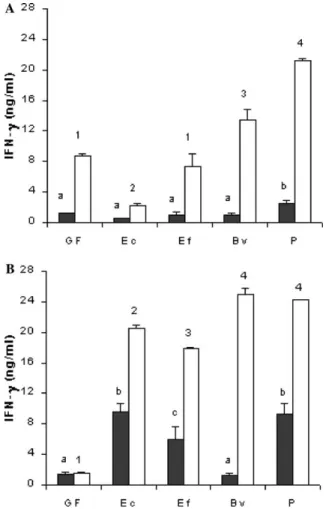
To determine if mono-association with bacteria would alter the response of germ-free mice to T. cruzi
antigens, spleen cells from germ-free and mono-associ-ated mice were cultured in the presence of T. cruzi
homogenate. As seen in Fig. 2A, except for the E. coli
mono-associated group, in vitro addition of T. cruzi
homogenate triggered IFN- production. Moreover,
Fig. 2A shows similar basal levels of IFN- in the super-natants of spleen cultures from germ-free and gnotobi-otic NIH mice, before the infection, except for the
Peptostreptococcus-associated group (P< 0.05). When spleen cells were cultured with T. cruzi antigens during 72 h, a signiWcant increase (P< 0.05) in IFN- production
was observed for germ-free animals but very diVerent responses were obtained for the diVerent bacterial asso-ciations. These responses to T. cruzi antigens ranged from a small increase observed for E. coli-associated mice to the production of high levels of IFN- by spleen cell from cultures of Peptostreptococcus-associated ani-mals (P< 0.05). Seven days after intraperitoneal infec-tion with T. cruzi (Fig. 2B), signiWcantly higher IFN-
levels (P< 0.05) were found in spleen cell culture super-natants from gnotobiotic mice stimulated with parasite antigens (P< 0.05). In germ-free animals, this increase in IFN- production after experimental infection was not observed, even in supernatants of spleen cells cultured in the presence of T. cruzi antigens.
Before experimental infection, basal TNF- produc-tion was similar in germ-free and gnotobiotic animals (Fig. 3A). Increased levels of this cytokine were observed in germ-free and gnotobiotic mice when spleen cells were stimulated with parasite antigens, and higher values were observed for E. coli, E. faecalis, and Peptostreptococcus -associated groups (P< 0.05). After infection (Fig. 3B), again, higher TNF- levels were found in supernatants of spleen cell cultures from the gnotobiotic mice stimulated with T. cruzi antigens when compared to their germ-free and B. vulgatus-associated counterparts (P< 0.05), partic-ularly for Peptostreptococcus-associated group.
Similar NO levels were observed in supernatants of spleen cultures of non-infected germ-free and gnotobi-otic animals (Table 1). No increase was found when anti-gens were added to the culture (data not shown). Seven
Fig. 1. Survival of Swiss/NIH mice, germ-free (䊊) or mono-associated with Escherichia coli (䊐), Enterococcus faecalis (䊏), Bacteroides vulga-tus (䉫), and Peptostreptococcus sp. (䉬) after intraperitoneal challenge with 5£103 trypomastigotes of Trypanosoma cruzi. Data of one
days after infection, an increase in NO production was observed only in spleen cell culture from E. coli- and
Peptostreptococcus-associated mice (P< 0.05) (Table 1). Before infection, background production of IL-10 was higher in supernatants of spleen cell cultures of germ-free and E. faecalis-associated mice than in the other groups (Fig. 4A). Furthermore, the presence of T. cruzi antigen in culture induced an increased IL-10 pro-duction, proportionally higher in the E. coli-associated animals. After infection (Fig. 4B), the same signiWcant diVerence (P< 0.05) between germ-free and E. faecalis -associated mice and the other groups was observed, but only when cells were stimulated with T. cruzi antigens.
Table 2 shows the impact of infection on the cytokine balance. After infection, the highest IFN-/IL-10 ratio was found in the E. coli mono-associated group. This
group also had the highest infected/non-infected IFN-
production ratio. Germ-free mice presented the lowest calculated ratios.
3.3. Immunoglobulin determinations
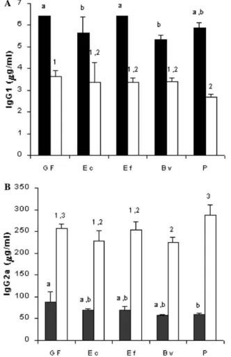
Since the ratio of IgG2a and IgG1 is usually an indica-tion of the prevalence of type 1 or type 2 responses in vivo, we assayed total and T. cruzi-speciWc IgG sub-classes in sera of mice. Higher concentrations of total
Fig. 2. IFN- production by spleen cells from Swiss/NIH mice, germ-free (GF) or mono-associated with Escherichia coli (EC), Enterococcus faecalis (EF), Bacteroides vulgatus (BV), and Peptostreptococcus sp. (P) before (A) and seven days after (B) intraperitoneal challenge with 5£103 trypomastigotes of Trypanosoma cruzi. Cells were cultured for
72 h in the absence (䊏) or presence (䊐) of T. cruzi antigen. Each bar represents the mean of one representative experiment of three per-formed (three mice/experiment; spleens from each animal were cul-tured individually in each experiment). Vertical lines represent standard deviations of the means. a,b,cDiVerent letters and 1,2,3,4diVerent
numbers between non-stimulated and stimulated groups, respectively, as evaluated by analysis of variance (P< 0.05).
Fig. 3. TNF- production by spleen cells from Swiss/NIH mice, germ-free (GF) or mono-associated with Escherichia coli (EC), Enterococcus faecalis (EF), Bacteroides vulgatus (BV), and Peptostreptococcus sp. (P) before (A) and seven days after (B) intraperitoneal challenge with 5£103 trypomastigotes of Trypanosoma cruzi. Cells were cultured for
72 h in the absence (䊏) or presence (䊐) of T. cruzi antigen. Each bar represents the mean of one representative experiment of three per-formed (three mice/experiment, spleens from each mouse were cul-tured individually in each experiment). Vertical lines represent standard deviations of the means. a,b,cDiVerent letters and 1,2,3diVerent
IgG1 were found, before the infection, in the serum of germ-free and E. faecalis-associated group than in the other gnotobiotic animals (P< 0.05). After the
experi-mental challenge, the levels of this immunoglobulin decreased. Similar values were found in germ-free, E. coli
and E. faecalis-associated groups, and lower concentra-tions (P< 0.05) were found for the other gnotobiotic groups (Fig. 5A). Similar concentrations of total IgG2a were observed in the serum of germ-free and gnotobiotic animals, before the infection. After the infection, levels of this immunoglobulin increased to similar values for all groups, except for the Peptostreptococcus-associated mice (Fig. 5B), which showed slightly higher levels of IgG2a.
Finally, Fig. 6 shows that both T. cruzi-speciWc IgG1 and IgG2a increased after infection. Although no statis-tical diVerences were found among groups before infec-tion, E. faecalis mono-associated mice presented higher levels of anti-T. cruzi IgG1 and IgG2a. The other mono-associated groups showed levels of IgG1 and IgG2a sim-ilar to those found in germ-free infected animals.
4. Discussion
Similar to infections with other intracellular patho-gens (Leishmania, Mycobacterium, and Listeria), where a strong Th1 response is protective whereas a Th2 response increases susceptibility to infection, several reports show that a Th1 response promotes protection to T. cruzi (Hoft et al., 2000; Michailowsky et al., 2001) and a Th2 response promotes susceptibility (Oliveira et al., 1996). Protection during the acute phase has been shown to be dependent on IFN-, and many reports describe activation of macrophages by this cytokine to produce NO and kill the obligate intracellular amasti-gote form of the parasite (Vespa et al., 1994). In addi-tion, TNF- provides a second signal for stimulation of NO production and anti-T. cruzi activity in IFN- -activated macrophages (Silva et al., 1995). On the other hand, the down-regulatory cytokines IL-10 and TGF-
are associated with susceptibility to infection by inhib-iting IFN--mediated macrophage activation ( Car-dillo et al., 1996).
In the present study, germ-free and gnotobiotic mice were used to determine the inXuence of four predominant bacterial components of the human indigenous intestinal microbiota on the survival, production of cytokines, and immunoglobulins during the course of experimental
Fig. 4. IL-10 production by spleen cells from Swiss/NIH mice, germ-free (GF) or mono-associated with Escherichia coli (EC), Enterococcus faecalis (EF), Bacteroides vulgatus (BV), and Peptostreptococcus sp. (P) before (A) and seven days after (B) intraperitoneal challenge with 5£103 trypomastigotes of Trypanosoma cruzi. Cells were cultured for
72 h in the absence (䊏) or presence (䊐) of T. cruzi antigen. Each bar represents the mean of one representative experiment of three per-formed (three mice/experiment, spleens from each mouse were cultured individually in each experiment). Vertical lines represent stan-dard deviations of the means. a,b,c,dDiVerent letters and 1,2,3diVerent
numbers indicate statistically signiWcant diVerence between non-stimu-lated and stimunon-stimu-lated groups, respectively, as evaluated by analysis of variance (P< 0.05).
Table 1
Nitrite production (M) in supernatants of spleen cell cultures from Swiss/NIH mice, germ-free or mono-associated with Escherichia coli, Enterococ-cus faecalis, Bacteroides vulgatus, and PeptostreptococEnterococ-cus sp. before and seven days after intraperitoneal challenge with 5£103 trypomastigotes of Trypanosoma cruzi
a,b DiVerent letters indicate statistically signiWcant diVerence between groups for the same infected or non-infected status, as evaluated by analysis of
variance (P< 0.05).
Association
Germ-free E. coli E. faecalis B. vulgatus Peptostreptococcus
Non-infected 4.98§1.23a 4.89
§1.06a 3.45
§0.10a 5.48
§0.10a 5.97
§0.50a
Infected 5.85§0.21a 16.51
§2.63b 7.07
§0.76a 7.68
§1.70a 17.78
Chagas disease. A less severe T. cruzi experimental infec-tion in conveninfec-tional mice than in germ-free animals was repeatedly described in diVerent reports published by our group along the last 15 years (Cintra et al., 1998; Furarah et al., 1991; Pedrosa et al., 1993; Santos et al., 1992; Silva et al., 1987). In the present study, a diVerence in survival was observed between gnotobiotic animals
mono-associ-ated with E. faecalis, B. vulgatus or Peptostreptococcus sp. and germ-free mice. This protection was not so clear in E. coli-associated animals. Additionally, a stronger Th1 response was found in all the gnotobiotic groups after infection. Thus, higher IFN-, TNF-, and NO produc-tions by spleen cell cultures, and higher serum levels of speciWc immunoglobulins of IgG2a isotype were
Fig. 5. Total IgG1 (A) and total IgG2a (B) in serum from Swiss/NIH mice, germ-free (GF) or mono-associated with Escherichia coli (EC), Enterococcus faecalis (EF), Bacteroides vulgatus (BV), and Peptostrep-tococcus sp. (P) before (䊏) and seven days after (䊐) intraperitoneal challenge with 5£103 trypomastigotes of Trypanosoma cruzi. Each
bar represents the mean of one representative experiment of three per-formed (three mice/experiment, sera from each mouse were assayed individually). Vertical lines represent standard deviations of the means. a,bDiVerent letters and 1,2,3diVerent numbers indicate
statisti-cally signiWcant diVerence between non-infected and infected groups, respectively, as evaluated by analysis of variance (P< 0.05).
Table 2
Impact of infection on cytokine production ratio
a IFN-/IL-10 in infected mice/IFN-/IL-10 in non-infected mice. b IFN- in infected mice/IFN- in non-infected mice ratio.
Association
Germ-free E. coli E. faecalis B. vulgatus Peptostreptococcus
IFN-/IL-10a 0.37
§0.10 44.28§8.09 5.14§1.33 6.74§0.13 3.60§0.62 IFN- I/NIb 0.17
§0.01 9.60§1.72 2.51§0.64 1.85§0.17 1.14§0.06
Fig. 6. Anti-Trypanosoma cruzi IgG1 (A) and anti-T. cruzi IgG2a (B) in serum from Swiss/NIH mice, germ-free (GF) or mono-associated with Escherichia coli (EC), Enterococcus faecalis (EF), Bacteroides vulgatus (BV), and Peptostreptococcus sp. (P) before (䊏) and seven days after (䊐) intraperitoneal challenge with 5£103 trypomastigotes
of T. cruzi. Each bar represents the mean of one representative experi-ment of three performed (three mice/experiexperi-ment, sera from each mouse were assayed individually). Vertical lines represent standard deviations of the means. aDiVerent letters and 1,2,3diVerent numbers
observed seven days after infection in gnotobiotic ani-mals when compared to their germ-free counterparts. Basal productions of IFN- by spleen cells were similar between germ-free and gnotobiotic mice before the infec-tion, and were increased (P< 0.05) only in gnotobiotic animals after the infectious challenge. Spleen cells from
T. cruzi-infected germ-free mice were unable to produce IFN- after antigen stimulation in vitro, as opposed to gnotobiotic animals. As can be seen in Table 2, all mono-associated mice had a ratio of production of IFN- after/ before infection higher than the germ-free one, particu-larly in E. coli-associated mice. Similar results were obtained for TNF- production after infection. In accor-dance with the higher IFN- and TNF- levels in spleen cell cultures from gnotobiotic mice infected with T. cruzi, an increase in NO production was also noted in these ani-mals, except for E. faecalis-associated mice. Only, cells from germ-free and E. faecalis-associated infected group produced IL-10 after in vitro stimulation with antigen. However, before infection, IL-10 was produced after in vitro stimulation by cells from all groups. No IL-4 was detected in cell cultures from any of the groups. Hence, a higher production of type 1 cytokines was found in cul-tures from infected gnotobiotic mice, but with some diVerence among mono-associated groups. This can be seen in Table 2, which shows that all mono-associated mice displayed a high IFN-/IL-10 ratio, indicating a strong pro-inXammatory response with a more pronounced phenomenon observed in E. coli-associated animals.
In accordance to the cytokine data, infected gnotobi-otic mice showed slightly higher levels of T. cruzi-speciWc IgG2a in sera, after infection. These results are in agree-ment with the classical work by MacDonald and Carter (1979) who showed that germ-free mice had a deWciency in mounting a cell-mediated immune response, com-pared to conventional controls. Interestingly, we also detected an increase in the levels of T. cruzi-speciWc IgG1 after infection. Although this observation may, in princi-ple, contradict the Th1/Th2 proWle, it has been shown that IgG1 production can be observed in IL-4-deprived mice and thus, would not be associated with a Th2 type response. On the contrary, this non-anaphylactic IgG1 would be induced by IL-12 (Faquim-Mauro et al., 1999). Nevertheless the measurements of total IgG levels in our study reXect a shift towards a Th1 response (Fig. 5A).
IFN- production by spleen cells from uninfected germ-free, B. vulgatus, E. faecalis-, and Peptostreptococ-cus-associated mice in response to T. cruzi antigen was higher than in the E. coli-associated group. This unpre-dicted observation might be explained by the lower stim-ulation by LPS in the germ-free and Gram positive associated mice, since exposure to low levels of LPS has been shown to decrease, at least partially, IFN- produc-tion to further LPS stimuli (Erroi et al., 1993; Henricson et al., 1990). According to Ropert et al. (2002), LPS can
induce tolerance to tGPI mucins and vice-versa (Ropert et al., 2002). Hence, it is possible that E. coli-associated mice are exposed to low levels of LPS from the entero-bacterium and were, therefore, less reactive to T. cruzi
antigens (which contain tGPI mucins). Heterogeneity and diVerence in LPS immunogenic potential between Gram negative bacteria are well known (Ogawa et al., 1997) and these diVerences could explain why B. vulga-tus, another Gram negative bacterium, did not induce tolerance to tGPI mucins.
Due to the higher production of protective cytokines by gnotobiotic animals in response to infection, protec-tion as measured by mortality was found in our experi-ments. This is not surprising, since the presence of these pro-inXammatory cytokines has been extensively associ-ated with resistance to T. cruzi (Hoft et al., 2000; Michai-lowsky et al., 2001). However, E. coli mono-associated mice were more susceptible to T. cruzi than the other mono-associated animals. Interestingly, cells from non-infected E. coli mono-associated mice produced less IFN- upon antigenic stimulation than the other groups (Fig. 2A), suggesting a decreased innate response to this stimulus in these animals that could be related either to a decreased IL-12 production or to a lack of NK cell activ-ity. This decreased response would impair the early con-trol of the parasite resulting in lower survival. We hypothesize that, due to the lack of background IFN-
production to T. cruzi antigens found before infection in these mice, early protective response to the parasite was impaired, hence the lower survival.
It is possible that priming of T cells by the indigenous microbiota inXuences the memory cell repertoire in con-ventional mice, hence inXuencing their immune response. Indeed, an exacerbated early IL-4 production in response to infection with Leishmania major has been associated with a CD4+ T cell population expressing the memory phenotype (possibly primed by E. coli) in BALB/c mice (Julia et al., 2000). However, more recent data have associated the early IL-4 production in response to the parasite to a naïve phenotype (Stetson et al., 2002), probably ruling out the role of the indige-nous microbiota in skewing the immune response during infection with L. major. The number of T cells expressing the memory phenotype in germ-free mice is also contro-versial: some authors Wnd that there are more cells expressing memory markers when mice are exposed to the indigenous microbiota (Inagaki et al., 1996; Lee et al., 1990; Price and Cerny, 1999), others do not ( Bon-orino et al., 1998; Dobber et al., 1992; Park et al., 2000). Hence, the extent to which the indigenous microbiota can change the host T cell repertoire and inXuence its response to pathogens is still not clear.
the Th1 or of the Th2 type (Wills-Karp et al., 2001; Yazdanbakhsh et al., 2002). However, our data show that the lack of exposition to antigenic stimuli from the indigenous microbiota does not induce an exacerbated immune response against a single invasive microorgan-ism, suggesting that there might be a threshold of anti-genic stimulation below which an inXammatory response is not fully mounted.
In conclusion, this study provides evidence that com-ponents of the indigenous microbiota play an important role in the development of an immune system that is competent to react against an acute infection. However, the experiments with gnotoxenic animals mono-associ-ated with bacterial strains representative of the predomi-nant gut microbiota of human showed that some strains may be more eVective than others in interfering in the Th1–Th2 balance.
Acknowledgments
This study was supported by grants and fellowships from the Conselho Nacional do Desenvolvimento Cient-íWco e Tecnológico (CNPq) and Fundação de Amparo à Pesquisa do Estado de Minas Gerais (FAPEMIG). The authors acknowledge Dr. Olindo Assis Martins Filho (Centro de Pesquisa René Rachou, FIOCRUZ, Belo Horizonte, MG, Brazil) for very helpful suggestions. The authors are grateful to Maria Gorete Barbosa Ribas for valuable technical help, and to Ronilda Maria de Paula (in memoriam), Maria Helena Alves de Oliveira and Antônio Mesquita Vaz for animal care.
References
Berg, R.D., 1996. The indigenous gastrointestinal microXora. Trends in Microbiology 4, 430–435.
Bonorino, C., Nardi, N.B., Zhang, X., Wysocki, L.J., 1998. Characteris-tics of the strong antibody response to mycobacterial Hsp70: a pri-mary, T cell-dependent IgG response with no evidence of natural priming or gamma delta T cell involvement. Journal of Immunol-ogy 161, 5210–5216.
Brener, Z., 1962. Therapeutic activity and criterion of cure on mice experimentally infected with Trypanosoma cruzi. Revista do Insti-tuto de Medicina Tropical de São Paulo 4, 389–396.
Cardillo, F., Voltarelli, J.C., Reed, S.G., Silva, J.S., 1996. Regulation of Trypanosoma cruzi infection in mice by IFN- and IL-10: the role of NK cells. Infection and Immunity 64, 128–134.
Cintra, I.P., Silva, M.E., Silva, M.E., Silva, M.E.C., Crocco-Afonso, L.C., Nicoli, J.R., Bambirra, E.A., Vieira, E.C., 1998. InXuence of dietary protein content on Trypanosoma cruzi infection in germfree and conventional mice. Revista do Instituto de Medicina Tropical de São Paulo 40, 355–362.
Dobber, R., Hertogh-Huijbregts, A., Rozing, J., Bottomly, K., Nagel-kerken, L., 1992. The involvement of the intestinal microXora in the expansion of CD4+ T cells with a naive phenotype in the periphery. Development Immunology 2, 141–150.
Duarte, R., Silva, A.M., Vieira, L.Q., Afonso, L.C.C., Nicoli, J.R., 2004. InXuence of normal microbiota on some aspects of the immune
response during experimental infection with Trypanosoma cruzi in mice. Journal of Medical Microbiology 53, 741–748.
Erroi, A., Fantuzzi, G., Mengozzi, M., Sironi, M., Orencole, S.F., Clark, B.D., Dinarello, C.A, Isetta, A., Gnocchi, P., Giovarelli, M., 1993. DiVerential regulation of cytokine production in lipopolysaccha-ride tolerance in mice. Infection and Immunity 61, 4356–4359. Faquim-Mauro, E.L., CoVman, R.L., Abrahamsohn, I.A., Macedo,
M.S., 1999. Mouse IgG1 antibodies comprise two functionally dis-tinct types that are diVerentially regulated by IL-4 and IL-12. Jour-nal of Immunology 163, 3572–3576.
Furarah, A.M., Crocco-Afonso, L.C., Silva, M.E.C., Silva, M.E., Silva, M.E., Bambirra, E.A., Vieira, E.C., Nicoli, J.R., 1991. Immune response of germ-free mice to experimental infection with Trypano-soma cruzi. Brazilian Journal of Medical and Biological Research 24, 1223–1231.
Gouet, P., Yvore, P., Naciri, M., Contrepois, M., 1984. InXuence of digestive Xora on parasite development and the pathogenic eVect of Eimeria ovinoidalis in the axenic, gnotoxenic and conventional lamb. Research in Veterinary Sciences 36, 21–23.
Green, L., Wagner, D., Glogowski, J., Skipper, P., Wishnok, J., Tannen-baum, S., 1982. Analysis of nitrate, nitrite and (15N) nitrate in bio-logical Xuids. Analytical Biochemistry 126, 131–138.
Harleman, J.H., Meyer, R.C., 1984. Life cycle of Isopora suis in gnoto-biotic and conventional piglets. Veterinary Parasitology 17, 27–39. Henricson, B.E., Benjamin, W.R., Vogel, S.N., 1990. DiVerential
cyto-kine induction by doses of lipopolysaccharide and monophospho-ryl lipid A that result in equivalent early endotoxin tolerance. Infection and Immunity 58, 2429–2437.
Hoft, D.F., Schnap, A.R., EickhoV, C.S., Roodman, S.T., 2000. Involve-ment of CD4+ Th1 cells in systemic immunity protective against
primary and secondary challenges with Trypanosoma cruzi. Infec-tion and Immunity 68, 197–204.
Inagaki, H., Suzuki, T., Nomoto, K., Yoshikai, Y., 1996. Increased sus-ceptibility to primary infection with Listeria monocytogenes in germ-free mice may be due to lack of accumulation of L-selectin+ CD44+ T
cells in sites of inXammation. Infection and Immunity 64, 3280–3287. Johnson, J., Reid, W.M., 1973. Ascaridia galli (Nematoda): develop-ment and survival in gnotobiotic chickens. Experidevelop-mental Parasitol-ogy 33, 95–99.
Julia, V., McSorley, S.S., Malherbe, L., Breittmayer, J.P., Girard-Pipau, F., Beck, A., Glaichenhaus, N., 2000. Priming by microbial antigens from the intestinal Xora determines the ability of CD4+ T cells to
rapidly secrete IL-4 in BALB/c mice infected with Leishmania major. Journal of Immunology 165, 5637–5645.
Lee, W.T., Yin, X.M., Vitetta, E.S., 1990. Functional and ontogenetic analysis of murine CD45Rhi and CD45Rlo CD4+ T cells. Journal of Immunology 144, 3288–3295.
MacDonald, T.T., Carter, P.B., 1979. Requirement for a bacterial Xora before mice generate cells capable of mediating the delayed hyper-sensitivity reaction to sheep red blood cells. Journal of Immunology 122, 2624–2629.
MacFarland, L.V., 2000. Normal Xora: diversity and functions. Micro-bial Ecology in Health and Disease 12, 193–207.
Martins, W.A., Melo, A.L., Nicoli, J.R., Cara, D.C., Carvalho, M.A.R., Lana, M.A., Vieira, E.C., Farias, L.M., 2000. A method of decon-taminating Strongyloides venezuelensis larvae for the study of strongyloidiasis in germfree and conventional mice. Journal of Medical Microbiology 49, 387–390.
Michailowsky, V., Silva, N.M., Rocha, C.D., Vieira, L.Q., Lannes-Vie-ira, J., Gazzinelli, R.T., 2001. Pivotal role of interleukin-12 and interferon- axis in controlling tissue parasitism and inXammation in the heart and central nervous system during Trypanosoma cruzi infection. American Journal of Pathology 159, 1723–1733. National Research Council, 1996. Guide for the Care and Use of
Lab-oratory Animals. National Academy Press, Washington.
lipopolysac-charide in comparison with Porphyromonas gingivalis lipid A and Escherichia coli-type synthetic lipid A (compound 506). Vaccine 15, 1598–1605.
Oliveira, L.C.B., Lafaille, M.A.C., Lima, G.M.C.A., Abrahamsohn, I.A., 1996. Antigen-speciWc IL-4- and IL-10-secreting CD4+
lympho-cytes increase in vivo susceptibility to Trypanosoma cruzi infection. Cell Immunology 170, 41.
Owen, D., 1975. Eimeria falciformis (Eimer, 1870) in speciWc pathogen free and gnotobiotic mice. Parasitology 71, 293–303.
Park, S.H., Benlagha, K., Lee, D., Balish, E., Bendelac, A., 2000. Unaltered phenotype, tissue distribution and function of Valpha14(+) NKT cells in germ-free mice. European Journal of Immunology 30, 620–625. Pedrosa, M.L., Nicoli, J.R., Silva, M.E., Silva, M.E., Silva, M.E.C.,
Vie-ira, L.Q., Bambirra, E.A., VieVie-ira, E.C., 1993. The eVect of iron nutri-tional status on Trypanosoma cruzi infection in germ-free and conventional mice. Comparative Biochemistry and Physiology A 106, 813–821.
Phillips, B.P., Wolfe, P.A., 1959. The use of germ-free guinea pigs in studies on the microbial interrelationships in amoebiasis. Annals of the New York Academy of Sciences 78, 308–314.
Price, P.W., Cerny, J., 1999. Characterization of CD4+ T cells in mouse bone marrow. I. Increased activated/memory phenotype and altered TCR Vbeta repertoire. European Journal of Immunology 29, 1051–1056.
Przyjalkowski, Z., Wescott, R.B., 1969. Trichinella spiralis: establish-ment in gnotobiotic mice aVected by Bacillus mesentericus, Bacillus subtilis and Pseudomonas aeruginosa. Experimental Parasitology 25, 8–12.
Reid, W.M., Botero, H., 1967. Growth of the cestode Raillietina cesticillus in bacteria-free chickens. Experimental Parasitology 21, 149–153. Ropert, C., Ferreira, L.R., Campos, M.A., Procópio, D.O., Travassos,
L.R., Ferguson, M.A., Reis, L.F., Teixeira, M.M., Almeida, I.C., Gazzinelli, R.T., 2002. Macrophage signaling by glycosylphosphat-idylinositol-anchored mucin-like glycoproteins derived from Try-panosoma cruzi trypomastigotes. Microbes and Infection 4, 1015– 1025.
Rutter, J.M., Beer, R.J.S., 1975. Synergism between Trichuris suis and the microbial Xora of the large intestine causing dysentery in pigs. Infection and Immunity 11, 395–404.
Salkowski, C.A., Bartizal, K.F., Balish, M.J., Balish, E., 1987. Coloniza-tion and pathogenesis of Cryptococcus neoformans in gnotobiotic mice. Infection and Immunity 55, 2000–2005.
Santos, C.F., Silva, M.E., Silva, M.E., Silva, M.E.C., Nicoli, J.R., Crocco-Afonso, L.C., Santos, J.E., Bambirra, E.A., Vieira, E.C., 1992. EVect of essential fatty acid deWcient diet on experimental infection with Trypanosoma cruzi in germ-free and conventional mice. Brazilian Journal of Medical and Biological Research 25, 795–803.
Silva, M.E., Evangelista, E.A., Nicoli, J.R., Bambirra, E.A., Vieira, E.C., 1987. American trypanosomiasis (Chagas disease) in conventional and germ-free rats and mice. Revista do Instituto de Medicina Tropical de São Paulo 29, 284–288.
Silva, J.S., Vespa, G.N.R., Cardoso, M.A.G., Aliberti, J.C., Cunha, F.Q., 1995. Tumor necrosis factor alpha mediates resistance to Trypano-soma cruzi in mice by inducing nitric oxide production in infected IFN--activated macrophages. Infection and Immunity 63, 4862– 4867.
Stetson, D.B., Mohrs, M., Mallet-Designe, V., Teyton, L., Locksley, R.M., 2002. Rapid expansion and IL-4 expression by Leishmania-speciWc naive helper T cells in vivo. Immunity 17, 191–200. Torres, M.F., Uetanabaro, A.P.T., Costa, A.F., Alves, C.A., Farias,
L.M., Bambirra, E.A., Penna, F.J., Vieira, E.C., Nicoli, J.R., 2000. InXuence of bacteria from the duodenal microbiota of patients with symptomatic giardiasis on the pathogenicity of Giardia duodenalis in gnotoxenic mice. Journal of Medical Microbiology 49, 209–215. Vespa, G.N.R., Cunha, F.Q., Silva, J.S., 1994. Nitric oxide is involved in
the control of Trypanosoma cruzi induced parasitemia and directly kills parasite in vitro. Infection and Immunity 62, 5177–5182. Visco, R.J., Barnes, W.C., 1972. Eimeria tenella in bacteria-free and
conventionalized chicks. Journal of Parasitology 58, 323–331. Wescott, R.B., 1968. Experimental Nematospiroides dubius infection in
germ-free and conventional mice. Experimental Parasitology 22, 245–249.
Wescott, R.B., Todd, A.C., 1964. A comparison of the development of Nippostrongylus brasiliensis in germ-free and conventional mice. Journal of Parasitology 50, 138–143.
Wills-Karp, M., Santeliz, J., Karp, C.L., 2001. The germless theory of allergic disease revisiting the hygiene hypothesis. Nature Review of Immunology 1, 69–75.
Wilson, K.H., 1995. Ecological concepts in the control of pathogens. In: Roth, J.A. (Ed.), Virulence Mechanisms of Bacterial Pathogens. American Society for Microbiology, Washington, pp. 245–256. Yazdanbakhsh, M., Kremsner, P.G., van Ree, R., 2002. Allergy,