Acta Tropica 106 (2008) 27–38
Early infection with
Leishmania major
restrains pathogenic
response to
Leishmania amazonensis
and parasite growth
C.Z. Gonz´alez-Lombana
a, H.C. Santiago
a, J.P. Macedo
a, V.A.R. Seixas
a,
R.C. Russo
a, W.L. Tafuri
b, L.C.C. Afonso
c, L.Q. Vieira
a,∗aDepartamento de Bioqu´ımica e Imunologia, Instituto de Ciˆencias Biol´ogicas (ICB), Universidade Federal de Minas Gerais,
CP 486, 31270-901 Belo Horizonte, MG, Brazil
bDepartamento de Patologia Geral, Instituto de Ciˆencias Biol´ogicas (ICB), Universidade Federal de Minas Gerais,
CP 486, 31270-901 Belo Horizonte, MG, Brazil
cDepartamento de Ciˆencias Biol´ogicas, Instituto de Ciˆencias Exatas e Biol´ogicas and N´ucleo de Pesquisa em Ciˆencias Biol´ogicas,
Universidade Federal de Ouro Preto, Morro do Cruzeiro, 35400-000 Ouro Preto, MG, Brazil
Received 2 October 2007; received in revised form 26 November 2007; accepted 21 December 2007 Available online 15 January 2008
Abstract
Experimental models of infection withLeishmaniaspp. have provided knowledge of several immunological events involved in the resistance mechanism used by the host to restrain parasite growth. It is well accepted that concomitant immunity exists, and there is some evidence that it would play a major role in long-lasting acquired resistance to infection. In this paper, the resistance toLeishmania amazonensisinfection in C57BL/6 mice infected withLeishmania majorwas investigated. C57BL/6 mice, which spontaneously heal lesions caused by infection withL. major, were infected withL. amazonensisat different times before and afterL. major. We demonstrated that C57BL/6 mice previously infected withL. majorrestrain pathogenic responses induced byL. amazonensisinfection and decrease parasite burdens by one order of magnitude. Co-infected mice showed production of IFN-␥in lesions similar to mice infected solely withL. major, but higher TNF-␣and nitric oxide synthase (iNOS) mRNA expression was observed. Surprisingly, the restrained pathogenic response was not related to IL-10 production, as evidenced by lower levels of both mRNA, protein expression in lesions from co-infected mice and in co-infections in IL-10−/−mice. Examination of the
inflammatory infiltrate at the site of infection showed a reduced number of monocytes and lymphocytes inL. amazonensislesions. Additionally, differential production of the CCL3/MIP-1␣and CCL5/RANTES was observed. We suggest that the control of lesion progression caused byL. amazonensisin C57BL/6 mice pre-infected withL. majoris related to the induction of a down-regulatory environment at the site of infection with
L. amazonensis.
© 2008 Elsevier B.V. All rights reserved.
Keywords: Protozoan parasites; Cutaneous leishmaniasis; Co-infection; Cellular recruitment
1. Introduction
Several experimental models have been used to determine the susceptibility and resistance mechanisms triggered by infection withLeishmaniaparasites.Leishmania majorinfection has been considered as a unique model of host resistance and susceptibil-ity toLeishmania. Most mouse strains are considered resistant to
∗Corresponding author at: Departamento de Bioqu´ımica e Imunologia, ICB,
Universidade Federal de Minas Gerais, CP 486, 30161-970 Belo Horizonte, MG, Brazil. Tel.: +55 31 3409 2656; fax: +55 31 3409 2614.
E-mail address:lqvieira@icb.ufmg.br(L.Q. Vieira).
L. majorinfection but a few strains, such as BALB/C and CBA/J develop a progressive, nonhealing disease. However, L. major
resistant mice are susceptible to infection withLeishmania ama-zonensis(Afonso and Scott, 1993; Lemos de Souza et al., 2000). It is postulated that in protective immunity, usually acquired during the latent and chronic phase following cutaneous infec-tion withLeishmania, CD4+ T cells, IL-12, IFN-␥and inducible nitric oxide synthase (iNOS) maintain the immune pressure against parasite growth (Afonso and Scott, 1993; Soong et al., 1996; Jones et al., 1998; Mukbel et al., 2007). Impairment of these responses during latency has been shown to promote parasite growth, pathology and the reappearance of lesions (Stenger et al., 1996; Mendez et al., 2004). In fact, susceptibility
to L. amazonensis infection has been described not only as associated with defective IFN-␥production (Afonso and Scott, 1993) but also with specific impairment of the cell-mediated immune response at the early stages of infection (Ji et al., 2003) and deficient production of TNF-␣by macrophages (Gomes et al., 2003). So, unlike infection withL. major, there is a delayed expression of multiple inflammatory mediators during L. amazonensisinfection, which are correlated with reduced T-cell responsiveness.
Several experimental strategies trigger protection against infection with Leishmania, such as vaccination and cross-immunity (Alexander and Phillips, 1978, 1980; Coelho et al., 2003; Vanloubbeeck and Jones, 2004; Uzonna et al., 2004; Ji et al., 2005; Dondji et al., 2005; Hernandez et al., 2006). Antigen persistence appears to be a critical factor for the maintenance of protective immunity (Belkaid et al., 2001; Uzonna et al., 2001), and this has been the basis for the use of live parasites to immunize humans (Khamesipour et al., 2005) Some experimen-tal studies have shown relative protection againstL. amazonensis
infection after vaccination with live parasites (Veras et al., 1999; Vanloubbeeck and Jones, 2004). In one report, previous infection withL. majorrendered C3H mice more resistant toL. amazo-nensisthan na¨ıve controls (Veras et al., 1999; Vanloubbeeck and Jones, 2004). However, it was not clear from these studies what were the mechanisms involved in this resistance. In the present work, we attempt to more fully define the degree of protection generated by the use of previous infection withL. majorand the immunological mechanisms involved in this protection.
2. Materials and methods
2.1. Mice and parasites
Female C57BL/6 mice were purchased from CEBIO (Instituto de Ciˆencias Biol´ogicas, UFMG Belo Horizonte, MG, Brazil). Mice deficient in IL-10 (IL-10−/−) by homologue recombination in the C57BL/6 background (originally from Jackson Laboratories, B6.129P2-Il10tm1Cgn) were bred at the Gnotobiology facility (Instituto de Ciˆencias Biologi-cas). All mice were 8-week old at the time of infection. L. major (WHO MHOM/IL/80/Friedlin) and L. amazonensis
(IFLA/BR/67/PH8) promastigotes were grown to the sta-tionary phase in Grace’s insect cell culture medium (GIBCO BRL, Grand Island, NY, USA) supplemented with 20% heat-inactivated fetal bovine serum (FBS, Cultilab, Campinas, SP, Brazil), 2 mMl-glutamine, 100 U of penicillin and 100g of streptomycin/mL. Infective stage metacyclic promastig-otes of parasites were isolated from stationary culture (5–6 days old) (Spath and Beverley, 2001) and washed twice in phosphate-buffered saline pH 7.3 (PBS) before they were used for infection. Freeze-thawed leishmanial antigen was obtained from stationary-phase promastigotes that were washed twice in PBS, resuspended in PBS and subjected to seven freeze-thaw cycles consisting of−70 and 37◦C. The protein concentration was determined by the Lowry assay (Lowry et al., 1951) the final concentration was adjusted to 1 mg/mL, and aliquots were stored at−70◦C before use.
2.2. Infections
Mice were inoculated subcutaneously with 1×10⁴ stationary-phase L. major or L. amazonensis promastigotes into one hind footpad. Simultaneously or at different time points after infection, mice were infected subcutaneously with 1×10⁴parasites of the other Leishmaniaspecies in 40l de PBS in the contralateral footpad. Lesion size was monitored weekly with dial micrometer and the results were expressed as the difference in thickness between infected footpads and non-infected footpads of control mice. The parasite burden was determined by performing a limiting dilution analysis as previously described (Vieira et al., 1996). In brief, the homogenates of infected lesions were serially diluted (1:10 or 1:4 serial dilutions) in Grace’s insect culture medium plus 20% FBS and observed 5–7 days later for growth of promastigotes. Parasite numbers are expressed as the negative log10dilution at
which promastigote growth was observed. Results are expressed as mean log10of the inverse of the titer.
2.3. Histopathological analysis
L. amazonensis-infected footpads were collected at 10 weeks postL. amazonensisinfection, fixed in 10% phosphate-buffered formalin and embedded in paraffin. Tissues were sectioned at 5M thickness, mounted onto slides and stained with hema-toxylin and eosin. Histological examinations were performed under a light microscope in an Olympus microscopy using 100–400-fold magnification.
2.4. Cytokine production by lymph nodes and spleens
Spleen and draining lymph node cells from infected mice were harvested and made into single cell suspensions. In brief, the cells were washed, resuspended at 5×10⁶/mL of
com-plete RPMI 1640 medium (Gibco Laboratories) supplemented with 10% heat-inactivated FBS, 25 mM HEPES, 0.05 mM 2-mercaptoethanol, 2 mMl-glutamine, 100 U/mL penicillin and 100g/mL streptomycin, and plated at 1 mL per well in 24-well tissue culture plates (Costar, New York, NY, USA.). Cells were re-stimulated in the presence of 50g/mLLeishmania anti-gen freeze-thawed lysates of promastigotes ofL. amazonensis
orL. major, respectively. Culture supernatants were collected after 72 h and analyzed for their IFN-␥and IL-4 content by cap-ture enzyme-linked immunosorbent assay (ELISA) (sensitivity 80–150 pg/mL). IL-10 production was measured using ELISA kit (R&D systems, Minneapolis, MN, USA), as indicated by the manufacturer (sensitivity 31.25 pg/mL).
2.5. Measurement of chemokines and cytokines in footpads by ELISA
Concentration of the chemokines CCL2 (MCP-1), CCL3 (MIP-1␣), CCL5 (RANTES) and CXCL10 (IP-10) and the cytokines IL-10 and TGF-1 in L. amazonensis-infected footpads was measured at different times postL. amazonensis
and 10 mM de Na2HPO4, pH 7.2) containing anti-proteases
(0.1 mM phenylmethylsulfonyl fluoride, 0.1 mM benzethonium chloride, 10 mM EDTA and 20 mM KI aprotinin A/100 mL) and 0.05% Tween-20. Homogenized tissues were kept on ice for 30 min and subsequently centrifuged (3000×gfor 10 min). Supernatants recovered were diluted in PBS (1:4) and tested for each chemokine or cytokine using commercially available antibodies and according to the procedures supplied by the manufacturer (R&D Systems). All samples were assayed in duplicates. The threshold of sensitivity for each cytokine or chemokine was 7.5 pg/mL.
2.6. Detection of mRNA by reverse transcription and real-time reverse-transcribed polymerase chain reaction (PCR) in footpad
At different time points after L. amazonensis infection, total RNA was isolated fromL. amazonensis-infected footpads for real-time polymerase chain reaction (RT-PCR) as previ-ously described (Cummings and Tarleton, 2004). Briefly, RNA was isolated from footpads using Trizol (Invitrogen, Carlsbad, CA, USA) followed by a phenol–chloroform–isoamyl alco-hol affinity extraction. Purity of RNA was confirmed by a ratio of readings at 260/280 nm. 1g of RNA was reverse-transcribed into cDNA. The amplification of specific mRNA expression was achieved by RT-PCR using specific primer sequences for specific primer sequences for IFN-␥, TNF-␣, iNOS, and IL-10 (Giulietti et al., 2001). The reactions were performed on an ABI PRISM 7900HT sequence detection system (Applied Biosystems) using 20% of the cDNA reac-tion in a total volume of 20L of PCR mixture. All PCR reactions were performed in duplicate using a SYBR Green PCR Master Mix according to the manufacturer’s recommen-dations. The relative quantification of products was determined by the comparative threshold cycle method to determine fold increases in product. Each gene of interest was normalized to the hypoxanthine phosphoribosyl transferase (HPRT for-ward, GTTGGATACAGGCCAGACTTTGTT; HPRT reverse, GATTCAACTTGCGCTCATCTTAGGC), based onCT cal-culation. The fold change in expression was represented as relative to na¨ıve controls. Five footpads were used per group for each time point.
2.7. Statistical procedures
Results are expressed as mean±S.D. To determine whether differences were statistically significant, the Student’st-test was performed using a two-tailed distribution with unpaired sam-ples. Differences were considered to be statistically significant ifp< 0.05.
3. Results
3.1. Lesion progression due to L. amazonensis is controlled by C57BL/6 mice previously infected with L. major
To evaluate the effect of pre-infection with L. major on the course of infection with L. amazonensis, C57BL/6 mice
were injected with 1×10⁴stationary-phaseL. major promastig-otes and challenged withL. amazonensisin the contralateral or the same footpad at different time points. As seen in Fig. 1, lesion sizes following L. amazonensis infection were signifi-cantly reduced when mice were infected withL. majorfor 3 (A) or 15 (B) weeks. L. amazonensisloads were reduced by pre-vious infection withL. major(Fig. 1E and F),L. amazonensis
numbers were smaller at 6 and 10 weeks afterL. amazonensis
infection in mice pre-infected withL. majorfor 15 weeks, when lesions were already different. Interestingly, in mice challenged 3 weeks after the first infection, L. amazonensisloads did not differ at 6 weeks after challenge, but at 10 weeks a reduction in parasitism of about 25 times was found, when compared to animals infected solely withL. amazonensis(1.4 log10,Fig. 1E,
p< 0.05). Noteworthy, at this time point, lesions were very differ-ent in size between the two groups, and mice previously infected withL. majorhad almost completely healed theirL. amazonen-sislesions. To exclude the possibility that the smaller lesions in mice previously infected with L. major mice were due to a switch from a cutaneous form into a visceral form of infec-tion, we determined the parasite load in spleens and draining lymph nodes. Parasite burdens in draining lymph nodes were comparable in both groups (1.6 log10±0.7 and 2.4 log10±0.5).
Moreover, no parasites were recovered from spleens from either group of mice. Simultaneous infections in the same footpad or in different footpads did not interfere in the lesion sizes or para-site burdens, when compared with mice solely infected withL. amazonensis(Fig. 1C, D, G and H). In addition, lesions caused byL. majorwere not affected by the subsequent or simultaneous infection with L. amazonensis, as shown inFig. 2A–D. These results indicate that C57BL/6 mice pre-infected withL. major
were able to control lesion progression byL. amazonensisand reduce, albeit not completely control, parasite burdens.
3.2. Control of lesion progression of L. amazonensis infection by mice previously infected with L. major is associated with reduced inflammatory infiltrate
Fig. 1. C57BL/6 mice pre-infected withL. majorcontrol progression ofL. amazonensislesions. C57BL/6 mice infected with 1×10⁴stationary-phaseL. major
orL. amazonensiswere infected simultaneously or challenged at different time points after infection with the otherLeishmaniaspecies. Lesion sizes of theL. amazonensis-infected footpads were monitored over time and expressed as the difference between the footpad thickness of the infected foot and the footpad thickness of the uninfected foot, taken prior to challenge. The values are the mean±S.D. of lesion sizes. (A and B) C57BL/6 mice were infected in the right hind footpad with 1×10⁴stationary-phaseL. majorpromastigotes. Three (A) or fifteen (B) weeks after infection, mice were challenged in the contralateral footpad with 1×10⁴stationary-phaseL. amazonensispromastigotes (labeledLm/La). (C) C57BL/6 mice were infected in the right hind footpad with 1×10⁴stationary-phaseL. amazonensispromastigotes and challenged in the contralateral footpad with 1×10⁴stationary-phaseL. majorpromastigotes 3 weeks later (La/Lm). (D) C57BL/6
mice were infected simultaneously in the same footpad (La-Lm SF) or in different footpad (La-Lm DF) with 1×10⁴stationary-phaseL. majorandL. amazonensis
Fig. 2. Concomitant infection withL. majorandL. amazonensisdoes not alterL. majorinfection. C57BL/6 mice infected previously with 1×10⁴stationary-phase
L. majorpromastigotes were infected simultaneously or challenged later withL. amazonensis. (A) Lesion sizes in theL. major-infected footpad in C57BL/6 mice infected withL. majorand challenged withL. amazonensis3 weeks later (Lm/La) or (B) infected simultaneously withL. majorandL. amazonensispromastigotes in different footpads (La-Lm DF). Control groups consisted of mice infected only withL. major(Lm). Lesion size was monitored over time with a dial micrometer and was expressed as the difference between the footpad thickness of the infected foot and the footpad thickness of the uninfected foot, taken prior to challenge. The values are the mean±S.D. of lesion sizes. (C) Estimated number of parasites in theL. major-infected footpads at 13 weeks post-infection by limiting dilution analysis. (D) Estimated number of parasites in theL. major-infected footpads at 15 weeks post-infection by limiting dilution analysis. *A statistically significant difference (p< 0.05, by Student’st-test). The results were representative of one experiment of three separate experiments performed with five mice per group.
Fig. 3. Histological aspects of lesions from mice pre-infected withL. majorand challenged withL. amazonensis. C57BL/6 mice were infected in the right hind footpad with 1×10⁴stationary-phaseL. majorpromastigotes. Three weeks later mice were infected in the contralateral footpad with 1
×104stationary-phaseL.
density in selected foci within the lesion tissue appeared high (Fig. 3C and D, arrows) and thus parasite burdens were coherent with the quantification by limiting dilution.
3.3. IL-10 has no effect on control of lesion progression of L. amazonensis induced by previous infection with L. major
IL-10 has been implicated in the control of lesion sizes and to promote persistence of parasites duringL. majorinfections (Belkaid et al., 2001). To determine whether IL-10 plays a role on the regulation of lesions or persistence of parasites in mice previously infected with L. major and challenged with
L. amazonensis, we compared infection outcome in C57BL/6 mice genetically deficient in the gene for IL-10 (IL-10−/−) and wild-type mice. L. amazonensis-infected IL-10−/− and C57BL/6 mice developed similar lesion evolution (Fig. 4A). When IL-10−/−mice were pre-infected withL. majorand sub-sequently withL. amazonensis, they controlled lesions caused
Fig. 4. Course of mixed infection withL. amazonensisandL. majorin IL-10−/−
mice. (A) Mean lesion size produced by 1×10⁴ L. amazonensisstationary promastigotes in IL-10−/−mice infected previously (Lm/La KO) or not (La KO)
in the contralateral footpad with 104L. majorstationary promastigotes. The
courses of infection of C57BL/6 mice infected previously (Lm/La WT) or not (La WT) in the contralateral footpad with 104L. majorare also shown. Results are
presented as mean±S.D. between infected and uninfected footpads. (B) Parasite burden fromL. amazonensislesions at 10 weeks after infection. *A statistically significant difference (p< 0.05, by Student’st-test). The results presented are of one experiment representative of two performed independently, with five mice per group.
byL. amazonensissimilarly to WT animals. Importantly, both IL-10−/−and C57BL/6 mice infected withL. amazonensisalone developed persistent lesions and high tissue parasitism. Pre-infected IL-10−/− mice not only developed smaller lesions, similarly to C57BL/6 WT mice, but also displayed a reduction of almost 2 log on footpad parasite loads, when compared to non-pre-infected IL-10−/−controls. Pre-infected WT mice had a reduction of approximately 1 log, when compared to non-pre-infected WT.
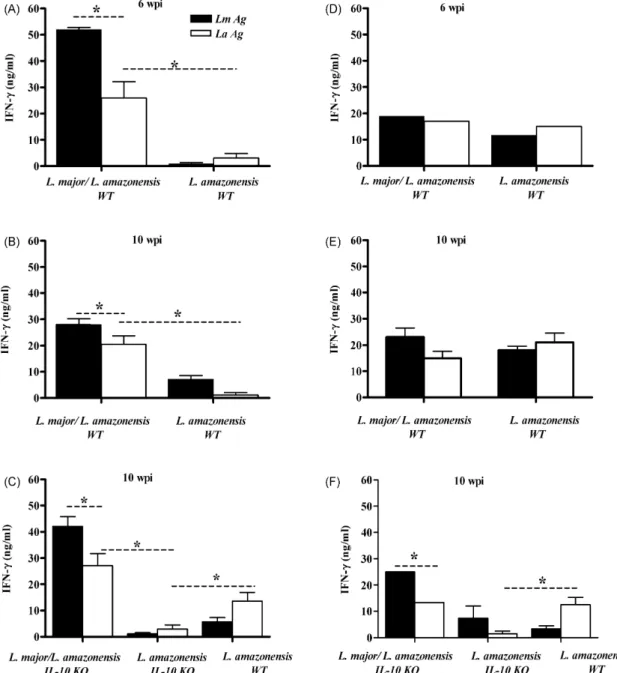
3.4. IFN-γproduction by mice previously infected with L. major is higher than in mice solely infected with L. amazonensis
Since susceptibility and pathogenesis to cutaneous leish-maniasis may be associated with the relative levels of IFN-␥
and 4 production in mice, we assessed these cytokines. IL-10−/−and C57BL/6 mice pre-infected withL. majorand later infected with L. amazonensiswere sacrificed 6 or 10 weeks afterL. amazonensisinfection and cells from spleens and lymph nodes drainingL. amazonensislesions were cultured in vitro for 72 h following stimulation with freeze-thawed promastigotes as described in Section2(Fig. 5). The analysis of IFN-␥production by spleen cells at 6 and 10 weeks post-infection demonstrated that cells from C57BL/6 mice previously infected withL. major
(stimulated either with L. major antigen or L. amazonensis
antigen) produced higher amounts IFN-␥ than C57BL/6 mice infected solely with L. amazonensis (Fig. 5A and B). IFN-␥
production by spleen cells stimulated withL. amazonensis anti-gen was smaller than that of cells stimulated with L. major
antigen. IFN-␥production by draining lymph nodes from both groups of WT mice was comparable at 6 and 10 weeks (Fig. 5D and E). In IL-10−/− mice previously infected withL. major, the detected IFN-␥ levels by both spleen and draining lymph node cells were higher when compared to animals infected only withL. amazonensis(Fig. 5C and F). IL-4 production was not detected by ELISA. In previous work, we have tried to detect IL-4 extensively by ELISA, ELISPOT and antibody isotype in sera from C57BL/6 mice infected withL. amazonensisand have failed (Hernandez et al., 2006). Our current data is in accordance with our previous data. These data indicate that the pre-infection withL. majorincreases IFN-␥production by spleen cells, but IFN-␥response is not affected in draining node lymph of theL. amazonensislesions.
3.5. iNOS, inflammatory cytokines and chemokines in lesions by L. amazonensis in pre-infected mice
Several studies addressed the early response toLeishmania
pro-Fig. 5. IFN-␥production by spleen cells from mice pre-infected withL. majorand challenged withL. amazonensisis higher thanL. amazonensis-infected mice. Spleen (A, B and C) and lymph node cells (D, E and F) cells from C57BL/6 (WT) and IL-10−/−(IL-10 KO) mice pre-infected withL. majorand challenged withL. amazonensis3 weeks later were stimulated in vitro in the presence ofL. major(LmAg) orL. amazonensis(LaAg) antigen and culture supernatants were harvested 72 h later and assayed for IFN-␥. Results in (C) are from pooled lymph nodes. IFN-␥was measured by ELISA at 6 and 10 weeks postL. amazonensis-infection (wpi). The values are the mean±S.D. *A statistically significant difference (p< 0.05, by Student’st-test). One of three experiments (five mice per experiment) is shown.
tein expression levels in foot tissues at different points afterL. amazonensisinfection. Total mRNAs were isolated from foot-pad tissues and cytokines gene expression was examined by real-time PCR. Coherently with draining lymph node data, the expression of IFN-␥in the lesion did not differ between pre-infected or not pre-pre-infected animals (Fig. 6A). On the other hand, the expression of iNOS and TNF were found up-regulated inL. major-previously infected animals when compared to con-trols infected only withL. amazonensis(Fig. 6B and C). iNOS transcripts were also higher in the pre-infected mice at 10 weeks post-infection (Fig. 6B). Another striking difference was observed in the IL-10 expression: IL-10 transcripts in lesions from pre-infected mice were approximately 15- and 142-fold
lower than the levels inL. amazonensislesions from mice with-out previous infection at 3 and 10 weeks post-infection with
L. amazonensis, respectively. Interestingly, expression of IL-10 mRNA was barely detectable at 6 weeks post-infection with
L. amazonensis (Fig. 6D). Levels of IL-10 protein were also consistently lower in extracts from L. amazonensislesions of pre-infected mice than in L. amazonensis lesions from mice infected just withL. amazonensis(Fig. 7A).
Fig. 6. IFN-␥(A), iNOS (B), TNF-␣(C) and IL-10 (D) message levels in lesions from mice pre-infected withL. majorand challenged withL. amazonensis. C57BL/6 mice were pre-infected withL. majorand challenged withL. amazonensis3 weeks later. The hindL. amazonensis-infected footpad was assayed for mRNA expression. Total RNA was isolated directly from theL. amazonensis-infected footpad at the indicated times during the infection, reverse-transcribed, and analyzed by real-time RT-PCR. The target genes were normalized to the endogenous control, and the values shown are the fold increase relative to expression in na¨ıve footpad. Each bar is the mean±S.D. of five individual footpads from mice from pre-infected mice withL. major(Lm/La) or solely infected withL. amazonensis(La). *A statistically significant difference (p< 0.05, by Student’st-test). One of three experiments (five mice per experiment) is shown.
lesions from mice pre-infected withL. majorwas different from lesions from mice infected just withL. amazonensis. Chemokine levels were assessed in homogenates of footpads infected with
L. amazonensis by ELISA (Fig. 7). Infection with L. ama-zonensisup-regulated CCL2/MCP-1 when compared to na¨ıve mice at both 6 and 10 weeks after infection (Fig. 7B) in a manner not influenced by pre-infection withL. major. On the other hand, no significant up-regulation of CXCL10/IP-10 was
found (Fig. 7C). In contrast, different levels were observed for CCL5/RANTES and CCL3/MIP-1␣ between experimen-tal groups (Fig. 7D and E). While at 6 weeks of infection withL. amazonensisCCL5/RANTES protein was significantly higher in lesions fromL. majorpre-infected mice, at 10 weeks lesions from pre-infected mice showed less CCL5/RANTES than lesions from mice infected only with L. amazonensis
(Fig. 7D). At 6 weeks of infection, CCL3/MIP-1␣levels inL.
amazonensislesions from pre-infected mice were smaller at than those from their counterparts infected just withL. amazonensis
(Fig. 7E). Low levels of CCL5/RANTES and CCL3/MIP-1␣
were observed at 3 weeks post-infection withL. amazonensis
(Fig. 7D and E).
Although several reports have demonstrated that IL-10 con-tributes to the suppression of the immunity to Leishmania
infection (Fiorentino et al., 1991; Rocha et al., 1999; Belkaid et al., 2001; Kane and Mosser, 2001; Buxbaum and Scott, 2005; Anderson et al., 2007), TGF-1 also plays a role in suppres-sion of the immune response (Barral-Netto et al., 1992). Thus, we also assessed the levels of TGF-1 (as protein in lesions) during of development of lesions. An analysis during infec-tion revealed an early increase in TGF-1 after infection with
L. amazonensis, although no differences were observed in the TGF-1 levels among groups at 3 and 10 weeks post-infection and na¨ıve mice. In contrast, there were differences between the two groups at 6 weeks post-infection, when pre-infected mice showed a higher level of TGF-1 than mice without previously infection (Fig. 7F).
4. Discussion
Several studies have demonstrated that the host response to leishmaniasis is determined by the species or strain of Leishma-nia(Barral et al., 1983; Calabrese and da Costa, 1992; Afonso and Scott, 1993; Soong et al., 1996; Anderson et al., 2005). In fact, L. amazonensis infections in most L. major resistant mouse strains result in the development of chronic lesions con-taining persistent parasite loads (Afonso and Scott, 1993; Soong et al., 1996; Jones et al., 2000; Ramer et al., 2006). Neverthe-less, several papers have demonstrated cross-immunity between
Leishmania species either using Leishmania antigens or live promastigotes and, although someLeishmaniaspecies may con-fer protection against others (Alexander and Phillips, 1978; Alexander, 1982; Alexander and Kaye, 1985; De Rossell et al., 1987; Coelho et al., 2003; Vanloubbeeck and Jones, 2004; Mukbel et al., 2006), at present, the mechanism responsible for this cross-protection is not yet known. In this work we stud-ied co-infections withL. amazonensisandL. majorin C57BL/6 mice.
A dramatic alteration in the course of infection byL. amazo-nensiswas observed in mice that had been previously inoculated withL. major. Hence, lesions were smaller in these mice, regard-less of the time of the previous challenge withL. amazonensis. This process may be related in part with enhanced parasite killing, as evidenced by a reduction in parasite burdens inL. amazonensislesions from C57BL/6 mice pre-infected withL. major, both by parasite quantification and histological analysis. This reduction in parasite burden is in sharp contrast with the one reported byVanloubbeeck and Jones (2004), who showed a difference of four orders of magnitude between groups similar to ours. In that study, the authors used C3Heb/FeJ mice. C3H/HeN and C3H/HeJ mice have been shown to have higher NK activ-ity than C57BL/6 mice, and also higher IFN-␥and lower IL-4 production in response to infection withL. major(Scharton and Scott, 1993). If the same is true for C3Heb/FeJ mice, a higher NK
cell response may explain the larger differences in parasite bur-dens found byVanloubbeeck and Jones (2004). The reduction we found in the parasite burden inL. amazonensislesions can-not be attributed to dissemination of the parasite to other tissues (Barral et al., 1986; Ramos-Santos et al., 2000; Vanloubbeeck et al., 2005) since, at 10 weeks of infection, the number of par-asites recovered from of draining lymph nodes in pre-infected infected mice was not higher than those in mice not pre-infected withL. major. Protection conferred byL. majorwas dependent on the time of infection. Hence, lesions due toL. amazonensis, as well as parasite numbers, were not altered ifL. majorwas given simultaneously or after infection with L. amazonensis. These data were indicative of a role of priming of the immune system withL. majorprior to infection withL. amazonensisin order to obtain the protective effect observed.
We observed higher production of IFN-␥by spleen cells from
L. major pre-infected mice in weeks 6 and 10 after infection withL. amazonensis. However, IFN-␥production by draining lymph node cells and IFN-␥mRNA in lesions were not different between pre-infected mice and their controls. Hence, although pre-infection withL. majorcontributes to the increase of sys-temic antigen-specific production of IFN-␥, the response toL. amazonensisat the site of infection did not seem to dramatically affect the production of IFN-␥. However, higher levels of tran-scripts for TNF-␣and iNOS were found in pre-infected mice, as well as lower levels of IL-10. Therefore, the smaller numbers of parasites found could be explained by a higher local level of iNOS, induced by activation by IFN-␥(which is similar in both groups) and TNF-␣(which is higher in the pre-infected group) (Green et al., 1990). Consequently, the synergism of these two cytokines could have caused enhanced killing in the pre-infected group. It has been shown that addition of TNF-␣to macrophages infected withL. amazonensisovercomes the defective killing of these parasites in vitro (Gomes et al., 2003). The lower levels of IL-10 may also have favored the higher expression of iNOS (Gazzinelli et al., 1992; Kane and Mosser, 2001; Jones et al., 2002).
Impaired microbicidal activity would not, clearly, be solely dependent of the expression of IL-10 as described forL. major
(Anderson et al., 2005), since the parasites persist even in absence of this cytokine (IL-10−/− mice), as shown here. Clearly, IL-10−/−mice use other mechanisms to control lesions and parasite burdens, one of them being the up-regulation of IFN-␥shown here.
Control of parasites was not complete in pre-infected mice. In this setting, we asked how pre-infected mice controlled pro-gressiveL. amazonensislesions without markedly reducing the number of parasites. Examination of the inflammatory infiltrate at the site of parasite inoculation revealed reduced recruitment of monocytes and lymphocytes intoL. amazonensislesions. Also, histological analysis showed that in lesions from pre-infected mice, most infected macrophages accumulated in limited areas of deep dermis. Hence, lesion progression in pre-infected mice is markedly associated with an alteration of cell recruitment.
be related with lesion and parasite burden reduction in BALB/c mice infected withL. amazonensis (Kane and Mosser, 2001). In IL-10−/−in the C57BL/6 background, however, lower para-site burdens and higher Th1 response than the wild type were found, even though lesion sizes were similar between the two groups (Jones et al., 2002). Our data do not argue for a role of IL-10 in the control of lesions sizes in co-infected mice, since lesion progression was similar in IL-10−/− mice pre-infected withL. major if compared to wild-type controls, as well as in mice infected solely withL. amazonensis. In addition, IL-10 lev-els were smaller inL. majorpre-infected wild-type mice when compared with levels found in wild-type mice solely infected withL. amazonensis, again speaking against a role for IL-10 in controlling lesion sizes. These results are consistent with other data previously published (Jones et al., 2002).
Increased expression of chemokines is the earliest response of macrophages toL. majorinfection (Racoosin and Beverley, 1997; Ji et al., 2003; Dasgupta et al., 2003). In humans, CCL2/MCP-1 was associated with healing of disease (Ritter et al., 1996), however, we found that CCL2/MCP-1, as well as CXCL10/IP-10 levels were similar in both groups; in fact CXCL10/IP-10 was not up-regulated in the footpads in response to infection. These results are consistent with the fact that sim-ilar levels of IFN-␥were found in footpads from both groups, since CCL2/MCP-1 and CXCL10/IP-10 are induced by IFN-␥
(Ritter and Moll, 2000; Muzio et al., 2000; Zaph and Scott, 2003; Steigerwald and Moll, 2005). The sustained and high produc-tion of CCL2/MCP-1 after 3 weeks might be responsible for the maintenance of macrophages in lesions in both groups (Ritter et al., 1996; Ritter and Moll, 2000). In contrast to CCL2/MCP-1 and CXCLCCL2/MCP-10/IP-CCL2/MCP-10, we observed differential production of the other-chemokines (CCL3/MIP-1␣and CCL5/RANTES). CCL3/MIP-1␣ levels were lower in pre-infected mice at 6 weeks post-infection than in mice infected just withL. ama-zonensis. This chemokine is associated with granulomatous condition (Lukacs et al., 1993) and some authors suggest that this chemokine is responsible for the increased cellularity of granulo-matous lesions (Hogaboam et al., 1997). Thus, it is possible that the increased cellularity in mice infected only withL. amazonen-sismight be the outcome of the increased CCL3/MIP-1␣levels. CCL5/RANTES was slightly increased in pre-infected mice at 6 weeks of infection, but was dramatically increased at 10 weeks in mice infected just withL. amazonensis. At 10 weeks of infection lesions were significantly larger inL. amazonensis-infected mice than in pre-infected mice. The delayed and reduced expression of CCL5/RANTES described in mice infected withL. amazo-nensis(Ji et al., 2003) is likely overcome by pre-infection withL major. It is tempting to speculate that reduced CCL3 and CCL5 expression in pre-infected mice could be related with the clus-tered distribution of the infected macrophages in lesions at 10 weeks of infection.
In summary, the findings presented here indicate that pre-infection with L. major can significantly modify the lesion progression caused by L. amazonensis without complete par-asite control. The concomitant infection withL. majorandL. amazonensisresults in the induction of a down-regulatory envi-ronment in the site of infection withL. amazonensis, site which
seems to be related with differential chemokines expression and altered composition of the cellular infiltrate.
Acknowledgements
This work was supported by CAPES, FAPEMIG grant CBB87 and by CNPq grant 350567/1995-6. RCR is a CAPES fellow, all other authors are CNPq fellows. We are indebted to Mr. Antonio Mesquita Vaz for expert animal care.
References
Afonso, L.C.C., Scott, P., 1993. Immune responses associated with suscepti-bility of C57BL/10 mice toLeishmania amazonensis. Infect. Immun. 61, 2952–2959.
Alexander, J., 1982. A radioattenuatedLeishmania majorvaccine markedly increases the resistance of CBA mice to subsequent infection with Leishma-nia mexicana mexicana. Trans. R. Soc. Trop. Med. Hyg. 76, 646–649. Alexander, J., Kaye, P.M., 1985. Immunoregulatory pathways in murine
leishmaniasis: different regulatory control duringLeishmania mexicana mexicana and Leishmania major infections. Clin. Exp. Immunol. 61, 674–682.
Alexander, J., Phillips, R.S., 1978.Leishmania tropicaandLeishmania mexi-cana: cross-immunity in mice. Exp. Parasitol. 45, 93–100.
Alexander, J., Phillips, R.S., 1980.Leishmania mexicanaandLeishmania tropica major: adoptive transfer of immunity in mice. Exp. Parasitol. 49, 34–40. Aliberti, J.C., Machado, F.S., Souto, J.T., Campanelli, A.P., Teixeira, M.M.,
Gazzinelli, R.T., Silva, J.S., 1999. beta-Chemokines enhance parasite uptake and promote nitric oxide-dependent microbiostatic activity in murine inflam-matory macrophages infected withTrypanosoma cruzi. Infect. Immun. 67, 4819–4826.
Anderson, C.F., Mendez, S., Sacks, D.L., 2005. Nonhealing infection despite Th1 polarization produced by a strain ofLeishmania majorin C57BL/6 mice. J. Immunol. 174, 2934–2941.
Anderson, C.F., Oukka, M., Kuchroo, V.J., Sacks, D., 2007. CD4(+)CD25(−)Foxp3(−) Th1 cells are the source of IL-10-mediated immune suppression in chronic cutaneous leishmaniasis. J. Exp. Med. 204, 285–297.
Arnoldi, J., Moll, H., 1998. Langerhans cell migration in murine cutaneous leishmaniasis: regulation by tumor necrosis factor alpha, interleukin-1 beta, and macrophage inflammatory protein-1 alpha. Dev. Immunol. 6, 3–11. Barral, A., Badaro, R., Barral-Netto, M., Grimaldi Jr., G., Momem, H., Carvalho,
E.M., 1986. Isolation ofLeishmania mexicana amazonensisfrom the bone marrow in a case of American visceral leishmaniasis. Am. J. Trop. Med. Hyg. 35, 732–734.
Barral, A., Petersen, E.A., Sacks, D.L., Neva, F.A., 1983. Late metastatic leish-maniasis in the mouse. A model for mucocutaneous disease. Am. J. Trop. Med. Hyg. 32, 277–285.
Barral-Netto, M., Barral, A., Brownell, C.E., Skeiky, Y.A., Ellingsworth, L.R., Twardzik, D.R., Reed, S.G., 1992. Transforming growth factor-beta in leish-manial infection: a parasite escape mechanism. Science 257, 545–548. Belkaid, Y., Hoffmann, K.F., Mendez, S., Kamhawi, S., Udey, M.C., Wynn,
T.A., Sacks, D.L., 2001. The role of interleukin (IL)-10 in the persistence of
Leishmania majorin the skin after healing and the therapeutic potential of anti-IL-10 receptor antibody for sterile cure. J. Exp. Med. 194, 1497–1506. Bhattacharyya, S., Ghosh, S., Dasgupta, B., Mazumder, D., Roy, S., Majum-dar, S., 2002. Chemokine-induced leishmanicidal activity in murine macrophages via the generation of nitric oxide. J. Infect. Dis. 185, 1704–1708.
Buxbaum, L.U., Scott, P., 2005. Interleukin 10- and Fcgamma receptor-deficient mice resolveLeishmania mexicanalesions. Infect. Immun. 73, 2101–2108. Calabrese, K.S., da Costa, S.C., 1992. Enhancement ofLeishmania amazonensis
infection in BCG non-responder mice by BCG-antigen specific vaccine. Mem. Inst. Oswaldo Cruz 87 (Suppl. 1), 49–56.
A.P., 2003. Immune responses induced by the Leishmania ( Leishma-nia)donovaniA2 antigen, but not by the LACK antigen, are protective against experimental Leishmania (Leishmania) amazonensis infection. Infect. Immun. 71, 3988–3994.
Cummings, K.L., Tarleton, R.L., 2004. Inducible nitric oxide synthase is not essential for control ofTrypanosoma cruziinfection in mice. Infect. Immun. 72, 4081–4089.
Dasgupta, B., Roychoudhury, K., Ganguly, S., Akbar, M.A., Das, P., Roy, S., 2003. Infection of human mononuclear phagocytes and macrophage-like THP1 cells withLeishmania donovaniresults in modulation of expression of a subset of chemokines and a chemokine receptor. Scand. J. Immunol. 57, 366–374.
De Rossell, R.A., Bray, R.S., Alexander, J., 1987. The correlation between delayed hypersensitivity, lymphocyte activation and protective immunity in experimental murine leishmaniasis. Parasite Immunol. 9, 105–115. Dondji, B., Perez-Jimenez, E., Goldsmith-Pestana, K., Esteban, M.,
Mahon-Pratt, D., 2005. Heterologous prime-boost vaccination with the LACK antigen protects against murine visceral leishmaniasis. Infect. Immun. 73, 5286–5289.
Fiorentino, D.F., Slotnik, A., Mosmann, T.R., Howard, M., O’Garra, A., 1991. IL-10 inhibits cytokine production by activated macrophages. J. Immunol. 147, 3815–3822.
Gazzinelli, R.T., Oswald, I.P., James, S.L., Sher, A., 1992. IL-10 inhibits parasite killing and nitrogen oxide production by IFN-gamma-activated macrophages. J. Immunol. 148, 1792–1796.
Giulietti, A., Overbergh, L., Valckx, D., Decallonne, B., Bouillon, R., Mathieu, C., 2001. An overview of real-time quantitative PCR: applications to quantify cytokine gene expression. Methods 25, 386–401.
Gomes, I.N., Calabrich, A.F., Tavares, R.S., Wietzerbin, J., De Freitas, L.A., Veras, P.S., 2003. Differential properties of CBA/J mononuclear phagocytes recovered from an inflammatory site and probed with two different species ofLeishmania. Microbes Infect. 5, 251–260.
Green, S.J., Crawford, R.M., Hockmeyer, J.T., Meltzer, M.S., Nacy, C.A., 1990.
Leishmania major amastigotes initiate the l-arginine-dependent killing mechanism in IFN-gamma-stimulated macrophages by induction of tumor necrosis factor-alpha. J. Immunol. 145, 4290–4297.
Hernandez, M.X., Barcante, T.A., Vilela, L., Tafuri, W.L., Afonso, L.C., Vieira, L.Q., 2006. Vaccine-induced protection againstLeishmania amazonensisis obtained in the absence of IL-12/23p40. Immunol. Lett. 105, 38–47. Hogaboam, C.M., Chensue, S.W., Steinhauser, M.L., Huffnagle, G.B., Lukacs,
N.W., Strieter, R.M., Kunkel, S.L., 1997. Alteration of the cytokine pheno-type in an experimental lung granuloma model by inhibiting nitric oxide. J. Immunol. 159, 5585–5593.
Ji, J., Masterson, J., Sun, J., Soong, L., 2005. CD4+CD25+ regulatory T cells restrain pathogenic responses duringLeishmania amazonensisinfection. J. Immunol. 174, 7147–7153.
Ji, J., Sun, J., Soong, L., 2003. Impaired expression of inflammatory cytokines and chemokines at early stages of infection withLeishmania amazonensis. Infect. Immun. 71, 4278–4288.
Jones, D., Elloso, M.M., Showe, L., Williams, D., Trinchieri, G., Scott, P., 1998. Differential regulation of the interleukin-12 receptor during the innate immune response toLeishmania major. Infect. Immun. 66, 3818– 3824.
Jones, D.E., Ackermann, M.R., Wille, U., Hunter, C.A., Scott, P., 2002. Early enhanced Th1 response afterLeishmania amazonensisinfection of C57BL/6 interleukin-10-deficient mice does not lead to resolution of infection. Infect. Immun. 70, 2151–2158.
Jones, D.E., Buxbaum, L.U., Scott, P., 2000. 4-independent inhibition of IL-12 responsiveness duringLeishmania amazonensisinfection. J. Immunol. 165, 364–372.
Kane, M.M., Mosser, D.M., 2001. The role of IL-10 in promoting disease progression in leishmaniasis. J. Immunol. 166, 1141–1147.
Khamesipour, A., Dowlati, Y., Asilian, A., Hashemi-Fesharki, R., Javadi, A., Noazin, S., Modabber, F., 2005. Leishmanization: use of an old method for evaluation of candidate vaccines against leishmaniasis. Vaccine 23, 3642–3648.
Lemos de Souza, V., Ascencao Souza, J., Correia Silva, T.M., Sampaio Tavares Veras, P., Rodrigues de-Freitas, L.A., 2000. DifferentLeishmaniaspecies
determine distinct profiles of immune and histopathological responses in CBA mice. Microbes Infect. 2, 1807–1815.
Lukacs, N.W., Kunkel, S.L., Strieter, R.M., Warmington, K., Chensue, S.W., 1993. The role of macrophage inflammatory protein 1 alpha in Schisto-soma mansoni egg-induced granulomatous inflammation. J. Exp. Med. 177, 1551–1559.
Lowry, O.H., Osebrough, N.J., Farr, A.L., Randall, R.J., 1951. Protein mea-surement with the Folin phenol reagent. J. Biol. Chem. 193, 265– 275.
Mendez, S., Reckling, S.K., Piccirillo, C.A., Sacks, D., Belkaid, Y., 2004. Role for CD4(+) CD25(+) regulatory T cells in reactivation of persistent leish-maniasis and control of concomitant immunity. J. Exp. Med. 200, 201– 210.
Mukbel, R., Petersen, C.A., Jones, D.E., 2006. Soluble factors fromLeishmania major-specific CD4+ T cells and B cells limitL. amazonensisamastigote survival within infected macrophages. Microbes Infect. 8, 2547–2555. Mukbel, R.M., Patten Jr., C., Gibson, K.A.T.H., Ghosh, M.O.U.S., Petersen,
C.H.R.I., Jones, D.E., 2007. Macrophage killing ofLeishmania amazonensis
amastigotes requires both nitric oxide and superoxide. Am. J. Trop. Med. Hyg. 76, 669–675.
Muzio, M., Bosisio, D., Polentarutti, N., D’amico, G., Stoppacciaro, A., Mancinelli, R., van’t Veer, C., Penton-Rol, G., Ruco, L.P., Allavena, P., Man-tovani, A., 2000. Differential expression and regulation of toll-like receptors (TLR) in human leukocytes: selective expression of TLR3 in dendritic Cells. J. Immunol. 164, 5998–6004.
Racoosin, E.L., Beverley, S.M., 1997.Leishmania major: promastigotes induce expression of a subset of chemokine genes in murine macrophages. Exp. Parasitol. 85, 283–295.
Ramer, A.E., Vanloubbeeck, Y.F., Jones, D.E., 2006. Antigen-responsive CD4+ T cells from C3H mice chronically infected withLeishmania amazonensis
are impaired in the transition to an effector phenotype. Infect. Immun. 74, 1547–1554.
Ramos-Santos, C., Hernandez-Montes, O., Sanchez-Tejeda, G., Monroy-Ostria, A., 2000. Visceral leishmaniosis caused byLeishmania(L.)mexicanain a Mexican patient with human immunodeficiency virus infection. Mem. Inst. Oswaldo Cruz 95, 733–738.
Ritter, U., Korner, H., 2002. Divergent expression of inflammatory dermal chemokines in cutaneous leishmaniasis. Parasite Immunol. 24, 295–301. Ritter, U., Moll, H., 2000. Monocyte chemotactic protein-1 stimulates the killing
ofLeishmania majorby human monocytes, acts synergistically with IFN-gamma and is antagonized by IL-4. Eur. J. Immunol. 30, 3111–3120. Ritter, U., Moll, H., Laskay, T., Brocker, E., Velazco, O., Becker, I., Gillitzer,
R., 1996. Differential expression of chemokines in patients with localized and diffuse cutaneous American leishmaniasis. J. Infect. Dis. 173, 699–709. Rocha, P.N., Almeida, R.P., Bacellar, O., de Jesus, A.R., Filho, D.C., Filho, A.C., Barral, A., Coffman, R.L., Carvalho, E.M., 1999. Down-regulation of Th1 type of response in early human American cutaneous leishmaniasis. J. Infect. Dis. 180, 1731–1734.
Santiago, H.C., Oliveira, C.F., Santiago, L., Ferraz, F.O., de Souza, D.G., De Fre-itas, L.A., Afonso, L.C., Teixeira, M.M., Gazzinelli, R.T., Vieira, L.Q., 2004. Involvement of the chemokine RANTES (CCL5) in resistance to experimen-tal infection withLeishmania major. Infect. Immun. 72, 4918–4923. Scharton, T.M., Scott, P., 1993. Natural killer cells are a source of interferon
gamma that drives differentiation of CD4+ T cell subsets and induces early resistance toLeishmania majorin mice. J. Exp. Med. 178, 567–577. Soong, L., Xu, J.C., Grewal, I.S., Kima, P., Sun, J., Longley, B.J., Ruddle, N.H.,
Mahon-Pratt, D., Flavell, R.A., 1996. Disruption of CD40-CD40 ligand interactions results in an enhanced susceptibility toLeishmania amazonensis
infection. Immunity 4, 263–273.
Spath, G.F., Beverley, S.M., 2001. A lipophosphoglycan-independent method for isolation of infectiveLeishmaniametacyclic promastigotes by density gradient centrifugation. Exp. Parasitol. 99, 97–103.
Steigerwald, M., Moll, H., 2005.Leishmania majormodulates chemokine and chemokine receptor expression by dendritic cells and affects their migratory capacity. Infect. Immun. 73, 2564–2567.
Uzonna, J.E., Spath, G.F., Beverley, S.M., Scott, P., 2004. Vaccination with phosphoglycan-deficientLeishmania majorprotects highly susceptible mice from virulent challenge without inducing a strong Th1 response. J. Immunol. 172, 3793–3797.
Uzonna, J.E., Wei, G., Yurkowski, D., Bretscher, P., 2001. Immune elimination of Leishmania major in mice: implications for immune memory, vaccination, and reactivation disease. J. Immunol. 167, 6967– 6974.
Vanloubbeeck, Y., Ackermann, M.R., Jones, D.E., 2005. Late cutaneous metas-tases in C3H SCID mice infected withLeishmania amazonensis. J. Parasitol. 91, 226–228.
Vanloubbeeck, Y., Jones, D.E., 2004. Protection of C3HeB/FeJ mice against
Leishmania amazonensischallenge after previousLeishmania major infec-tion. Am. J. Trop. Med. Hyg. 71, 407–411.
Vasquez, R.E., Soong, L., 2006. CXCL10/gamma interferon-inducible protein 10-mediated protection againstLeishmania amazonensisinfection in mice. Infect. Immun. 74, 6769–6777.
Veras, P., Brodskyn, C., Balestieri, F., Freitas, L., Ramos, A., Queiroz, A., Bar-ral, A., Beverley, S., Barral-Netto, M., 1999. A dhfr-ts-Leishmania major
knockout mutant cross-protects againstLeishmania amazonensis. Mem. Inst. Oswaldo Cruz 94, 491–496.
Vieira, L.Q., Goldschmidt, M., Nashleanas, M., Pfeffer, K., Mak, T., Scott, P., 1996. Mice lacking the TNF receptor p55 fail to resolve lesions caused by infection withLeishmania major, but control parasite replication. J. Immunol. 157, 827–835.