Rodrigues RN et al. / Compressive neuropathy of the inferior calcaneal nerve
Radiol Bras. 2015 Nov/Dez;48(6):368–372 368
Original Article
Compressive neuropathy of the first branch of the lateral
plantar nerve: a study by magnetic resonance imaging
*
Neuropatia compressiva do primeiro ramo do nervo plantar lateral: estudo por ressonância magnética
Rodrigues RN, Lopes AA, Torres JM, Mundim MF, Silva LLG, Silva BRC. Compressive neuropathy of the first branch of the lateral plantar nerve: a study by magnetic resonance imaging. Radiol Bras. 2015 Nov/Dez;48(6):368–372.
Abstract
R e s u m o
Objective: To assess the prevalence of isolated findings of abnormalities leading to entrapment of the lateral plantar nerve and respective branches in patients complaining of chronic heel pain, whose magnetic resonance imaging exams have showed complete selective fatty atrophy of the abductor digiti quinti muscle.
Materials and Methods: Retrospective, analytical, and cross-sectional study. The authors selected magnetic resonance imaging of hindfoot of 90 patients with grade IV abductor digiti quinti muscle atrophy according to Goutallier and Bernageau classification. Patients presenting with minor degrees of fatty muscle degeneration (below grade IV) and those who had been operated on for nerve decompression were excluded.
Results: A female prevalence (78.8%) was observed, and a strong correlation was found between fatty muscle atrophy and plantar fasciitis in 21.2%, and ankle varices, in 16.8% of the patients.
Conclusion: Fatty atrophy of the abductor digiti quinti muscle is strongly associated with neuropathic alterations of the first branch of the lateral plantar nerve. The present study showed a significant association between plantar fasciitis and ankle varices with grade IV atrophy of the abductor digiti quinti muscle.
Keywords: Abductor digiti quinti muscle; Baxter; Lateral plantar nerve; Inferior calcaneal nerve; Atrophy.
Objetivo: Avaliar a prevalência de achados isolados que causam compressão do primeiro ramo do nervo plantar lateral em pacientes com queixa de dor crônica no calcanhar, cujos exames de ressonância magnética mostraram atrofia gordurosa seletiva completa do músculo abdutor do quinto dedo.
Materiais e Métodos: Estudo retrospectivo, analítico e transversal. Selecionamos exames de ressonância magnética do retropé de 90 pacientes que apresentavam atrofia muscular grau IV do abdutor do quinto dedo utilizando a classificação de Goutallier e Bernageau. Foram excluídos do estudo pacientes com níveis menores de degeneração muscular (abaixo do grau IV).
Resultados: Houve predomínio do sexo feminino de 78,8% e alto índice de concordância da atrofia gordurosa do músculo abdutor do quinto dedo com fasciite plantar e varizes no tornozelo, respectivamente, encontrados em 21,2% e 16,8% dos pacientes.
Conclusão: Atrofia gordurosa do músculo abdutor do quinto dedo está fortemente associada a alterações neuropáticas do primeiro ramo do plantar lateral. Nosso estudo mostrou associação significativa entre a fasciite plantar e varizes do tornozelo com atrofia grau IV do abdutor do quinto dedo.
Unitermos: Músculo abdutor quinto dedo; Baxter; Nervo plantar lateral; Nervo calcâneo inferior; Atrofia.
* Study developed at Axial Medicina Diagnóstica, Belo Horizonte, MG, Brazil. 1. MDs, Musculoskeletal Radiologists, Axial Medicina Diagnóstica, Belo Horizonte, MG, Brazil.
2. MD, Orthopedist, Head of the Foot Surgery Group at Santa Casa de Belo Horizonte, Professor at Faculdade de Ciências Médicas de Minas Gerais (FCMMG), Belo Horizonte, MG, Brazil.
3. MDs, Radiologists, Axial Medicina Diagnóstica, Belo Horizonte, MG, Brazil. 4. MD, Physician Assistant, Axial Medicina Diagnóstica, Belo Horizonte, MG, Brazil. Mailing Address: Dra. Rogéria Nobre Rodrigues. Rua Gonçalves Dias, 2867, Santo Agostinho. Belo Horizonte, MG, Brazil, 30140-093. E-mail: [email protected].
Received December 23, 2013. Accepted after revision May 7, 2015.
concerned with the relevant role played by imaging meth-ods in the improvement of the diagnosis in the musculosk-eletal system(1–15).
Thalamic pain presents a wide spectrum of differential diagnosis including plantar fasciitis, fat pad involvement, stress fracture, enthesopathy and inflammatory arthropathy(16). One
of the most common causes of chronic pain is entrapment of the first branch of the lateral plantar nerve, a condition that is known as Baxter’s neuropathy(17,18). It is believed that
approximately 20% of cases of pain in the medial region of the heel are associated with neuropathy of that nerve(19–22).
Generally, the nerve to the abductor digiti quinti (ADQ) muscle originates as first branch of the lateral plantar nerve (82.1%) that divides itself at the level of the medial malleolus,
Rogéria Nobre Rodrigues1, Alexia Abuhid Lopes1, Jardélio Mendes Torres2, Marina Franco Mundim3,
Lênio Lúcio Gavio Silva3, Breno Rabelo de Carvalho e Silva4
INTRODUCTION
but presents with some anatomical variations. In 11.7% of cases, such nerve may present as a direct branch from the posterior tibial nerve, or even originate from a common branch with the posterior branch to the lateral plantar nerve and with the medial calcaneal branch (4.1%) or in a branch in common with the posterior branch to the plantar square (2.1%)(23). The nerve follows the medial pathway along the
long plantar ligament to the lateral, between the abductor hallucis muscle and the medial calcaneal tuberosity, insert-ing into the proximal aspect of the ADQ muscle (Figure 1). The nerve to the ADQ is mixed and originates motor nerves to the ADQ and, occasionally, to the short flexor of the digits and plantar square muscle, as well as sensory branches to the calcaneal periosteum, long plantar ligament and adjacent skin(24). Any situation determining increased
volume in the region of the nerve might cause a focal com-pressive effect with consequential neuropathy.
At clinical evaluation, the symptoms may not be distin-guished from plantar fasciitis and, frequently, both condi-tions overlap(25). ADQ muscle weakness may be present in chronic cases, with decreased fifth toe abduction strength de-termined by the muscle degeneration.
Magnetic resonance imaging (MRI) may be used to de-tect alterations associated with ADQ muscle denerva-tion(18,26). The presence of such a muscle atrophy observed
at MRI reflects a chronic compression of the inferior calca-neal nerve and contributes to the clinical diagnosis of Baxter’s neuropathy(19).
The literature suggests two possible sites of nerve en-trapment which could result in Baxter’s neuropathy, as fol-lows: the first one, in patients with altered biomechanics, such as excessive pronation, since the nerve may be com-pressed in the movement of lateral rotation between the plan-tar square and abductor hallucis muscles(24); and second,
either the nerve may be compressed as it passes anteriorly to the medial calcaneal tuberosity, or interfere mechanically with the plantar calcaneal spur(17,25,27,28) (Figure 2).
The present study is aimed at evaluating the prevalence of MRI findings associated with compression of the first branch of the lateral plantar nerve in patients with chronic heel pain manifesting by complete selective atrophy of the ADQ muscle.
MATERIALS AND METHODS
Retrospective, analytical, and cross-sectional study of 90 patients with diagnosis of. grade IV atrophy of ADQ muscle (according to Goutallier and Bernageau classification(29)) with
mean age of 49.2 years, submitted to hindfoot MRI in a high field 1.5 T apparatus with fast spin echo, sagittalT1-weighted sequences and proton density (PD) with fat suppression, and axial and coronal/oblique PD T2-weighted sequences with fat suppression. After intravenous paramagnetic contrast injection, coronal and sagittal T1-weighted sequences with fat suppres-sion were acquired. Only patients classified with grade IV atrophy of ADQ muscle (complete fatty muscle atrophy) were considered for evaluation. Amongst the evaluated patients,
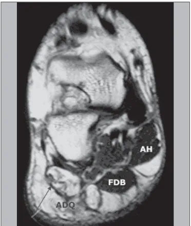
Figure 1. PM, medial plantar nerve; PL, lateral plantar nerve; AQD, abductor digiti quinti nerve.
Table 2—Frequency of MRI findings related to ADQ atrophy according to sex.
Plantar fasciitis Varicosities Trauma Tendinopathy Plantar lipoma
Male
11.5% 9.7% 19.5%
6.5% 0%
Female
37.8% 31.9% 11.2% 9.6% 1.9%
p-value
p < 0.01
p < 0.01
p < 0.01
p < 0.01
p < 0.01 21.2% were men with mean age of 42.1 years, and 78.8%
were women with mean age of 56.3 years.
RESULTS
Atrophy of the ADQ muscle was most prevalent in women – 71 cases (78.8%) –, with a high prevalence in this group at the age ranges from 40 to 50 years (45.9%) and 50–60 years (38.3%), and p < 0.01 in both groups as
dem-onstrated on Figure 3. On the other hand, no statistically significant difference was observed in the prevalence of at-rophy of the ADQ muscle, as demonstrated on Figure 4.
A strong correlation (p < 0.01) between grade IV
atro-phy of the ADQ muscle and plantar fasciitis and hindfoot varicosities in 21.2% and 16.8% of patients, respectively, con-sidering isolated factors determining neural compression (Table 1).
In the female group, plantar fasciitis and ankle varicosi-ties were determinant as statistically significant (p < 0.01),
while in the male group only the trauma factor was appar-ently reliable as an isolated factor of compressive neural injury (Table 2).
Table 1—Frequency of isolated MRI findings associated with ADQ atrophy.
Plantar fasciitis Varicosities Lateral ligament injury Medial ligament injury Tendinopathy Previous trauma Tarsal tunnel syndrome Plantar lipoma
Percentage
21.2% 16.8% 9.2% 1.8% 8.4% 7.2% 7.2% 0.9%
p-value
p < 0.01
p < 0.01
p > 0.01
p > 0.01
p > 0.01
p > 0.01
p > 0.01
p > 0.01
Figure 3. High prevalence of abduc-tor digit quinti atrophy in patients aged between 40 and 50 years (45.9%) and between 50 and 60 years (38.3%).
20–30 years 30–40 years 40–50 years 50–60 years 60–70 years
Figure 4. No statistically significant difference was observed as regards prevalence of ADQ muscle atrophy in male individuals.
DISCUSSION
MRI has shown to be an invaluable investigative method to detect muscle alterations associated with denervation. It is the most sensitive method to detect involvement of muscle tissues as compared with ultrasonography and computed tomography. Because of its noninvasive nature and capacity to demonstrate anatomical details, this method presents some advantages as compared with electromyography(30).
Acute and subacute muscle denervation is more appro-priately evaluated at fluid-sensitive MRI sequences such as PD/T2-weighted sequences with fat suppression or STIR se-quences, showing increased signal intensity within the muscle belly as compared with the normal muscle, corresponding to neurogenic muscle edema(30,31) (Figure 5). The enhance-ment of the muscle by gadolinium also occurs either in the acute or subacute phase of denervation(30). In compressive
Baxter’s neuropathy, muscle edema occurs selectively inside the ADQ muscle and potentially in the short flexor muscle of the toes and plantar square muscle, depending on the anatomical variation in the patient. Chronic denervation leads to muscle atrophy and subsequent irreversible fat infiltration. Such findings are clearly depicted at T1-weighted images without fat suppression(30,31) (Figure 6). Typically, atrophy
and fat infiltration occur homogeneously in the muscle belly. On the other hand, in the presence of double or redundant innervation, such changes either may not occur or occur heterogeneously(30).
It is estimated that in 20% of patients with chronic hell pain such condition is related to compression of the abduc-tor digiti quinti nerve(32). In a study evaluating the associa-tion between ADQ atrophy and MRI findings of potential
causes, there was a strong correlation between muscle atro-phy and plantar fasciitis and calcaneal spur. However, the patients considered in such study presented with any degree of ADQ muscle atrophy(33).
In the present study, the authors have selected only pa-tients with grade IV atrophy, unequivocally evidencing the presence of compressive neuropathy. Chronic plantar fasciitis and local varicosities represented the findings most frequently associated with entrapment of the abductor digiti quinti nerve. Initially, heel pain should be treated with conservative mea-sures, including the use of a nocturnal orthosis, therapeutic footwear, physical therapy, anti-inflammatory drugs and corticoid infiltration(16,34,35).
As the pain becomes chronic, over a period longer than six month and without any improvement with the conserva-tive treatment, the hypothesis of compression of the first branch of the lateral plantar nerve should be considered. In such cases, the patients may benefit from surgical decom-pression of the region(16,35–37) by endoscopic approach(16,36),
radiofrequency ablation techniques(34) or open surgery.
CONCLUSION
Atrophy of the ADQ muscle is strongly associated with neuropathic compression of the first branch of the lateral plan-tar nerve. MRI is considered to be a noninvasive and highly accurate diagnostic method to evaluate grade IV atrophy of ADQ muscle and other associated diseases.
Figure 5. Sagittal MRI DP-weighted sequence with fat suppression showing signs of plantar fasciitis and abnormality in the signal of ADQ muscle fibers, that is hyperintense, corresponding to a pattern of edema resulting from acute denerva-tion.
REFERENCES
1. Terazaki CRT, Trippia CR, Trippia CH, et al. Synovial chondro-matosis of the shoulder: imaging findings. Radiol Bras. 2014;47:38– 42.
2. Arend CF. The carpal boss: a review of different sonographic find-ings. Radiol Bras. 2014;47:112–4.
3. Arend CF. Sonography of the iliotibial band: spectrum of find-ings. Radiol Bras. 2014;47:33–7.
4. Nakamura SA, Lorenzato MM, Engel EE, et al. Incidental enchon-dromas at knee magnetic resonance imaging: intraobserver and interobserver agreement and prevalence of imaging findings. Radiol Bras. 2013;46:129–33.
5. Souza CG, Gasparetto EL, Marchiori E, et al. Pyogenic and tuber-culous discitis: magnetic resonance imaging findings for differen-tial diagnosis. Radiol Bras. 2013;46:173–7.
6. Machado BB, Lima CMAO, Junqueira FP, et al. Magnetic reso-nance imaging in intersection syndrome of the forearm: icono-graphic essay. Radiol Bras. 2013;46:117–21.
7. Alves MPT, Fonseca COP, Granjeiro JM, et al. Carpal tunnel syn-drome: comparative study between sonographic and surgical mea-surements of the median nerve in moderate and severe cases of dis-ease. Radiol Bras. 2013;46:23–9.
8. Simão MN, Helms CA, Richardson WJ. Magnetic resonance imag-ing findimag-ings of disc-related epidural cysts in nonsurgical and post-operative microdiscectomy patients. Radiol Bras. 2012;45:205–9. 9. Chojniak R, Grigio HR, Bitencourt AGV, et al. Percutaneous com-puted tomography-guided core needle biopsy of soft tissue tumors: results and correlation with surgical specimen analysis. Radiol Bras. 2012;45:259–62.
10. Cotta AC, Melo RT, Castro RCR, et al. Diagnostic difficulties in osteoid osteoma of the elbow: clinical, radiological and histopatho-logical study. Radiol Bras. 2012;45:13–9.
11. Tavares Júnior WC, Faria FM, Figueiredo R, et al. Bone attrition: a cause of knee pain in osteoarthritis. Radiol Bras. 2012;45:273–8. 12. Jacob Jr C, Barbosa DM, Batista PR, et al. Thoracolumbar burst fracture: what the radiologist should know. Radiol Bras. 2012;45: 101–4.
13. Moura MVT. Trapped periosteum in a distal femoral physeal in-jury: magnetic resonance imaging evaluation. Radiol Bras. 2012;45: 184–6.
14. Bayerl JS, Oliveira ARN, Peçanha PM, et al. Osteomyelitis of the wrist in a patient with disseminated paracoccidioidomycosis: a rare presentation. Radiol Bras. 2012;45:238–40.
15. Arend CF. Tenosynovitis and synovitis of the first extensor com-partment of the wrist: what sonographers should know. Radiol Bras. 2012;45:219–24.
16. Thomas JL, Christensen JC, Kravitz SR, et al. The diagnosis and treatment of heel pain: a clinical practice guideline-revision 2010. J Foot Ankle Surg. 2010;49(3 Suppl):S1–19.
17. Baxter DE, Thigpen CM. Heel pain – operative results. Foot Ankle. 1984;5:16–25.
18. Recht MP, Grooff P, Ilaslan H, et al. Selective atrophy of the ab-ductor digiti quinti: an MRI study. AJR Am J Roentgenol. 2007;189: W123–7.
19. Delfaut EM, Demondion X, Bieganski A, et al. Imaging of foot and
ankle nerve entrapment syndromes: from well-demonstrated to un-familiar sites. Radiographics. 2003;23:613–23.
20. Oztuna V, Ozge A, Eskandari MM, et al. Nerve entrapment in pain-ful heel syndrome. Foot Ankle Int. 2002;23:208–11.
21. Przylucki H, Jones CL. Entrapment neuropathy of muscle branch of lateral plantar nerve: a cause of heel pain. J Am Podiatry Assoc. 1981;71:119–24.
22. Baxter DE. Release of the nerve to the abductor digiti minimi. In: Kitaoka HB, editor. Master techniques in orthopaedic surgery. The foot and ankle. Philadelphia, PA: Lippincott Williams & Wilkins; 2002. p. 359.
23. del Sol M, Olave E, Gabrielli C, et al. Inervation of the abductor digiti minimi muscle of the human foot: anatomical basis of the entrapment of the abductor digiti minimi nerve. Surg Radiol Anat. 2002;24:18–22.
24. Rondhuis JJ, Huson A. The first branch of the lateral plantar nerve and heel pain. Acta Morphol Neerl Scand. 1986;24:269–79. 25. Offutt S, DeHeer P. How to address Baxter’s nerve entrapment.
Podiatry Today. 2004;17:52–8.
26. Alshami AM, Souvlis T, Coppieters MW. A review of plantar heel pain of neural origin: differential diagnosis and management. Man Ther. 2008;13:103–11.
27. Donovan A, Rosenberg ZS, Cavalcanti CF. MR imaging of entrap-ment neuropathies of the lower extremity. Part 2. The knee, leg, ankle, and foot. Radiographics. 2010;30:1001–19.
28. Louisia S, Masquelet AC. The medial and inferior calcaneal nerves: an anatomic study. Surg Radiol Anat. 1999;21:169–73.
29. Lippe J, Spang JT, Leger RR, et al. Inter-rater agreement of the Goutallier, Patte, and Warner classification scores using preopera-tive magnetic resonance imaging in patients with rotator cuff tears. Arthroscopy. 2012;28:154–9.
30. Kim SJ, Hong SH, Jun WS, et al. MR imaging mapping of skeletal muscle denervation in entrapment and compressive neuropathies. Radiographics. 2011;31:319–32.
31. Fleckenstein JL, Watumull D, Conner KE, et al. Denervated hu-man skeletal muscle: MR imaging evaluation. Radiology. 1993;187: 213–8.
32. Shon LC, Easley ME. Chronic pain. In: Myerson MS, editor. Foot and ankle disorders. Philadelphia, PA: WB Saunders; 2000. p. 815– 81.
33. Chundru U, Liebeskind A, Seidelmann F, et al. Plantar fasciitis and calcaneal spur formation are associated with abductor digiti minimi atrophy on MRI of the foot. Skeletal Radiol. 2008;37:505–10. 34. Cozzarelli J, Sollitto RJ, Thapar J, et al. A 12-year long-term
retro-spective analysis of the use of radiofrequency nerve ablation for the treatment of neurogenic heel pain. Foot Ankle Spec. 2010;3:338– 46.
35. Baxter DE, Pfeffer GB. Treatment of chronic heel pain by surgical release of the first branch of the lateral plantar nerve. Clin Orthop Relat Res. 1992;(279):229–36.
36. Mesmar M, Amarin Z, Shatnawi N, et al. Chronic heel pain due to the entrapment of the first branch of the lateral plantar nerve: analysis of surgical treatment. Eur J Orthop Surg Traumatol. 2010;20:563– 7.