Case Report
180
Pastore R, Lenza RM, Rodrigues FB, Tostes LV, Guerra NC, Crema E. Cecal diverticulitis or appendicitis. When should I suspect? A case report. J Coloproctol, 2011;32(2): 180-183.
ABSTRACT : The objective of this article was to report a case of cecal diverticulitis and point out the differential diagnosis of acute appendicitis. The clinical manifestations of these pathological conditions are similar, and the accurate diagnosis of cecal diverticulitis before the surgery is dificult. Therefore, most diagnoses are made during the surgery. Moreover, cecal diverticulum is uncommon in western countries, but it is prevalent in Asian people and their descendants. We report a case of a 55-year-old female patient, whose imaging exams (ultrasonography and computed tomography) and blood tests were not enough to diagnose the affection, requiring lapa-rotomy and pathological exams for the inal diagnosis. Some studies suggesting the best practice in case of diverticulum of the cecum were revised, as the diagnosis usually occurs during the surgery.
Keywords: appendicitis; diverticulitis; cecum; diverticulum.
RESUMO: O objetivo deste trabalho foi relatar um caso de diverticulite no ceco e chamar a atenção para o diagnóstico diferencial com apendicite aguda. As manifestações clínicas das duas afecções são semelhantes, diicultando o diagnóstico exato de diverticulite cecal, além de ser incomum, em nosso meio, o aparecimento de divertículo em cólon direito, sendo essa entidade mais comum em asiáticos e em seus descendentes. Relata-se atendimento a uma paciente de 55 anos, cujos exames de imagem (ultrassonograia e tomograia com -putadorizada) e de sangue não foram suicientes para o diagnóstico. Houve necessidade de realizar-se laparotomia exploradora e exames anatomopatológicos para a conirmação. Também foram revisados alguns trabalhos que sugerem qual a melhor conduta a ser tomada quando se encontra divertículo cecal no perioperatório, já que, na maioria das vezes, o diagnóstico é feito neste momento.
Palavras-chave: apendicite; diverticulite; ceco; divertículo.
Cecal diverticulitis or appendicitis. When should I suspect?
A case report
Ricardo Pastore1, Roberto da Mata Lenza2, Flávio Batista Rodrigues3, Lucas Vieira Tostes3, Natalia Cavasini Guerra3,
Eduardo Crema4
1Professor, Discipline of General Surgery and Surgical Technique at the Universidade Federal do Triângulo Mineiro
(UFTM) – Uberaba (MG), Brazil. 2Digestive Tract Surgeon and Physician at the Emergency Service at the UFTM –
Uberaba (MG), Brazil. 3Academicians of Medicine at the UFTM – Uberaba (MG), Brazil. 4Full Professor, Discipline of
Digestive Tract Surgery at the UFTM – Uberaba (MG), Brazil.
Study carried out at the Discipline of General Surgery, Universidade Federal do Triângulo Mineiro (UFTM) – Uberaba (MG), Brazil. Financing source: none.
Conlict of interest: nothing to declare.
Submitted on: 10/14/2010 Approved on: 11/18/2010
INTRODUCTION
Cecal diverticulitis is a rare condition1,2,3, with prevalence of 0.004 to 2.1%4, affecting more often the Asian people3,5,6 and their descendants1,7,8. The irst description was reported in 18633. The preoperative diagnosis is dificult, as its signs and symptoms can
be confused with the signs and symptoms of acute ap-pendicitis1,5,7,9-12. Consequently, the diagnosis is most
of the times during the surgery2,9,11-13 and conirmed only with an anatomopathological exam1.
CASE REPORT
M.A.C., female, 55 years old, came to the
Cecal diverticulitis or appendicitis. When should I suspect? A case report Ricardo Pastore et al.
181
J Coloproctol April/June, 2012
Vol. 32 Nº 2
a week. She said she had no nauseas, vomiting or al
-teration to bowel habits. She presented anorexia and
no fever since the beginning of this condition. The physical examination showed peritoneal reaction in the right iliac fossa (positive Blumberg sign).
Pelvic ultrasonography (US) showed fecalith in
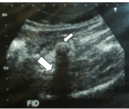
the right iliac fossa, with peritoneal reaction around it (Figure 1) and no collections. The report suggested appendicitis as the most probable diagnosis or focal diverticulitis near the cecum. Abdominal computed tomography (CT) showed a tubular shape posterolat-erally to the cecum. The lesion area was highlighted after the intravenous infusion of contrast medium
and calciied focus in its proximal segment, as well as densiication of surrounding mesenteric fat. No co -lonic diverticular formations with evidence of acute
inlammatory were observed. Then, based on CT, the
patient’s condition was compatible with acute appen-dicitis. Two complete blood tests were performed, which did not present alterations.
Infraumbilical median exploratory laparotomy was the selected method and a tumor mass was found in the cecum. Then, segmental colectomy was per-formed, with removal of the cecum and the mass in-volving it, as well as the appendix, which presented unaltered aspect. In addition, termino-terminal ileo-colic anastomosis was performed. The anatomopatho-logical exam showed ulcerated and abscessed diver-ticulum in the wall of the large bowel and contained by the peri-intestinal adipose tissue (Figure 2).
DISCUSSION
The (false) left colon diverticulosis occurs pre-dominantly in the sigmoid and affects the western population more often9,14,15, while the (true) right co-lon diverticulosis occurs predominantly in the cecum and affects the young population and descendants of Asians more often1,9. Cecal diverticulitis is rare in western population, but it is prevalent in Asian countries.6,7,14. The preoperative diagnosis is difi -cult9 and infrequent, despite de use of radiological imaging. The diagnostic certainty is obtained only with the anatomopathological exam16. The differen-tial diagnoses are: Crohn’s disease, actinomycosis, perforation by a strange body, amebiasis, carcinoid tumor, tuberculosis, gastroenteritis, ureteral colic,
ec-topic pregnancy, ovarian cyst rupture, pelvic inlam -matory disease and, especially, acute appendicitis2,17. The clinical presentation of cecal diverticulitis with fever and abdominal pain in the right lower quadrant is practically indistinguishable from acute appendici-tis1, but there are some differences: the pain in diver-ticulitis starts directly in the right iliac fossa, instead of starting vaguely in the periumbilical region, as it occurs in appendicitis. Diverticulitis is more insidi-ous and extended, and its systemic toxic signs are mild, with rare nauseas and vomiting7. A case has been reported of cecal diverticulitis initially caus-ing pain in the periumbilical region, and the patient presented recurrent abdominal pain for six months, without alteration to bowel habits or systemic toxic signs3, compatible with the clinical condition sug-gested for cecal diverticulitis. The blood test may show elevated white blood cell count1,9. However, in our case, no alteration was observed in the absolute number of leucocytes.
US and CT are very helpful, enabling the cor -rect diagnosis and preventing unexpected findings
Figure 1. Ultrasonography of the patient, showing fecalith (smaller arrow) in the right iliac fossa. The larger arrow shows the acoustic shadow of the dense material.
Figure 2. Picture of cecal diverticulum at the
Cecal diverticulitis or appendicitis. When should I suspect? A case report Ricardo Pastore et al.
182
J Coloproctol April/June, 2012
Vol. 32 Nº 2
during the surgery2. A study that analyzed 934 pa-tients18 with pain of undetermined nature in the right iliac fosse showed that US presented 100%
accuracy when distinguishing diverticulitis from
appendicitis. However, this is a limited exam, as it
depends on the examiner’s experience, a fact that becomes a problem, particularly in western coun-tries, where the experience with cecal diverticu-litis is low2. CT offers good cost-benefit ratio at the differential diagnosis of abdominal pain condi-tions involving suspicion of acute appendicitis19. Helical CT may suggest or define the diagnosis of
cecal diverticulitis18.
In this report, only ultrasonography suggested that it was cecal diverticulitis. When the diagnosis of cecal diverticulitis is secured, antibioticotherapy can be applied in patients without signs of peritoni-tis1,9,20,21. As the right colonic diverticulitis is benign, the conservative treatment with minimal surgical in-tervention should be the best therapeutic option10. Exploratory laparotomy is suggested in cases with-out diagnostic certainty1. However, the greatest di
-lemma is what to do when cecal diverticulitis is in-cidentally found during appendicectomy3. There is no standard procedure for the treatment of solitary cecal diverticulitis3. The surgical resection of di-verticulum is recommended9 plus colectomy, if the histopathological exam shows the presence of neo-plasm9. When the diagnosis is secured, the procedure of diverticulectomy combined with appendicectomy is suggested3. Otherwise, colectomy is suggested3. In this case, the second approach was selected, with segmental colectomy.
A successful clinical treatment was reported in a case whose diagnosis was made without laparotomy, but the patient had history of appendicectomy for 15 years and no pain at rapid decompression1. In addition, emergency colectomy is well accepted in the treat-ment of complicated diverticulitis10. Two cases have been reported in which right hemicolectomy was per-formed, without complications in both cases2,3. Lap-aroscopy could be applied for diagnostic purposes, but it involves the risk of not detecting diverticula in the posterior wall of the cecum22.
REFERENCES
1. Chedid AD, Domingues LA, Chedid MF, Villwock MM, Mondelo AR. Divertículo único do ceco: experiência de um hospital geral brasileiro. Arq Gastroenterol 2003;40(4):216-9.
2. Grifiths EA, Date RS. Acute presentation of a solitary caecal diverticulum: a caser report. J Med Case Reports 2007;1:129.
3. Kurer MA. Solitary caecal diverticulitis as an unusual cause of a right iliac fossa mass: a case report. J Med Case Reports 2007;1:132.
4. Barría C, Pujado B, Zepeda N, Beltrán MA. Diveriticulitis apendicular como causa de apendicectomía: reporte de un caso. Rev child Cir 60(2):154-7.
5. Fontes D, Luz MMP, Andrade Jr JCCG, Santos BMR, Andrade DC. Doença diverticular no apêndice cecal. Rev bras Coloproct 2006;23(1):25-7.
6. Poon RT, Chu KW. Inlammatory cecal masses in
patients presenting with appendicitis. World J Surg 1999;23(7):713-6.
7. Shyung LR, Lin SC, Shih SC, Kao CR, Chou SY. Decision making in right-sided diverticulitis. World J Gastroenterol 2003;9(3):606-8.
8. Ruiz-Tovar J, Reguero-Callejas ME, Gonzáles FP. Inlammation and perforation of a solitary divericulum of the cecum. A report of 5 cases and literature review. Rev Esp Enferm Dig 2006;98(11):875-80.
9. Karatepe O, Gulcicek OB, Adas G, Battal M, Ozdenkaya Y, Kurtulus I, et. al. Cecal diverticulitis mimicking acute appendicitis: a report of 4 cases. World J Emerg Surg 2008;3:16-4.
10. Leung WW, Lee JF, Liu SY, Mou JW, Ng SS, Yiu RY, et al. Critical appraisal on the role and outcome of emergency colectomy for uncomplicated right-sided colonic divericulitis. World J Surg 2007;31(2):383-7.
Cecal diverticulitis or appendicitis. When should I suspect? A case report Ricardo Pastore et al.
183
J Coloproctol April/June, 2012
Vol. 32 Nº 2
should I do? Ann R Coll Surg Engl 2006;88(7):672-4. 12. Grifiths EA, Bergin FG, Henry JA, Mudawi AM. Acute
inlammation of a congenital cecal diverticulum mimicking appendicitis. Med Sci Monit 2003; 9(12):CS107-9.
13. Papapolychroniadis C, Kaimakis D, Fotiadis P, Karamanlis E, Stefopoulou M, Kouskouras K, et al. Perforated diverticulum of the caecum. A dificult preoperative diagnosis. Report of 2 cases and review of the literature. Tech Coloproctol 2004;(Suppl l):116-8.
14. Hildebrand P, Kropp M, Stellmacher F, Roblick UJ, Bruch HP, Schwandner O. Surgery for right-sided colonic diverticulitis: results of a 10-year-observation period. Langenbecks Arch Surg 2007;392(2):143-7.
15. Paulino F, Roselli A, Martins U. Pathology of diverticular disease of the colon. Surgery 1971;69(1):63-9.
16. Nunes FC, Mattos MP, Silva AL. Divertículo do apêndice vermiforme. Rev Col Bras Cir 2004;31(5):342-3.
17. Rasmussen I, Enblad P. Acute solitary diverticulitis of the caecum. Case report. Acta Chir Scand 1988;154(5-6):399-401. 18. Chou YH, Chiou HJ, Tiu CM, Chen JD, Hsu CC, Lee CH, et
al. Sonography of acute right side colonic diverticulitis. Am J Surg 2001;181(2):122-7.
19. Rao PM, Rhea JT, Novelline RA, Mostafavi AA, McCabe CJ. Effect of computed tomography of the appendix on treatment of patients and use of hospital resources. N Engl J Med 1998;338(3):141-6.
20. Abogunrin FA, Arya N, Somerville JE. Case report solitary caecal diverticulitis – a rare cause of right iliac fossa pain. Ulster Med J 2005;74(2):132-3.
21. Jang HJ, Lim HK, Lee SJ, Lee WJ, Kim EY, Kim SH. Acute diverticulitis of the cecum and ascending colon: the value of thin-section helicoidal CT indings in excluding colonic carcinoma. ARJ Am J Roentgenol 2000;174(5):1397-402. 22. Sauerland S, Lefering R, Neugebauer EA. Laparoscopic
versus open surgery for suspected appendicitis. Cochrane Database Syst Rev 2004;(4):CD001546.
Correspondence to: Dr. Eduardo Crema
Disciplina de Cirurgia Geral
Universidade Federal do Triângulo Mineiro (UFTM) Avenida Frei Paulino, 30 – Abadia
