Submitted20 December 2015
Accepted 9 April 2016
Published10 May 2016
Corresponding author
Jie Zhao, medzhaojie@163.com
Academic editor
Tzi Bun Ng
Additional Information and Declarations can be found on page 15
DOI10.7717/peerj.1992
Copyright
2016 Li et al.
Distributed under
Creative Commons CC-BY 4.0
OPEN ACCESS
Cordycepin inhibits LPS-induced
inflammatory and matrix degradation
in the intervertebral disc
Yan Li1,*, Kang Li1,*, Lu Mao2, Xiuguo Han1, Kai Zhang1, Changqing Zhao1 and Jie Zhao1
1Shanghai Key Laboratory of Orthopedic Implants, Department of Orthopedics, Shanghai Ninth People’s Hospital, Shanghai JiaoTong University School of Medicine, Shanghai, China
2Spine Center, Zhongda Hospital, School of Medicine, Southeast University, Nanjing, China *These authors contributed equally to this work.
ABSTRACT
Cordycepin is a component of the extract obtained from Cordyceps militaris and has many biological activities, including anti-cancer, anti-metastatic and anti-inflammatory effects. Intervertebral disc degeneration (IDD) is a degenerative disease that is closely related to the inflammation of nucleus pulposus (NP) cells. The effect of cordycepin on NP cells in relation to inflammation and degeneration has not yet been studied. In our study, we used a rat NP cell culture and an intervertebral disc (IVD) organ culture model to examine the inhibitory effects of cordycepin on lipopolysaccharide (LPS)-induced gene expression and the production of matrix degradation enzymes (MMP-3, MMP-13, ADAMTS-4, and ADAMTS-5) and oxidative stress-associated factors (nitric oxide and PGE2). We found a protective effect of cordycepin on NP cells and IVDs against LPS-induced matrix degradation and macrophage infiltration. In addition, western blot and luciferase assay results demonstrated that pretreatment with cordycepin significantly suppressed the LPS-induced activation of the NF-κB pathway. Taken together, the results of our research suggest that cordycepin could exert anti-inflammatory and anti-degenerative effects on NP cells and IVDs by inhibiting the activation of the NF-κB pathway. Therefore, cordycepin may be a potential treatment for IDD in the future.
SubjectsMolecular Biology, Drugs and Devices, Orthopedics
Keywords Cordycepin, Intervertebral disc, Nucleus pulposus cells, Degeneration, Inflammation
INTRODUCTION
lipopolysaccharide (LPS) can induce gene upregulation and the production of various proinflammatory cytokines and matrix-degrading enzymes, including MMP-3, MMP-13, ADAMTS-4 and ADAMTS-5 in NP cells, thus causing a reduction in PG content and IDD (Ellman et al., 2012;Iwata et al., 2013). Moreover, proinflammatory cytokine such as interleukin-1 (IL-1β) and tissue necrosis factor-α (TNF-α) also play important roles in IDD (Wuertz & Haglund, 2013). Cytokines do not directly degrade the IVD like MMPs or ADAMTSs do; instead, they accelerate IDD by promoting the production of inflammatory substances by the disc cells (Kepler et al., 2013). As a TLR ligand, LPS can also initiate TLR signaling in NP cells, leading to the increased expression of proinflammatory cytokines and MMPs (Klawitter et al., 2014;Rajan et al., 2013). In addition to MMPs and ADAMTSs, cytokines can also induce chemokine ligand (CCL) expression in NP cells (Wang et al., 2013). According to previous research, CCLs can promote macrophage migration into the IVD, exacerbating the inflammatory state and causing pain (Wang et al., 2013;
You et al., 2013).
Cordyceps militarisis a traditional Chinese medicine that has been widely used for decades. Recent studies have demonstrated that bioactive components isolated from Cordyceps species have various pharmacological functions (Yue et al., 2013;Paterson, 2008). Cordycepin (3′-deoxyadenosine) is one of the most widely studied components of C. militaris and has diverse bioactivities, such as anti-cancer, anti-metastatic and anti-inflammatory effects (Nakamura, Shinozuka & Yoshikawa, 2015;Lee, Kim & Moon, 2010;Jeong et al., 2010). Many studies have shown that anti-inflammatory treatments is an effective therapy for treating IDDin vitro(Yang et al., 2015;Walter et al., 2015). However, the results may be quite different between in vitroandin vivo, and the precise delivery of therapeutic agents to IVDs without causing tissue damage and while still maintaining an appropriate concentration for a long time is very difficult. To address some aspects of this problem, the present study used bothin vitro and ex vivo models to investigate the inhibitory effect of cordycepin on LPS-induced inflammation and matrix decrease in intervertebral discs. Based on our findings, we suggest that cordycepin may be a potential treatment for IDD in the future.
MATERIALS AND METHODS
Reagents and animal ethics
NP cell isolation and culture
Nucleus pulposus (NP) cells were isolated from the lumbar spines of Sprague Dawley rats (6–8 weeks old, mixed male and female). The spines was separated between each of the lumbar discs, and then, a sterile scalpel blade was used to completely remove the nucleus pulposus. Before digestion with trypsin and collagenase, the nucleus pulposus was washed with PBS to remove other cells that may have been attached to the surface of nucleus pulposus. NP cells were cultured in complete medium (high-glucose DMEM with 10% FBS, 100 U/ml penicillin and 100µg/ml streptomycin) up to passage 2–3.
Cell viability assay
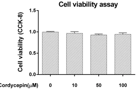
A commercial kit (Cell Counting Kit-8, CCK-8; Dojindo) was used to evaluate the potential cytotoxic effect of cordycepin. NP cells were plated in 96-well plates at a density of 5×10³ per well, incubated with various concentrations of cordycepin for 24 h, administered 10µl of CCK-8 solution and incubated for an additional 2 h. The optical density (OD) of each well was measured at 450 nm. The culture medium was used as a blank. Cell viability was calculated as follows: cell viability=[OD (with cordycepin)−OD (blank)]/[OD (without cordycepin)−OD (blank)].
ELISA assessments
NP cells were incubated with various concentrations of cordycepin for 2 h and then stimulated with 10µg/ml LPS for 24 h. The PGE2, MMP-3 and MMP-13 levels in the culture medium were measured using commercially available enzyme-linked immunosorbent assay kits according to the manufacturer’s instructions (R&D Systems).
Measurement of nitric oxide
Nitric oxide (NO) was measured by Griess reagent (Imamura et al., 2015). Briefly, NP cells were incubated with 100µM cordycepin for 2 h and then stimulated with 10µg/ml LPS (Sigma) for 24 h. Then, 50µl of each culture supernatant was incubated at room temperature for 15 min with 50µl of Griess reagent (Sigma) in a 96-well plate. The absorbance at 540 nm was measured. A standard curve was made using NaNO2to calculate the NO concentration of each sample.
Immunofluorescence
NP cells were stimulated with 10 µg/ml LPS in the presence or absence of cordycepin (100µM) in a 24-well plate for 5 days. Then, the cells were fixed with 4% paraformaldehyde for 30 min, treated with 0.1% Triton X-100 for 10 min, and blocked with 2% bovine serum albumin (Sigma) for 1 h. After being washed by PBS, the NP cells were incubated with an anti-collagen II antibody (1:50; Cell Signaling Technology) overnight at 4 ◦C and
then exposed to Alexa FluorR 594-conjugated secondary antibodies (1:100 dilution;
Cell migration assay
A total of 1×10⁵ RAW 264.7 cells were seeded on a Matrigel-coated polycarbonate membrane insert (8.0 µm pores) in a transwell apparatus (Costar) and maintained in 100µl of complete medium (high-glucose DMEM with 10% FBS and 1% antibiotic). NP cells were also cultured in complete medium with or without 10µg/ml LPS or 100µM cordycepin in the lower chamber for 24 h. Then, the inserts were washed with PBS, and the cells on the top surface of the insert were carefully removed by using a cotton swab. The cells on the bottom surface of the insert were fixed with 4% paraformaldehyde for 10 min, followed by staining with 0.1% crystal violet for 20 min, and then subjected to an inspection and cell count via microscopy. The cells were counted under 200×magnification, and the counts of 5 randomly chosen fields were averaged for each sample.
RNA isolation and PCR
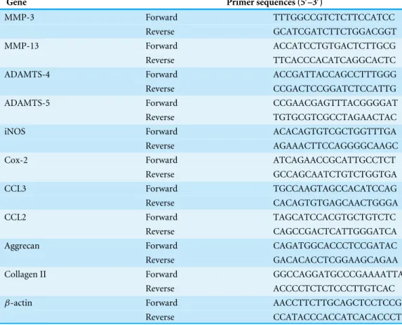
NP cells were incubated with various concentrations of cordycepin for 2 h and then stimulated with 10µg/ml LPS for 24 h. Then, the total RNA of the NP cells was isolated using TRIzol reagent (Invitrogen) following the manufacturer’s instructions. Reverse transcription was carried out from 1µg of RNA using the 1st Strand cDNA Synthesis Kit (TAKARA) for first-strand complementary DNA (cDNA) synthesis. The relative gene expression was determined by real-time PCR. Real-time PCR was performed using the SYBR Premix Ex Taq kit (TAKARA) with the ABI Prism 7500 Fast Real-Time PCR system (Applied Biosystems) according to the manufacturer’s instructions. The primers were designed and selected using BLAST. Gene expression was measured using the 2−11Ct method (Schmittgen & Livak, 2008). The primer sequences are summarized inTable 1and we usedβ-actin as the internal control.
Western blotting
For pathway related protein assays, NP cells were pretreated with various concentrations of cordycepin for 2 h and then stimulated with 10µg/ml LPS for 30 min. For collagen II and aggrecan protein assays, NP cells were cultured with various concentrations of cordycepin and 10 µg/ml LPS for 5 days. Then, all cells were washed twice with cold PBS, and the total protein was extracted using an RIPA lysis buffer. The protein concentration was quantified using a BCA Protein Assay Kit (Thermo Scientific), Samples (20µg of protein) were loaded into gel, separated by 10% SDS-PAGE and transferred onto polyvinylidene fluoride (PVDF) membranes (Millipore). The transfer membranes were blocked with 5% fat-free milk at room temperature for 1 h and then incubated with primary antibodies against ERK1/2, p-ERK1/2, JNK, p-JNK, p38, p-p38, IκBα, p-IκBα, p65, p-p65 (1:1,000, Cell Signaling Technology) aggrecan and collagen-II (1:1,000, Abcam) at 4 ◦C overnight.
Table 1 Sequences of the primers used in the polymerase chain reaction (PCR).
Gene Primer sequences (5′–3′)
MMP-3 Forward TTTGGCCGTCTCTTCCATCC
Reverse GCATCGATCTTCTGGACGGT
MMP-13 Forward ACCATCCTGTGACTCTTGCG
Reverse TTCACCCACATCAGGCACTC
ADAMTS-4 Forward ACCGATTACCAGCCTTTGGG
Reverse CCGACTCCGGATCTCCATTG
ADAMTS-5 Forward CCGAACGAGTTTACGGGGAT
Reverse TGTGCGTCGCCTAGAACTAC
iNOS Forward ACACAGTGTCGCTGGTTTGA
Reverse AGAAACTTCCAGGGGCAAGC
Cox-2 Forward ATCAGAACCGCATTGCCTCT
Reverse GCCAGCAATCTGTCTGGTGA
CCL3 Forward TGCCAAGTAGCCACATCCAG
Reverse CACAGTGTGAGCAACTGGGA
CCL2 Forward TAGCATCCACGTGCTGTCTC
Reverse CAGCCGACTCATTGGGATCA
Aggrecan Forward CAGATGGCACCCTCCGATAC
Reverse GACACACCTCGGAAGCAGAA
Collagen II Forward GGCCAGGATGCCCGAAAATTA
Reverse ACCCCTCTCTCCCTTGTCAC
β-actin Forward AACCTTCTTGCAGCTCCTCCG
Reverse CCATACCCACCATCACACCCT
Luciferase assay
In addition to western blotting, we also used a luciferase assay to investigate the effect of cordycepin on NF-κB activity. One day before transfection, NP cells were transferred to a 96-well plate at a density of 5×10³cells/well. NP cells were cotransfected with 20 ng of NF-κB luciferase reporter and 5 ng of pRL-TK plasmids for each well. Lipofectamine 2000 (Invitrogen) was used as a transfection reagent. After transfection, the cells were cultured for 24 h and then pretreated with cordycepin (10, 50 or 100µM) and/or stimulated with 10µg/ml LPS for 24 h. Then, the cells were harvested for the luciferase assay. A Dual-Luciferase Reporter Assay System (Promega) was used for firefly and renilla luciferase activity measurements.
Organ culture
for 7 days with or without 10µg/ml LPS and 100µM cordycepin. The culture medium was replaced daily. A total of 30 Sprague Dawley rats, including 90 lumbar segments, were used in the organ culture (30 lumbar segments for each group).
Dimethylmethylene blue assay
A DMMB assay was used to measure the proteoglycan (PG) content during 7 days of IVD organ culture. At specific time points, NP tissue was isolated from each cultured IVD and then digested with 5 mg/ml papain (sigma) prior to the DMMB assay. The amount of PG was normalized by the Total DNA of the NP tissue. The total DNA content was determined via an assay of total DNA using the PicoGreen kit (Molecular Probes). The DMMB assay was performed as previously described using chondroitin sulfate as a standard (Li et al., 2015). The PG level of each sample was expressed asµg PG /60 ng DNA (equal to 104cells).
Histological analysis
Discs were removed from the culture medium after 7 days and fixed in 4% paraformaldehyde. After fixation, the discs were decalcified in EDTA for 14 days. Serial sagittal sections of discs (5-µm thick) were obtained to prepare slides. NBT/DAPI staining was used to evaluate the cell viability as previously described (Lim et al., 2006). Live cells were defined as cells with both DAPI and NBT staining, and cells with DAPI staining alone were registered as dead. The live/dead cell ratio was calculated using fluorescence microscopy (OLYMPUS) at 400×magnification. We analyzed three sections from each IVD tissue, and for each section, we calculated three fields and took the average. The cell viability was calculated as follows: (cell viability =live cells in field/total cells in field)×100%.
Sagittal sections were also stained with hematoxylin and eosin (HE) and Safranin O-fast green to assess the degeneration of the IVD. Type II collagen and Aggrecan expression was detected using mouse monoclonal antibodies (1:200; Abcam) and a horseradish peroxidase-conjugated anti-mouse antibody (1:100; Dako), followed by color development with diaminobenzidine tetrahydrochloride (DAB, Dako). The results of the type II collagen and aggrecan staining were quantified as the IOD using the Image-Pro Plus 6.0 software. Cell and immunohistochemical staining was performed following standard histochemical protocols.
Statistical analysis
All of the experiments were repeated 3 times. The data are expressed as the mean±SD. A statistical analysis was performed with a one-way analysis of variance (ANOVA), followed by Duncan’s post hoc test using SPSS 19.0 (IBM, Inc.). A P-value of less than 0.05 was considered statistically significant.
RESULTS
Cell Viability assay in the cell culture model
Figure 1 Cell viability assay in the NP cell culture model.We used CCK-8 to measure the NP cell viability in a monolayer culture model. Cordycepin did not show any cytotoxicity at concentrations of 10–100µM.
Cordycepin regulates LPS-induced matrix-degrading enzymes and extracellular matrix-related gene expression in NP cells
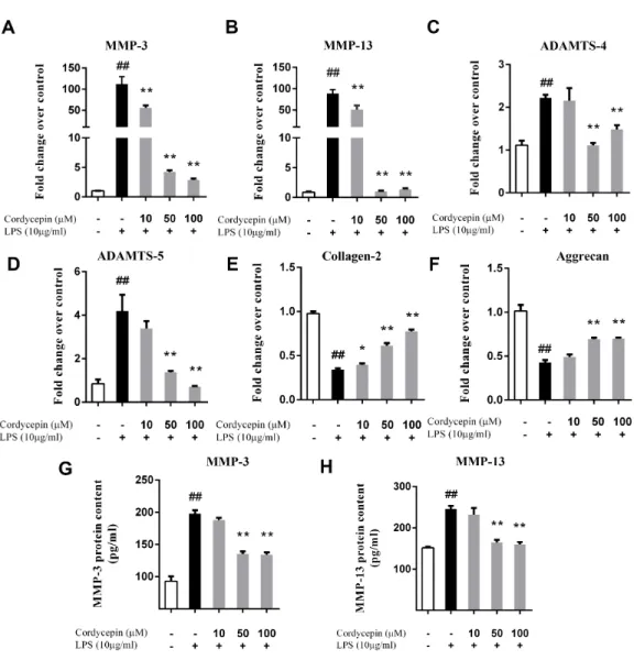
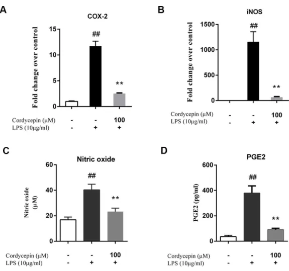
Monolayer cultures of NP cells were stimulated with 10µg/ml LPS and 0, 10, 50 or 100 µM cordycepin, followed by a PCR assay and ELISA to measure the mRNA and protein levels respectively of various matrix-degrading enzymes. Cordycepin markedly inhibited the mRNA expression of multiple MMPs (MMP-3 and MMP-13) and ADAMTSs (ADAMTS-4 and ADAMTS-5) in a concentration-dependent manner (Figs. 2A–2D). Cordycepin also counteracted the LPS-induced gene downregulation of collagen-2 and aggrecan especially at a concentration of 50 or 100µM. (Figs. 2Eand2F). The ELISA results showed that 50 and 100µM cordycepin significantly inhibited MMP-3 and MMP-13 protein production (Figs. 2Gand2H). Moreover, cordycepin also reversed the LPS-induced increased in the gene expression of Cox-2 and iNOS at a concentration of 100µM (Figs. 3Aand3B). Thus, cordycepin significantly inhibited PGE2 and NO production as induced by LPS in NP cells at a concentration of 100µM (Figs. 3Cand3D).
Cordycepin protects NP cells from LPS-induced matrix degradation
Figure 2 Cordycepin regulates the LPS-induced matrix-degrading enzymes and extracellular matrix related gene expression in NP cells.We used PCR (A–F) and an ELISA assay (G, H) to investigate the effect of cordycepin on the LPS-induced gene expression and matrix-degrading enzyme secretion of NP cells. (A–F) Real-time PCR indicated that cordycepin downregulated the LPS-induced gene over-expression of MMP-3, MMP-13, ADAMTS-4 and ADAMTS-5. Moreover, cordycepin also counteracted the LPS-induced gene downregulation of collagen-2 and aggrecan especially at concentration of 50 mM or 100µM. (G, H) An ELISA assay of MMP-3 and MMP-13 demonstrated that cordycepin inhibited LPS-induced MMPs secretion. The values are presented as the mean±standard deviation. *P <0.05 compared to the LPS group; **P<0.01 compared to the LPS group; ##P<0.01 compared to the control group.
Cordycepin inhibits the NP-mediated migration of macrophages after treatment with LPS
Figure 3 Cordycepin decreases the LPS-induced production of PGE2 and NO in NP cells.(A, B) Real-time PCR indicated that cordycepin reversed the LPS-induced increased gene expression of Cox-2 and iNOS. (C) The NO content was measured using the Griess reaction; the results showed that cordycepin inhibited LPS-induced NO production in NP cells. (D) An ELISA assay demonstrated that cordycepin inhibited the LPS-induced PGE2 production in NP cells. The values are presented as the mean±standard deviation. **P<0.01 compared to the LPS group; ##P<0.01 compared to the control group.
reversed the LPS-induced gene upregulation of chemokine ligand 2 (CCL2, or monocyte chemotactic protein-1 (MCP-1)), a well-characterized macrophage chemotactic factor that is expressed in NP cells (Yoshida et al., 2002).
Cordycepin inhibits the LPS-induced activation of the NF-κB pathway
in NP cells
Figure 4 Cordycepin reverses LPS-induced matrix degradation in NP cells.NP cells were treated with or without 100µM cordycepin and 10µg/ml LPS for 5 days and then fixed with 4% paraformaldehyde and used to perform fluorescence staining. (A, B) Collagen-II (Col-II) immunofluorescent staining showed that cordycepin reduced the LPS-induced collagen-II decrease in NP cells. (C, D) Western blotting results showed that cordycepin significantly reversed LPS-induced collagen-II and aggrecan loss. These results indicated that cordycepin could protect NP cells from LPS-induced matrix degradation. *P<0.05 compared to the LPS group; ##P<0.01 compared to the control group.
(Figs. 6B,6F–6H). A luciferase assay also showed that cordycepin could inhibit the LPS-induced activation of the NF-κB pathway in NP cells (Fig. 6C).
Cordycepin reverses the LPS-induced degeneration of IVDs in an organ culture model
The NBT/DAPI staining showed that the cell viability of all groups at seven days was still greater than 80%, confirming the reliability of the organ culture model (Figs. 7Aand7B).
Figure 8Ashows cultured IVD sections that were stained with HE and Safranin-O fast green. On the Safranin-O fast green sections, proteoglycans (PGs) stained red, and on the HE sections, they stained purple. After seven days of IVD organ culture, the presence of LPS resulted in severe PG loss compared to the effects of co-incubation with cordycepin (Fig. 8A).
Figure 5 Cordycepin inhibits LPS-induced CCL2 expression and macrophage migration in NP cells.
Macrophage migration was measured using a 24-well cell culture insert system. The results of crystal violet staining (A) and a positive cell count (B) showed that LPS promoted NP-mediated macrophage migration, which was inhibited by cordycepin. (C, D) Real-time PCR indicated that cordycepin inhibited the LPS-induced increased gene expression of CCL2 but not CCL3. The values are presented as the mean± stan-dard deviation. *P<0.05 compared to the LPS group; **P<0.01 compared to the LPS group; ##P<0.01 compared to the control group.
A DMMB assay was used to quantify the PG content of NP in an IVD culture. Similar to the histological results, cordycepin significantly attenuated the PG loss induced by LPS (Fig. 8D,P<0.01).
DISCUSSION
The present study provides, for the first time, evidence that cordycepin exhibits pharmacological anti-inflammation and anti-degeneration effects in LPS-induced NP cells and IVDs. Our results also show that cordycepin blocks the LPS-induced activation of the NF-κB pathway, but not the MAPK pathway in NP cells.
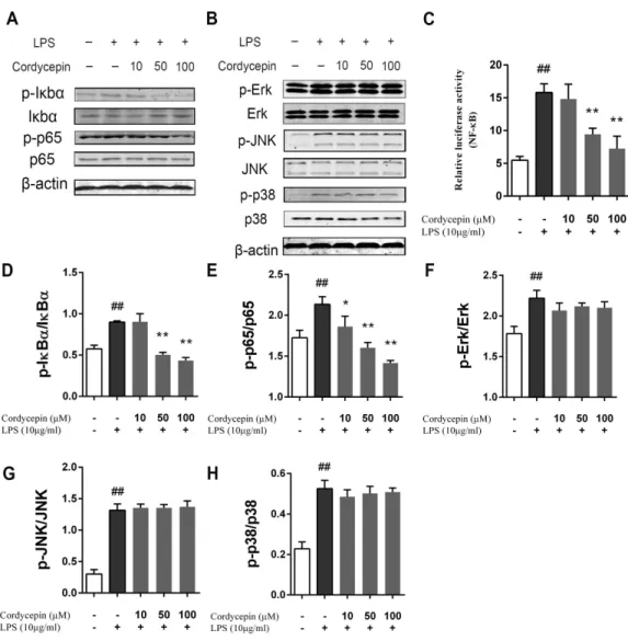
Figure 6 Effect of cordycepin on the LPS-induced activation of the NF-κB and MAPK pathways.NP
cells were pretreated with various concentrations of cordycepin for 2 h and then stimulated with 10µg/ml LPS for 24 h. Then, western blotting was performed to evaluate the mechanism of cordycepin on LPS-treated NP cells. (A) Cordycepin significantly inhibied the phosphorylation of IκBαand p65 induced by LPS. (B) Cordycepin did not influence the phosphorylation of ERK, p38 or JNK enhanced by LPS. (C) NP cells were transfected with a NF-κB luciferase reporter, and the NF-κB pathway activity was determined by luciferase assay using a commercially available kit. (D–H) Quantitative analysis of western blotting data show cordycepin significantly inhibited the activation of the NF-κB pathway induced by LPS at concentra-tions of 50 and 100µM. The values are presented as the mean±standard deviation. *P<0.05 compared to the LPS group; **P<0.01 compared to the LPS group; ##P<0.01 compared to the control group.
Figure 7 Cell viability assay in the IVD organ culture model.(A) NBT/DAPI staining on the 7th day of IVD organ culture. (B) Cell viability of the organ culture model. Cell viability=(NBT positive cell number/DAPI positive cell number)×100%.
correlated with disc degeneration (Shi et al., 2015;Kozaci et al., 2006). Our study shows that cordycepin can both prevent LPS-induced collagen-II and aggrecan loss and promote their synthesis.
Many studies have revealed that NO and PGE2 play important roles in both the regulation of cellular metabolism in discs under mechanical stress conditions and the pathology of intervertebral disc degeneration (Wang et al., 2011;Takada et al., 2012;Hou et al., 2014). According to previous reports, PGE2 increases the excitability of rat sensory neurons and is involved in the development of sciatica in herniated disc disease (England, Bevan & Docherty, 1996). In addition, NO contributes to the development of radiculopathy by mediating protein nitration, moreover, the symptoms can be relieved by its suppression (Lee et al., 2013). Macrophages also play a critical role in the inflammatory response that is associated with degenerative disc disease and LBP (Gawri et al., 2014;Gruber et al., 2015). Macrophages could upregulate many cytokines expression including PGE2, and the interaction of macrophages and IVDs induce MMP-3 production which may cause extracellular matrix resorption (Haro et al., 2000). Previous studies have suggested that chemokine ligand 2 (CCL2) and chemokine ligand 3 (CCL3) are important mediators of macrophage infiltration into disc tissue (Gawri et al., 2014;Gruber et al., 2015). In our research, LPS upregulated the gene expression of CCL2 and CCL3 in NP cells, thereby promoting macrophage migration in NP cells and in a RAW 264.7 cell co-culture model. However, cordycepin reversed LPS-induced CCL2 gene upregulation of NP cells and inhibit macrophage migration, thus reducing the early-stage inflammation of NP cells. We also demonstrated that cordycepin could inhibit LPS-induced iNOS and Cox-2 gene overexpression and reduce the production of NO and PGE2 in NP cells.
(He et al., 2010). In our study, cordycepin inhibited the NF-κB pathway by inhibiting the phosphorylation of IκBα and p65 in LPS-stimulated NP cells. In contrast, the phospho-rylation of Erk, JNK and p38 of the MAPK pathway did not significantly change. This result was consistent to the data reported previously (Jeong et al., 2010;Ren et al., 2012).
Given that the in vitroexperiment showed encouraging results, we further study the potential therapeutic effects of cordycepin in an organ culture model. Although the permeability of the annulus fibrous and endplate is very limited, our previous and other studies have confirmed that LPS and bioactive molecules can penetrate into the nucleus pulposus and exert bioactive effects (Li et al., 2015;Kim et al., 2013). Our results clearly indicate that high concentrations of LPS (10 µg/ml) can induce the degeneration of cultured IVDs in a relatively short period (seven days), while cordycepin can significantly reduce the LPS-induced PG loss in cultured IVD. A similar study reported that cordycepin may suppress IL-1β-stimulated catabolic enzyme, COX-2 and iNOS gene expression in chondrocytes, exerting chondroprotective effect and interfering with the inflammatory response (Hu et al., 2014). Considering the similarity between chondrocytes and NP cells, this previous study support our results to some extent. However, we focus more on the early stage of inflammation of NP cells in the present study, while the chondrocytes used in the previous study were from subjects with advanced-stage osteoarthritis.
Previous study have reported fibrin-genipin annulus fibrosus sealant as a drug delivery system for IDD, and it can maintain the bioactivity of the drug for over 20 days (Likhitpanichkul et al., 2015). Based on our study, cordycepin needs to be directly deliver to IVDs before exerting its anti-degenerative effects. Future investigations are required to determine if such a system can effectively deliver cordycepin and allow it to exert its effects in IVDs.
In conclusion, our results show that cordycepin exhibits a strong anti-inflammatory and anti-catabolic effect by inhibiting LPS-induced NF-κB activation in NP cells. Our organ culture model also demonstrates an anti-degeneration effect of cordycepin in IVDs. Cordycepin may be a potential new agent for treating IDD in the future.
ACKNOWLEDGEMENTS
The authors thank the staff of the Shanghai Key Laboratory of Orthopedic Implants.
ADDITIONAL INFORMATION AND DECLARATIONS
Funding
This work was supported by grants from the National Natural Science Foundation of China (81272038). The funders had no role in study design, data collection and analysis, decision to publish, or preparation of the manuscript.
Grant Disclosures
Competing Interests
The authors declare there are no competing interests.
Author Contributions
• Yan Li conceived and designed the experiments, performed the experiments, analyzed the data, contributed reagents/materials/analysis tools, wrote the paper, prepared figures and/or tables, reviewed drafts of the paper.
• Kang Li performed the experiments, analyzed the data, contributed reagents/material-s/analysis tools, wrote the paper, prepared figures and/or tables, reviewed drafts of the paper.
• Lu Mao contributed reagents/materials/analysis tools.
• Xiuguo Han performed the experiments, analyzed the data, contributed reagents/mate-rials/analysis tools, prepared figures and/or tables.
• Kai Zhang performed the experiments, contributed reagents/materials/analysis tools, wrote the paper.
• Changqing Zhao wrote the paper, prepared figures and/or tables.
• Jie Zhao conceived and designed the experiments, reviewed drafts of the paper.
Animal Ethics
The following information was supplied relating to ethical approvals (i.e., approving body and any reference numbers):
All of the animal work was conducted according to relevant national and international guidelines and was approved by the Animal Experimental Ethical Committee of Shanghai Ninth People’s Hospital (Approval number: 2013-47).
Data Availability
The following information was supplied regarding data availability: Figshare:https://figshare.com/s/53cc3745ecacea5242d5.
DOI10.6084/m9.figshare.2056077.
REFERENCES
Bachmeier BE, Nerlich A, Mittermaier N, Weiler C, Lumenta C, Wuertz K, Boos N. 2009.Matrix metalloproteinase expression levels suggest distinct enzyme roles during lumbar disc herniation and degeneration.European Spine Journal 18:1573–1586DOI 10.1007/s00586-009-1031-8.
Berenbaum F. 2004.Signaling transduction: target in osteoarthritis.Current Opinion in Rheumatology16:616–622 DOI 10.1097/01.bor.0000133663.37352.4a.
Ellman MB, Kim JS, An HS, Chen D, KC R, An J, Dittakavi T, van Wijnen AJ, Cs-Szabo G, Li X, Xiao G, An S, Kim SG, Im HJ. 2012.Toll-like receptor adaptor signaling molecule MyD88 on intervertebral disk homeostasis:in vitro, ex vivo studies.Gene 505:283–290DOI 10.1016/j.gene.2012.06.004.
AMP-protein kinase A cascade.Journal of Physiology495(Pt 2):429–440 DOI 10.1113/jphysiol.1996.sp021604.
Gawri R, Rosenzweig DH, Krock E, Ouellet JA, Stone LS, Quinn TM, Haglund L. 2014. High mechanical strain of primary intervertebral disc cells promotes secretion of inflammatory factors associated with disc degeneration and pain.Arthritis Research & Therapy16: Article R21DOI 10.1186/ar4449.
Gendron C, Kashiwagi M, Lim NH, Enghild JJ, Thøgersen IB, Hughes C, Caterson B, Nagase H. 2007.Proteolytic activities of human ADAMTS-5: comparative studies with ADAMTS-4.Journal of Biological Chemistry282:18294–18306 DOI 10.1074/jbc.M701523200.
Gilbert AM, Bursavich MG, Lombardi S, Georgiadis KE, Reifenberg E, Flannery CR, Morris EA. 2007.5-((1H-pyrazol-4-yl)methylene)-2-thioxothiazolidin-4-one inhibitors of ADAMTS-5.Bioorganic & Medicinal Chemistry Letters17:1189–1192 DOI 10.1016/j.bmcl.2006.12.020.
Gruber HE, Hoelscher GL, Ingram JA, Bethea S, Cox M, Hanley Jr EN. 2015. Proinflam-matory cytokines modulate the chemokine CCL2 (MCP-1) in human annulus cells in vitro: CCL2 expression and production.Experimental and Molecular Pathology 98(1):102–105DOI 10.1016/j.yexmp.2014.12.002.
Haro H, Crawford HC, Fingleton B, MacDougall JR, Shinomiya K, Spengler DM, Matrisian LM. 2000.Matrix metalloproteinase-3-dependent generation of a macrophage chemoattractant in a model of herniated disc resorption.Journal of Clinical Investigation105:133–141DOI 10.1172/JCI7090.
Hart LG, Deyo RA, Cherkin DC. 1995.Physician office visits for low back pain. Frequency, clinical evaluation, and treatment patterns from a US national survey. Spine20:11–19DOI 10.1097/00007632-199501000-00003.
He W, Zhang MF, Ye J, Jiang TT, Fang X, Song Y. 2010.Cordycepin induces apoptosis by enhancing JNK and p38 kinase activity and increasing the protein expression of Bcl-2 pro-apoptotic molecules.Journal of Zhejiang University Science B11:654–660. Hou G, Lu H, Chen M, Yao H, Zhao H. 2014.Oxidative stress participates in age-related
changes in rat lumbar intervertebral discs.Archives of Gerontology and Geriatrics 59:665–669DOI 10.1016/j.archger.2014.07.002.
Hu P, Chen W, Bao J, Jiang L, Wu L. 2014.Cordycepin modulates inflammatory and catabolic gene expression in interleukin-1beta-induced human chondrocytes from advanced-stage osteoarthritis: anin vitrostudy.International Journal of Clinical and Experimental Pathology 7:6575–6584.
Iwata M, Ochi H, Asou Y, Haro H, Aikawa T, Harada Y, Nezu Y, Yogo T, Tagawa M, Hara Y. 2013.Variations in gene and protein expression in canine chondrodys-trophic nucleus pulposus cells following long-term three-dimensional culture.PLoS ONE8:e63120DOI 10.1371/journal.pone.0063120.
Jeong JW, Jin CY, Kim GY, Lee JD, Park C, Kim GD, Kim WJ, Jung WK, Seo SK, Choi IW, Choi YH. 2010.Anti-inflammatory effects of cordycepin via suppression of inflammatory mediators in BV2 microglial cells.International Immunopharmacology 10:1580–1586DOI 10.1016/j.intimp.2010.09.011.
Kepler CK, Ponnappan RK, Tannoury CA, Risbud MV, Anderson DG. 2013.The molecular basis of intervertebral disc degeneration.The Spine Journal13:318–330 DOI 10.1016/j.spinee.2012.12.003.
Kim JS, Ellman MB, Yan D, An HS, Kc R, Li X, Chen D, Xiao G, Cs-Szabo G, Hoskin DW, Buechter DD, Van Wijnen AJ, Im HJ. 2013.Lactoferricin mediates anti-inflammatory and anti-catabolic effects via inhibition of IL-1 and LPS activity in the intervertebral disc.Journal of Cellular Physiology228:1884–1896
DOI 10.1002/jcp.24350.
Kim H, Naura AS, Errami Y, Ju J, Boulares AH. 2011.Cordycepin blocks lung injury-associated inflammation and promotes BRCA1-deficient breast cancer cell killing by effectively inhibiting PARP.Missouri Medicine17:893–900.
Klawitter M, Hakozaki M, Kobayashi H, Krupkova O, Quero L, Ospelt C, Gay S, Hausmann O, Liebscher T, Meier U, Sekiguchi M, Konno S, Boos N, Ferguson SJ, Wuertz K. 2014.Expression and regulation of toll-like receptors (TLRs) in human intervertebral disc cells.European Spine Journal23:1878–1891
DOI 10.1007/s00586-014-3442-4.
Kozaci LD, Guner A, Oktay G, Guner G. 2006.Alterations in biochemical components of extracellular matrix in intervertebral disc herniation: role of MMP-2 and TIMP-2 in type II collagen loss.Cell Biochemistry and Function24:431–436 DOI 10.1002/cbf.1250.
Kuslich SD, Ulstrom CL, Michael CJ. 1991.The tissue origin of low back pain and sciatica: a report of pain response to tissue stimulation during operations on the lumbar spine using local anesthesia.Orthopedic Clinics of North America22:181–187. Leckie SK, Bechara BP, Hartman RA, Sowa GA, Woods BI, Coelho JP, Witt WT,
Dong QD, Bowman BW, Bell KM, Vo NV, Wang B, Kang JD. 2012.Injection of AAV2-BMP2 and AAV2-TIMP1 into the nucleus pulposus slows the course of intervertebral disc degeneration in anin vivorabbit model.The Spine Journal 12:7–20DOI 10.1016/j.spinee.2011.09.011.
Lee EJ, Kim WJ, Moon SK. 2010.Cordycepin suppresses TNF-alpha-induced invasion, migration and matrix metalloproteinase-9 expression in human bladder cancer cells. Phytotherapy Research24:1755–1761DOI 10.1002/ptr.3132.
Le Maitre CL, Freemont AJ, Hoyland JA. 2004.Localization of degradative enzymes and their inhibitors in the degenerate human intervertebral disc.Journal of Pathology 204:47–54DOI 10.1002/path.1608.
Li Y, Li K, Hu Y, Xu B, Zhao J. 2015.Piperine mediates LPS induced inflammatory and catabolic effects in rat intervertebral disc.International Journal of Clinical and Experimental Pathology 8:6203–6213.
Likhitpanichkul M, Kim Y, Torre OM, See E, Kazezian Z, Pandit A, Hecht AC, Iatridis JC. 2015.Fibrin-genipin annulus fibrosus sealant as a delivery system for anti-TNFalpha drug.The Spine Journal 15:2045–2054
DOI 10.1016/j.spinee.2015.04.026.
Lim TH, Ramakrishnan PS, Kurriger GL, Martin JA, Stevens JW, Kim J, Mendoza SA. 2006.Rat spinal motion segment in organ culture: a cell viability study.Spine 31:1291–1297DOI 10.1097/01.brs.0000218455.28463.f0.
Nakamura K, Shinozuka K, Yoshikawa N. 2015.Anticancer and antimetastatic effects of cordycepin, an active component of Cordyceps sinensis.Journal of Pharmacological Sciences127:53–56DOI 10.1016/j.jphs.2014.09.001.
Patel KP, Sandy JD, Akeda K, Miyamoto K, Chujo T, An HS, Masuda K. 2007. Aggrecanases and aggrecanase-generated fragments in the human intervertebral disc at early and advanced stages of disc degeneration.Spine32:2596–2603 DOI 10.1097/BRS.0b013e318158cb85.
Paterson RR. 2008.Cordyceps: a traditional Chinese medicine and another fungal therapeutic biofactory?Phytochemistry69:1469–1495
DOI 10.1016/j.phytochem.2008.01.027.
Pockert AJ, Richardson SM, Le Maitre CL. 2009.Modified expression of the ADAMTS enzymes and tissue inhibitor of metalloproteinases 3 during human intervertebral disc degeneration.Arthtitis and Rheumatism60:482–491DOI 10.1002/art.24291. Rajan NE, Bloom O, Maidhof R, Stetson N, Sherry B, Levine M, Chahine NO.
2013.Toll-Like Receptor 4 (TLR4) expression and stimulation in a model of intervertebral disc inflammation and degeneration.Spine38:1343–1351 DOI 10.1097/BRS.0b013e31826b71f4.
Ren Z, Cui J, Huo Z, Xue J, Cui H, Luo B, Jiang L, Yang R. 2012.Cordycepin sup-presses TNF-α-induced NF-κB activation by reducing p65 transcriptional activity, inhibiting Iκ Bαphosphorylation, and blocking IKKγ ubiquitination.International Immunopharmacology14:698–703 DOI 10.1016/j.intimp.2012.10.008.
Schmittgen TD, Livak KJ. 2008.Analyzing real-time PCR data by the comparative C(T) method.Nature Protocols3:1101–1108DOI 10.1038/nprot.2008.73.
Schwarzer AC, Aprill CN, Derby R, Fortin J, Kine G, Bogduk N. 1994.The relative contributions of the disc and zygapophyseal joint in chronic low back pain.Spine 19:801–806DOI 10.1097/00007632-199404000-00013.
Tak PP, Firestein GS. 2001.NF-kappaB: a key role in inflammatory diseases.Journal of Clinical Investigation107:7–11DOI 10.1172/JCI11830.
Takada T, Nishida K, Maeno K, Kakutani K, Yurube T, Doita M, Kurosaka M. 2012. Intervertebral disc and macrophage interaction induces mechanical hyperalgesia and cytokine production in a herniated disc model in rats.Arthtitis and Rheumatism 64:2601–2610DOI 10.1002/art.34456.
Tortorella MD, Burn TC, Pratta MA, Abbaszade I, Hollis JM, Liu R, Rosenfeld SA, Copeland RA, Decicco CP, Wynn R, Rockwell A, Yang F, Duke JL, Solomon K, George H, Bruckner R, Nagase H, Itoh Y, Ellis DM, Ross H, Wiswall BH, Murphy K, Hillman Jr MC, Hollis GF, Newton RC, Magolda RL, Trzaskos JM, Arner EC. 1999.Purification and cloning of aggrecanase-1: a member of the ADAMTS family of proteins.Science284:1664–1666DOI 10.1126/science.284.5420.1664.
Vo NV, Hartman RA, Yurube T, Jacobs LJ, Sowa GA, Kang JD. 2013.Expression and regulation of metalloproteinases and their inhibitors in intervertebral disc aging and degeneration.The Spine Journal 13:331–341DOI 10.1016/j.spinee.2012.02.027. Walter BA, Purmessur D, Likhitpanichkul M, Weinberg A, Cho SK, Qureshi
SA, Hecht AC, Iatridis JC. 2015.Inflammatory kinetics and efficacy of anti-inflammatory treatments on human nucleus pulposus cells.Spine40:955–963 DOI 10.1097/BRS.0000000000000932.
Wang J, Tian Y, Phillips KL, Chiverton N, Haddock G, Bunning RA, Cross AK, Shapiro IM, Le Maitre CL, Risbud MV. 2013.Tumor necrosis factor alpha- and interleukin-1beta-dependent induction of CCL3 expression by nucleus pulposus cells promotes macrophage migration through CCR1.Arthtitis and Rheumatism65:832–842 DOI 10.1002/art.37819.
Wang IC, Ueng SW, Lin SS, Niu CC, Yuan LJ, Su CI, Chen CH, Chen WJ. 2011. Effect of hyperbaric oxygenation on intervertebral disc degeneration: an in vitrostudy with human lumbar nucleus pulposus.Spine36:1925–1931 DOI 10.1097/BRS.0b013e3181feebde.
Weiler C, Nerlich AG, Zipperer J, Bachmeier BE, Boos N. 2002.SSE Award Competition in Basic Science: expression of major matrix metalloproteinases is associated with intervertebral disc degradation and resorption.European Spine Journal11:308–320 DOI 10.1007/s00586-002-0472-0.
Wuertz K, Haglund L. 2013.Inflammatory mediators in intervertebral disk degeneration and discogenic pain.Global Spine Journal3:175–184DOI 10.1055/s-0033-1347299. Yang H, Cao C, Wu C, Yuan C, Gu Q, Shi Q, Zou J. 2015.TGF-βl Suppresses
Inflammation in Cell Therapy for Intervertebral Disc Degeneration.Scientific Reports 5: Article 13254DOI 10.1038/srep13254.
Yoshida M, Nakamura T, Kikuchi T, Takagi K, Matsukawa A. 2002.Expression of monocyte chemoattractant protein-1 in primary cultures of rabbit intervertebral disc cells.Journal of Orthopaedic Research20:1298–1304
You C, Zhu K, Liu X, Xi C, Zhang Z, Xu G, Yan J. 2013.Tumor necrosis factor-alpha-dependent infiltration of macrophages into the dorsal root ganglion in a rat disc herniation model.Spine38:2003–2007DOI 10.1097/BRS.0b013e3182a84701. Yue K, Ye M, Zhou Z, Sun W, Lin X. 2013.The genus Cordyceps: a chemical and