Printed version ISSN 0001-3765 / Online version ISSN 1678-2690 http://dx.doi.org/10.1590/0001-3765201720160836
www.scielo.br/aabc
Inhibitory effects on the production of inflammatory mediators and reactive oxygen
species by Mori folium in lipopolysaccharide-stimulated macrophages and zebrafish
DA HYE KWON1,2
, JIN WOO JEONG1,2
, EUN OK CHOI1,2
, HYE WON LEE3
, KI WON LEE4
, KI YOUNG KIM4
, SUNG GOO KIM4
, SU HYUN HONG1
, GI-YOUNGKIM5
, CHEOL PARK6
, HYE-JIN HWANG7
, CHANG-GUE SON8 and
YUNG HYUN CHOI1,2
1
Department of Biochemistry, Dongeui University, College of Korean Medicine, 176 Yangjeong-ro, Busanjin-gu, 47227, Busan, Republic of Korea
2
Anti-Aging Research Center, Dongeui University, 176 Eomgwangno Busanjin-gu, 47340, Busan, Republic of Korea 3
KM Convergence Research Division, Korea Institute of Oriental Medicine, 1672 Yuseong-daero, Yuseong-gu, 34054, Daejeon, Republic of Korea 4
Bio-Port Korea INC, Marine Bio-industry Development Center, 27 Hoenggye-ri, Ilgwang-myeon, 46048, Gijang-gun, Republic of Korea
5
Laboratory of Immunobiology, Department of Marine Life Sciences, Jeju National University, 102 Jejudaehak-ro, 63243, Jeju, Republic of Korea
6
Department of Molecular Biology, College of Natural Sciences & Human Ecology, Dongeui University, 176 Eomgwangno Busanjin-gu, 47340, Busan, Republic of Korea 7
Department of Food and Nutrition, College of Natural Sciences & Human Ecology, Dongeui University, 176 Eomgwangno Busanjin-gu, 47340, Busan, Republic of Korea 8
Liver and Immunology Research Center, Daejeon Oriental Hospital of Oriental Medical College of Daejeon University, 176-9 Daeheung-ro, Jung-gu, 34929, Daejeon, Republic of Korea Manuscript received on December 1, 2016; accepted for publication on January 18, 2017
ABSTRACT
Mori folium, the leaf of Morus alba L. (Moraceae), has been traditionally used for various medicinal
purposes from ancient times to the present. In this study, we examined the effects of water extract of Mori folium (WEMF) on the production of inflammatory mediators, such as nitric oxide (NO) and prostaglandin
E2 (PGE2), and reactive oxygen species (ROS) in lipopolysaccharide (LPS)-stimulated murine RAW 264.7
macrophages. Our data indicated that WEMF significantly suppressed the secretion of NO and PGE2 in
RAW 264.7 macrophages without any significant cytotoxicity. The protective effects were accompanied by
a marked reduction in their regulatory gene expression at the transcription level. WEMF attenuated LPS-induced intracellular ROS production in RAW 264.7 macrophages. It inhibited the nuclear translocation of the nuclear factor-kappa B p65 subunit and the activation of mitogen-activated protein kinases in LPS-treated RAW 264.7 macrophages. Furthermore, WEMF reduced LPS-induced NO production and ROS
accumulation in zebrafish. Although more efforts are needed to fully understand the critical role of WEMF in the inhibition of inflammation, the findings of the present study may provide insights into the approaches for Mori folium as a potential therapeutic agent for inflammatory and antioxidant disorders.
Key words: Mori folium, inflammation, ROS, macrophage, zebrafish.
Correspondence to: Yung Hyun Choi E-mail: [email protected]
INTRODUCTION
Inflammation is a primary protective response of the body involving the activation of immune
system processes. The inflammatory response
is a highly regulated self-limiting process for identifying and destroying invading pathogens and restoring normal tissue structure and function (Conti et al. 2004, Freire and Van Dyke 2013). However, an excessive inflammatory response has been recognized as the main cause of chronic
inflammation, such as in cardiovascular disease, rheumatoid arthritis, inflammatory bowel disease,
Alzheimer’s disease, and even cancer (Amin et al. 1999, Freire and Van Dyke 2013).
When macrophages are over-activated by inflammatory stimulants, including the gram-negative bacterial endotoxin lipopolysaccharides (LPS), the cells induce the production of inflammatory mediators, including nitric
oxide (NO) and prostaglandin E2 (PGE2), and
inflammatory cytokines along with the activation of
several signaling pathways, such as nuclear
factor-kappa B (NF-κB) and mitogen-activated protein
kinases (MAPKs) signaling (Kaminska 2005, Lu et al. 2011, Rigoglou and Papavassiliou 2013, Muralidharan and Mandrekar 2013). Excessive
production of these inflammatory mediators and
cytokines further provoke deleterious consequences in the pathogenesis of many inflammatory diseases (McDaniel et al. 1996, Muralidharan and Mandrekar 2013).
Another important component of inflammation is oxidative stress, which reflects the imbalance
between the production of reactive oxygen species (ROS) and the ability of the biological system to remove them (Brüne et al. 2013, Mills and O'Neill
2016). The overproduced ROS by activated
macrophages acts as an important contributor to the
manifestation of inflammation (Varga et al. 2013,
Mills and O'Neill 2016), and it is also involved in
the production of inflammatory mediators in
LPS-stimulated macrophages (Haddad and Land 2002). Consequently, the suppression of the production
of inflammatory factors by blocking macrophage
activation emerges as a potential therapeutic
approach to relieve the progression of inflammatory
and oxidative disorders (Cunha et al. 2008, Zhang and Wang 2014). For this reason, the development of anti-inflammatory and/or antioxidant agents, which can control the activation of macrophages, is necessary for the prevention and treatment of various diseases.
Recent data have convincingly pointed out that natural resources have been widely and safely consumed over centuries and that most of them have a wide range of diverse biological activities with
few side effects (Bocanegra et al. 2009, Abuajah et
al. 2015). Among these natural resources, Morus alba L. that belongs to the Moraceae family, is one of the most valuable and rich in natural ingredients
plant. This tree is widely distributed in Eastern
Asia countries including Korea, China and Japan but now is cultivated throughout worldwide.
The leaf of plant, Mori folium (Supplementary
Material, Figure S1), has been used in traditional medicine for the treatment of various diseases (Yang et al. 2014, Chan et al. 2016). Mori folium exhibits a variety of pharmacological activities,
such as microbial (Tirupathi et al. 2011),
anti-tumor (Deepa et al. 2013), anti-obesity (Sugimoto et al. 2009, Ann et al. 2015), anti-hypotensive (Kobayashi et al. 2010), neuroprotective (Xiang et al. 2010), anti-diabetic (Naowaboot et al. 2009), and immunomodulatory potentials (Kwon et al. 2016). In addition, Mori folium possesses free radical-scavenging activities (Kim and Jang 2011, Iqbal et al. 2012, Raman et al. 2016), which may explain its antioxidant ability (Khan et al. 2013, Kim et al. 2014, Raman et al. 2016). Previous studies, including our recent data, indicated that the extracts and components of Mori folium have
strong anti-inflammatory properties (Hong et al.
et al. 2016). Despite these encouraging studies, the
effects and molecular mechanisms responsible for the anti-inflammatory and antioxidant potentials of Mori folium have remained elusive. Therefore, we investigated in this study the anti-inflammatory
and antioxidant actions of the water extract of Mori folium (WEMF) in LPS-stimulated RAW 264.7 macrophage cells by measuring its ability to inhibit
NO, PGE2, and ROS production. Moreover, we
confirmed the protective effects of WEMF on NO and ROS generation in zebrafish larvae.
MATERIALS AND METHODS
PREPARATION AND FINGERPRINTING OF WEMF
The dried leaves of M. alba were obtained from
Bio-Port Korea, Inc. (Busan, Republic of Korea), and WEMF was prepared as previous described (Jeong et al. 2016). WEMF was dissolved in a 100 mg/ml concentration with distilled water, and the stock solution was then diluted with culture medium to the desired concentration prior
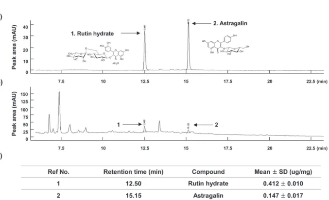
to use. To confirmation the reproducibility of WEMF, we conducted fingerprinting using a
high-performance liquid chromatography (HPLC)-based compositional analysis with two main reference compounds, namely, rutin hydrate and astragalin. All analyses were performed using an Agilent 1100
series HPLC instrument (Agilent Technologies,
San Jose, CA, USA) as previously reported (Lee et
al. 2014). The representative sample chromatogram
and quantitative analysis are illustrated in Figure 1.
CELL CULTURE AND CELL VIABILITY ASSAY
The RAW 264.7 murine macrophage cell line
was obtained from the Korean Cell Line Bank (Seoul, Republic of Korea) and cultured at 37°C
in 5% CO2 containing Dulbeccos modified
Eagle’s medium (WelGENE Inc., Daegu, Republic of Korea) supplemented with 10% fetal bovine serum (WelGENE Inc.), 100 U/ml of penicillin, and 100 mg/ml of streptomycin
(Sigma-Aldrich Chemical Co., St. Louis, MO, USA). A colorimetric
3-[4,5-dimethylthiazol-2-yl]-2,5-diphenyltetrazolium bromide (MTT,
Sigma-Aldrich Chemical Co.) assay was performed to measure cell viability. In brief, RAW 264.7 cells were treated with various concentrations of WEMF for 24 h or pretreated with WEMF for 1 h before stimulation with 500 ng/ml LPS (Sigma-Aldrich Chemical Co.) for 24 h. After incubation,
the medium was discarded, and MTT solution (5
mg/mL in phosphate-buffered saline, PBS) was added to each well and incubated for another 3 h
at 37°C. The medium was removed and dimethyl
sulfoxide (Sigma-Aldrich Chemical Co.) was
added to dissolve the formazan dye. The optical
density was then read at 560 nm using a microplate spectrophotometer (Molecular Devices, Sunnyvale, CA, USA) to determine cell viability (Oh et al. 2015).
MEASUREMENT OF NO PRODUCTION IN RAW 264.7 MACROPHAGES
The production of NO in culture supernatants
was assayed using Griess reagent (Sigma-Aldrich Chemical Co.). For this assay, the supernatant was collected and mixed with the same volume of Griess reagent for 10 min at room temperature in the dark. Absorbance was measured at 540 nm on a microplate reader, and NO concentrations were calculated by referencing a standard curve generated by known concentrations of sodium nitrite (Lee et al. 2015a).
MEASUREMENT OF PGE2 PRODUCTION IN RAW 264.7 MACROPHAGES
To measure the production of PGE2, the cells were cultured under the same conditions as those for
the NO measurement assay. The levels of PGE2
USA), according to the manufacturer’s instructions
(Wang et al. 2015).
RNA ISOLATION AND REVERSER TRANSCRIPTION POLYMERASE CHAIN REACTION (RT-PCR)
Total RNA was isolated from cells using TRIzol reagent (Invitrogen Life Technologies, Carlsbad,
CA, USA) according to the manufacturer’s
instructions and reverse transcribed using the
M-MLV reverse transcriptase kit (BioNEER,
Daejeon, Republic of Korea) to produce cDNAs.
RT-generated cDNAs encoding iNOS and COX-2 genes were amplifi ed by PCR using the desired primers (BioNEER). Following amplifi cation, the
PCR products were separated by 1.5% agarose gel
electrophoresis, stained with ethidium bromide,
and visualized by ultraviolet illumination. In a
parallel experiment, glyceraldehyde 3-phosphate
dehydrogenase was used as an internal control.
PROTEIN EXTRACTION AND WESTERN BLOT ANALYSIS
The cells were collected and resuspended in an extraction lysis buff er [25 mM Tris-Cl (pH 7.5),
250 mM NaCl, 5 mM ethylene diaminetetra acetic acid, 1% NP-40, 1 mM pheny-methylsulfonyl
fl uoride, and 5 mM dithiothreitol] for 30 min at
4°C. In a parallel experiment, nuclear and cytosolic proteins were separated using NE-PER nuclear and cytosolic extraction reagents (Pierce Biotechnology, Rockford, IL, USA) according to the manufacturer’s protocol. Equal amounts of protein from each sample were separated by sodium dodecyl sulfate (SDS)-polyacrylamide gel electrophoresis at 90 V for 2 h and transferred onto polyvinylidene fluoride membranes (Millipore, Bedford, MA,
USA). Thereafter, the membranes were incubated
overnight at 4°C with the corresponding primary antibodies purchased from Santa Cruz Biotechnology, Inc. (Santa Cruz, CA, USA) and
Cell Signaling Technology, Inc. (Boston, MA, USA). Then, the membranes were incubated with
the appropriate secondary antibodies conjugated to horseradish peroxidase (Amersham Co., Arlington Heights, IL, USA)) at room temperature for 2 h. Using an enhanced chemiluminescence (ECL, Amersham Co.) detection system, immunoreactive bands were detected.
IMMUNOFLUORESCENT STAINING FOR NF-κB P65 IN RAW 264.7 MACROPHAGES
The NF-κB p65 nuclear translocalization was detected by an immunofluorescence assay using a fluorescence microscope. For this study, RAW
246.7 cells were pretreated with WEMF for 1 h
and then stimulated with LPS for 1 h. The cells were fixed with 3.7% paraformaldehyde
(Sigma-Aldrich Chemical Co.) in PBS for 10 min at 4°C,
permeabilized with 0.4% Triton X-100 in PBS for
10 min, and blocked with 5% bovine serum albumin
for 1 h. The cells were probed with anti-p65 NF-κB antibody (Santa Cruz Biotechnology, Inc.) overnight at 4°C and then incubated with fluorescein
isothiocyanate-conjugated donkey anti-rabbit IgG (Jackson ImmunoResearch Laboratories Inc., West Grove, PA, USA) for 2 h at room temperature.
The position of the cell nucleus was determined
with 4,6-diamidino-2-phenylindole (DAPI, Sigma-Aldrich Chemical Co.) solution (1 mg/ml) for 15
min. After washing the cells with PBS, fluorescence was visualized using a fluorescence microscope
(Carl Zeiss, Oberkochen, Germany).
MEASUREMENT OF ROS GENERATION IN RAW 264.7 MACROPHAGES
To measure the ROS levels, the cells were washed twice with PBS and lysed with 1% Triton X-100 in PBS for 10 min at 37°C. The cells were then stained with 10 μM 2’,7’-dichlorofluorescein
diacetate (DCF-DA, Molecular Probes, Eugene, OR, USA) for 20 min at room temperature in the
dark. The green fluorescence emitted by DCF was
recorded at 515 nm using a flow cytometer (Becton
Dickinson, San Jose, CA, USA), and 10,000 events were counted per sample (Eom et al. 2015). Image analysis for the generation of intracellular ROS
was acquired using a fluorescence microscope.
ZEBRAFISH EMBRYO AND LARVAE MAINTENANCE
Adult zebrafish were obtained from Dr. Hyo-Jong
Lee, College of Pharmacy, Inje University (Gimhae, Republic of Korea) and maintained at 28.5°C with a 14:10 h light/dark cycle in a recirculating tank
system using local tap water (pH 7.27.6, salinity 0.03%0.04%). The embryos were obtained from
natural spawning within 30 min and maintained at
a density of about 50 embryos per 100 mm2 in a
Petri dish containing media, as previously reported
(Wijesinghe et al. 2014). The entire study design
and experimental procedures were approved by the Dongeui University Animal Care and Use Committee (Busan, Republic of Korea).
MEASUREMENT OF NO AND ROS PRODUCTION IN ZEBRAFISH LARVAE
Approximately three days post-fertilization (dpf), embryos (n = 25) were transferred to individual wells of a 24-well plate and maintained in embryo media containing sterile distilled water (vehicle
control), 800 μg/ml WEMF (final concentration), 10 μg/ml LPS (final concentration), or 800 μg/ml WEMF for 1 h followed by treatment with 10 μg/ml
LPS, except the larvae in the control group, for up to
4 dpf. The generation of NO and ROS in zebrafish larvae was analyzed using fluorescent probe dyes, 4-amino-5-methylamino-2’7’ difluorofluorescein
diacetate (DAF-FM-DA, Molecular Probes) and DCF-DA, respectively. After 4 dpf, the larvae were transferred into 24-well plates and incubated
with DAF-FM DA (5 μM) and DCF-DA (20 μg/
USA). The images of stained larvae were observed for NO and ROS generation under a fl uorescence microscope, and fl uorescence intensity of individual larvae was quantifi ed at an excitation wavelength
of 485 nm and an emission wavelength of 535 nm using a spectrophotometer and ImageJ 1.46r software (Wayne Rasband, National Institutes of
Health, Bethesda, MD, USA), respectively. The
generation of NO and ROS was calculated by
comparing the fl uorescence intensity of treatment
larvae with that of the controls (Wijesinghe et al. 2014).
STATISTICAL ANALYSIS
All data are presented as mean ± standard deviation
(SD). Signifi cant diff erences among groups were
determined using the unpaired Student’s t-test. A value of p<0.05 was accepted as an indication
of statistical signifi cance. All the fi gures shown here refl ect the data obtained from at least three
independent experiments.
RESULTS
CYTOTOXIC EFFECTS OF WEMF AND LPS ON RAW 264.7 MACROPHAGES
To exclude the cellular toxicity caused by WEMF
treatment, RAW 264.7 cells were treated with
WEMF and/or LPS for 24 h. The MTT assay showed that WEMF of up to 1,000 μg/ml in the
presence or absence of 100 ng/ml LPS was not
cytotoxic (Figure 2). Therefore, we selected 800 μg/ml WEMF as the maximum concentration for
further experiments in RAW 264.7 cells.
WEMF SUPPRESSED LPS-INDUCED NO AND PGE2 PRODUCTION IN RAW 264.7 MACROPHAGES
To determine the inhibitory properties of WEMF
on LPS-induced NO and PGE2 production in
RAW 264.7 cells, the cells were pretreated with the indicated concentrations of WEMF for 1 h and then stimulated with 100 ng/ml LPS for another
24 h. The levels of NO and PGE2 in the culture supernatants were determined by Griess reaction assay and ELISA, respectively. As indicated in Figure 3a and b, stimulation with LPS markedly
induced the production of NO and PGE2 compared
with not stimulating with LPS. However, WEMF
signifi cantly inhibited NO and PGE2 secretion in RAW 264.7 cells in a concentration-dependent manner.
WEMF ATTENUATES LPS-INDUCED INOS AND COX-2 EXPRESSION IN RAW 264.7 MACROPHAGES We next investigated if the inhibitory eff ects of
WEMF on NO and PGE2 production were related
to the regulation of the expression of their synthesis enzymes, iNOS and COX-2, respectively. As shown in Figure 3c and d, WEMF concentration-dependently inhibited the protein and mRNA expression of iNOS and COX-2 in the
LPS-stimulated RAW 264.7 cells. These data indicate
that WEMF suppresses NO and PGE2 production
by reducing the expression of their encoding genes.
Figure 3 - Inhibition of NO and PGE2 production by WEMF in LPS-stimulated RAW 264.7 macrophages. The cells were pretreated with the indicated concentrations of WEMF for 1 h prior to incubation with 100 ng/ml LPS for 24 h. The levels of NO (a) and PGE2 (b) in culture media were measured by Griess assay and a commercial ELISA kit, respectively. Each value indicates the mean ± SD and is representative of the results obtained from three independent experiments (#p<0.05 compared with the control; *p<0.05 compared with cells cultured with 100 ng/ml LPS). (c) The total RNAs were isolated from cells grown under the same conditions as those in Figure 3 and prepared for the RT-PCR analysis of the iNOS and COX-2 mRNA expression using the indicated primers. (d) Cell lysates were prepared for Western blot analysis, with antibodies specifi c for murine iNOS and COX-2, and for an ECL detection system. The experiment was repeated three times and similar results were obtained. GAPDH and actin were used as the internal controls for the RT-PCR and Western blot analysis, respectively.
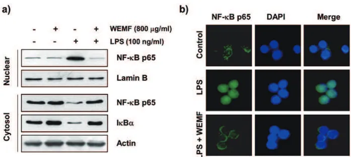
WEMF BLOCKS LPS-INDUCED NF-κB NUCLEAAR TRANSLOCATION IN RAW 264.7 MACROPHAGES
As active NF-κB translocates to the nucleus where
it activates its target genes including, iNOS,
COX-2, and pro-infl ammatory cytokines, by binding to
their promoter regions (Lu et al. 2011, Rigoglou and Papavassiliou 2013), we investigated whether or not WEMF attenuates the LPS-induced nuclear
translocation of NF-κB in RAW 264.7 cells. The immunoblotting data using cytoplasmic and
nuclear extracts indicated that WEMF pretreatment
inhibited the NF-κB p65 subunit nuclear
accumulation, which was associated with the
attenuation of IκBα degradation in LPS-stimulated
RAW 264.7 cells (Figure 4a). Consistent with
these results, immunocytochemistry analysis
also indicated that NF-κB p65 was normally
sequestered in the cytoplasm following stimulation with LPS. However, the LPS-mediated nuclear
translocation of NF-κB was considerably blocked by pretreatment with WEMF (Figure 4b). These
results indicated that WEMF attenuated the
transcriptional activation of NF-κB, which controls the expression of pro‐infl ammatory genes in
LPS-treated RAW 264.7 cells.
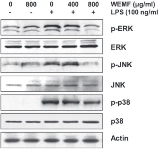
WEMF REDUCES THE ACTIVATION OF MAPKS IN LPS-STIMULATED RAW 264.7 MACROPHAGES
As the activation of MAPKs is crucial for
LPS-stimulated NF-κB activation and the subsequent
expression (Kaminska 2005, Muralidharan and Mandrekar 2013), we analyzed the phosphorylation of MAPKs, such as extracellular signal-regulated kinase (ERK), c-Jun N-terminal kinase (JNK), and p38 MAPK, by Western blot analysis. As shown in Figure 5, stimulation with LPS resulted in the marked phosphorylation of ERK, JNK, and p38 MAPK, and their total expressions did not have any significant change. However, WEMF concentration-dependently blocked the
LPS-induced phosphorylation of three MAPKs. The
result showed that WEMF suppressed MAPKs signaling pathway to reduce inflammatory responses in LPS-induced RAW 264.7 cells.
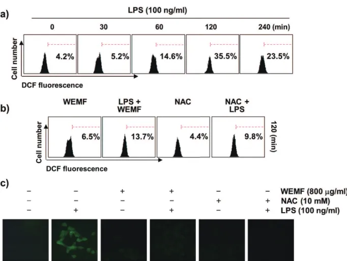
WEMF SUPPRESSES LPS-INDUCED ACCUMULATION OF ROS IN RAW 264.7 MACROPHAGES
As oxidative stress is partly involved in the initiation of inflammation (Brüne et al. 2013, Mills and O'Neill 2016), we examined whether or not WEMF
could reduce the LPS-induced generation of ROS
in RAW 264.7 cells using DCF-DA staining. The results of the fl ow cytometric assay indicated that
the accumulation of intracellular ROS was observed at 0.5 h, and that the levels continued to increase up to 2 h by LPS treatment (Figure 6a). However, the increase in LPS-stimulated ROS production was markedly attenuated by pretreatment with WEMF (Figure 6b and c). As a positive control, the ROS scavenger N-acetyl-l-cysteine (NAC) also effectively attenuated LPS-induced ROS generation, but WEMF itself did not contribute
to the ROS generation. This fi nding suggests that
the anti-inflammatory potential of WEMF may
be associated with its antioxidant eff ects on RAW
264.7 cells.
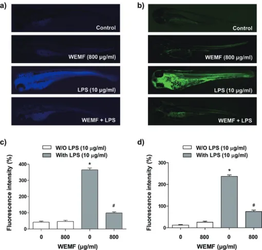
WEMF DOWNREGULATES LPS-INDUCED NO AND ROS PRODUCTION IN ZEBRAFISH
To confi rm the in vivo protective eff ects of WEMF
on LPS-induced NO generation, we visualized
DAF-FM-DA staining in a zebrafish model. As
shown in Figure 7a and b, the control, which was
not treated with LPS or WEMF, and WEMF alone groups generated a clear image, thus indicating that
WEMF alone did not aff ect the basal NO levels. However, stimulation of the zebrafi sh larvae with LPS markedly generated fl uorescence image, thus
suggesting that NO generation took place in the
presence of LPS and WEMF reduced the
LPS-stimulated elevation of NO production. We also
evaluated the inhibitory eff ect of WEMF on
LPS-induced ROS accumulation. Microphotographs
of DCF-DA staining revealed excessive ROS
accumulation after LPS stimulation. By contrast,
when the zebrafi sh larvae were treated with WEMF prior to LPS administration, an eff ective reduction
in the generation of ROS was observed (Figure 7c
and d), thus suggesting that WEMF also inhibited
LPS-stimulated ROS production in vivo.
DISCUSSION
Infl ammation is a host defense mechanism against
pathogenic challenges, and multiple events are involved in the development of inflammation. During infection by gram-negative bacterial LPS,
membrane-bound pattern recognition receptor Toll-like receptor 4 (TLR4) is a critical driver of immune
responses (Aderem and Ulevitch 2000, Nikaido
2003). The activation of TLR4 pathway leads to
intracellular signaling pathways that culminate in the activation of several intracellular signaling
pathways, including NF-κB and MAPKs. The
consequent activation of macrophages promotes inflammation through the aberrant production of pro-inflammatory mediators and cytokines that recruit additional immune cells to sites of infection or tissue injury (Aderem and Ulevitch 2000, Nikaido 2003). Regarding the importance of pro-inflammatory cytokines in inflammatory
responses, the pro-infl ammatory mediators of NO
and PGE2 play crucial roles in the development
of chronic inflammatory disease (Rocca and FitzGerald 2002, Kim et al. 2005). In addition,
LPS exerts its infl ammatory eff ects by inducing
the expression of iNOS and COX-2, which directly
stimulates the high production of NO and PGE2,
respectively. Therefore, a compound capable
of preventing the release of pro-inflammatory mediators or downregulating iNOS or COX-2 expression by the inactivation of macrophages may
possess anti-infl ammatory activities. In this study, we observed that WEMF signifi cantly attenuated
the LPS-induced increase in NO and PGE2, which
are representative pro-inflammatory mediators, in RAW 264.7 macrophages by down-regulating iNOS and COX-2 expression at both the protein and
mRNA levels without cytotoxicity. These indicated the inhibitory eff ect of WEMF on NO and PGE2
production was mainly due to the inhibitions of iNOS and COX-2 mRNA and protein expressions.
Thus, the results support WEMF being a promising
target for inhibiting the early steps in infl ammatory
pathways.
NF-κB has been shown to play an important role in various infl ammatory states as a key transcription factor for many infl ammation-associated enzymes and cytokine genes, which contain NF-κB binding
motifs within their respective promoters (Lu et al.
2011, Rigoglou and Papavassiliou 2013). NF-κB, a
dimer of p65 and p50 subunits, is normally retained in the cytoplasm because of its association with
its endogenous inhibitor, IκB-α. Once activated by infl ammatory stimulants, including LPS, IκB-α
is rapidly phosphorylated and degraded through
a proteasome-mediated pathway, followed by
a nuclear translocation of NF-κB, thus resulting in the transcriptional induction of infl
ammation-associated genes (Nikaido 2003, Rigoglou and Papavassiliou 2013). MAPKs, a family of serine/ threonine protein kinases including ERK, JNK, and p38 MAPK, are also directly involved in controlling signaling events that contribute to the production of
pro-infl ammatory factors in activated macrophages through the activation of NF-κB (Kaminska 2005, Muralidharan and Mandrekar 2013). Therefore,
pharmacologic agents that effectively modulate
NF-κB and MAPKs activation are promising
candidates for treating various inflammatory
diseases. To investigate the molecular mechanism
of WEMF-mediated inhibition of inflammatory
substances, its eff ect on the activation of MAPKs and NF-κB was evaluated. In the present study,
we found that WEMF strongly suppressed the
translocation of activated NF-κB to the nucleus and degradation of IκB-α was also inhibited. These findings indicate that WEMF inhibits NF-κB activation by suppressing IκB-α degradation and translocation of NF-κB from the cytosol into the
nucleus in LPS-induced RAW 264.7 macrophages.
The current study also demonstrated that WEMF
concentration-dependently diminished the phosphorylation of ERK, JNK, and p38 MAPK by
LPS treatment. These data suggest that suppression
of MAPKs phosphorylation by WEMF might also be involved in the inhibition of the LPS-induced
production of proinfl ammatory substances by RAW 264.7 macrophages. Taken together, our results
indicate that WEMF suppresses inflammatory
mediators, NO and PGE2 by inhibiting NF-κB B
and MAPKs signaling pathways.
Oxidative stress, which represents the over-production of ROS, is strongly associated with
other pathological statuses, including infl ammation
Figure 7 - Protective eff ect of WEMF on LPS-induced NO and ROS generation in zebrafi sh larvae. The zebrafi sh larvae were treated with 800 μg/ml WEMF and 10 μg/ml LPS for 24 h or pretreated with 800 μg/ml WEMF for 1 h prior to incubation with 10 μg/ml LPS for 24 h (a and b). The levels of NO and ROS generation were observed under a fl uorescence microscope after staining with DAF-FM-DA and DCF-DA, respectively (c and d). The fl uorescence intensities of NO and ROS levels in individual zebrafi sh larvae were quantifi ed. The values represented the means ± SD of three independent experiments (#
(Brüne et al. 2013, Mills and O'Neill, 2016). Moreover, during chronic inflammation, ROS amplifies inflammatory signals in macrophages
through the activation of NF-κB signaling pathway and the over-expression of inflammation-associated genes (Kauppinen et al. 2013, Tan et al. 2016).
Furthermore, MAPKs are also redox sensitive, and ROS targets the cysteines within the proteins and alters the kinase activation, which further activates redox-sensitive MAPKs (Rahman and MacNee
1998, Fu et al. 2009). Thus, we investigated the inhibitory effect of WEMF on LPS-induced ROS
generation and found that ROS accumulation was
significantly reduced after pretreatment with WEMF
in LPS-stimulated RAW 264.7 macrophages.
Therefore, the WEMF-mediated inhibition of
ROS generation might be attributed to its ability to scavenge free radicals, and could potentially inhibit
the NF-κB and MAPKs-dependent expression of pro-inflammatory mediators, thereby resulting in an anti-inflammatory efficacy. We further investigated the protective effect of WEMF against LPS-induced
NO and ROS generation using DAF-FM-DA and DCF-DA staining in zebrafish as an alternative in vivo animal model system. Consistent with previous results (Wijesinghe et al. 2014, Lee et al. 2015b, Cheong et al. 2016), a dramatic increase in
the fluorescence signals was observed in the
LPS-exposed group unlike in the unstimulated control group, thus indicating that NO and ROS generation
took place during LPS treatment in the zebrafish
larvae. However, similar to our in vitro results, a
significant reduction in the amount of NO and ROS was observed in zebrafish treated with WEMF prior
to LPS treatment, thus indicating the strong
anti-inflammatory and antioxidant potentials of WEMF.
CONCLUSIONS
The present results demonstrated that WEMF exerted potent anti-inflammatory and antioxidant effects in RAW 264.7 macrophages and zebrafish.
In LPS-stimulated RAW 264.7 macrophages, WEMF markedly attenuated the production of pro-inflammatory mediators and accumulation
of ROS. These effects of WEMF were associated
with the suppression of LPS-induced nuclear
translocalization of NF-κB and MAPKs activation. WEMF also significantly prevented the elevation
of NO and ROS levels in an LPS-stimulated
zebrafish model. Based on the results of this study, WEMF could have a beneficial effect to enhance anti-inflammatory and antioxidant treatment.
ACKNOWLEDGMENTS
This work was supported by the High Value-added Food Technology Development Program (314043-3), Ministry of Agriculture, Food and Rural Affairs,
Republic of Korea.
REFERENCES
ABUAJAH CI, OGBONNA AC AND OSUJI CM. 2015. Functional components and medicinal properties of food: a review. J Food Sci Technol 52: 2522-2529.
ADEREM A AND ULEVITCH RJ. 2000. Toll-like receptors in the induction of the innate immune response. Nature 406: 782-787.
AMIN AR, ATTUR M AND ABRAMSON SB. 1999. Nitric oxide synthase and cyclooxygenases: distribution, regulation, and intervention in arthritis. Curr Opin Rheumatol 11: 202-209.
ANN JY, EO H AND LIM Y. 2015. Mulberry leaves (Morus alba L.) ameliorate obesity-induced hepatic lipogenesis, fibrosis, and oxidative stress in high-fat diet-fed mice. Genes Nutr 10: 46.
BOCANEGRA A, BASTIDA S, BENEDI J, RODENAS S AND SANCHEZ-MUNIZ FJ. 2009. Characteristics and nutritional and cardiovascular-health properties of seaweeds. J Med Food 12: 236-258.
BRÜNE B, DEHNE N, GROSSMANN N, JUNG M, NAMGALADZE D, SCHMID T, VON KNETHEN A AND WEIGERT A. 2013. Redox control of inflammation in macrophages. Antioxid Redox Signal 19: 595-637. CHAN EW, LYE PY AND WONG SK. 2016. Phytochemistry,
pharmacology, and clinical trials of Morus alba. Chin J Nat Med 14: 17-30.
paniculata, Angelica sinensis and Morus alba ethyl acetate fractions. J Ethnopharmacol 122: 68-75.
CHEONG SH, YANG HW, KO EY, AHN G, LEE W, KIM D, JEON YJ AND KIM K. 2016. Anti-inflammatory effects of trans-1,3-diphenyl-2,3-epoxypropane-1-one in zebrafish embryos in vivo model. Fish Shellfish Immunol 50: 16-20.
CONTI B, TABAREAN I, ANDREI C AND BARFAI T. 2004. Cytokines and fever. Front Biosci 9: 1433-1449.
CUNHA TM, VERRI WA JR, SCHIVO IR, NAPIMOGA MH, PARADA CA, POOLE S, TEIXEIRA MM, FERREIRA SH AND CUNHA F. 2008. Crucial role of neutrophils in the development of mechanical inflammatory hypernociception. J Leukoc Biol 83: 824-832.
DEEPA M, SURESHKUMAR T, SATHEESHKUMAR PK AND PRIYA S. 2013. Antioxidant rich Morus alba leaf extract induces apoptosis in human colon and breast cancer cells by the downregulation of nitric oxide produced by inducible nitric oxide synthase. Nutr Cancer 65: 305-310. EOM SA ET AL. 2015. Protective effects of PEP-1-Catalase
on stress-induced cellular toxicity and MPTP-induced Parkinson's disease. BMB Rep 48: 395-400.
FREIRE MO AND VAN DYKE TE. 2013. Natural resolution of inflammation. Periodontol 2000 63: 149-164.
FU P, BIRUKOVA AA, XING J, SAMMANI S, MURLEY JS, GARCIA JG, GRDINA DJ AND BIRUKOV KG. 2009. Amifostine reduces lung vascular permeability via suppression of inflammatory signalling. Eur Respir J 33: 612-624.
HADDAD JJ AND LAND SC. 2002. Redox signaling-mediated regulation of lipopolysaccharide-induced proinflammatory cytokine biosynthesis in alveolar epithelial cells. Antioxid Redox Signal 4: 179-193. HONG CH, HUR SK, OH OJ, KIM SS, NAM KA AND LEE
SK. 2002. Evaluation of natural products on inhibition of inducible cyclooxygenase (COX-2) and nitric oxide synthase (iNOS) in cultured mouse macrophage cells. J Ethnopharmacol 83: 153-159.
IQBAL S, YOUNAS U, SIRAJUDDIN, CHAN KW, SARFRAZ RA AND UDDIN K. 2012. Proximate composition and antioxidant potential of leaves from three varieties of Mulberry (Morus sp.): a comparative study. Int J Mol Sci 13: 6651-6664.
JEONG JW ET AL. 2016. Mori folium inhibits interleukin-1β-induced expression of matrix metalloproteinases and inflammatory mediators by suppressing the activation of NF-κB and p38 MAPK in SW1353 human chondrocytes. Int J Mol Med 37: 452-460.
KAMINSKA B. 2005. MAPK signalling pathways as molecular targets for anti-inflammatory therapy-from molecular mechanisms to therapeutic benefits. Biochim Biophys Acta 1754: 253-262.
KAUPPINEN A, SUURONEN T, OJALA J, KAARNIRANTA K AND SALMINEN A. 2013. Antagonistic crosstalk between NF-κB and SIRT1 in the regulation of inflammation and metabolic disorders. Cell Signal 25: 1939-1948.
KHAN MA ET AL. 2013. A comparative study on the antioxidant activity of methanolic extracts from different parts of Morus alba L. (Moraceae). BMC Res Notes 6: 24. KIM DS, KANG YM, JIN WY, SUNG YY, CHOI G AND
KIM HK. 2014. Antioxidant activities and polyphenol content of Morus alba leaf extracts collected from varying regions. Biomed Rep 2: 675-680.
KIM GN AND JANG HD. 2011. Flavonol content in the water extract of the mulberry (Morus alba L.) leaf and their antioxidant capacities. J Food Sci 76: C869-873.
KIM SF, HURI DA AND SNYDER SH. 2005. Inducible nitric oxide synthase binds, S-nitrosylates, and activates cyclooxygenase-2. Science 310: 1966-1970.
KOBAYASHI Y, MIYAZAWA M, KAMEI A, ABE K AND KOJIMA T. 2010. Ameliorative effects of mulberry (Morus alba L.) leaves on hyperlipidemia in rats fed a high-fat diet: induction of fatty acid oxidation, inhibition of lipogenesis, and suppression of oxidative stress. Biosci Biotechnol Biochem 74: 2385-2395.
KWON DH ET AL. 2016. The immunomodulatory activity of Mori folium, the leaf of Morus alba L., in RAW 264.7 macrophages in vitro. J Cancer Prev 21: 144-151.
LEE JS, KIM HG, HAN JM, KIM DW, YI MH, SON SW, KIM YA, LEE JS, CHOI MK AND SON CG. 2014. Ethanol extract of Astragali Radix and Salviae Miltiorrhizae Radix, Myelophil, exerts anti-amnesic effect in a mouse model of scopolamine-induced memory deficits. J Ethnopharmacol 153: 782-792.
LEE H, PYO MJ, BAE SK, HEO Y, KIM CG, KANG C AND KIM E. 2015a. Improved therapeutic profiles of PLA2-free bee venom prepared by ultrafiltration method. Toxicol Res 31: 33-40.
LEE SH, YANG HW, DING Y, WANG Y, JEON YJ, MOON SH, JEON BT AND SUNG SH. 2015b. Anti-inflammatory effects of enzymatic hydrolysates of velvet antler in RAW 264.7 cells in vitro and zebrafish model. EXCLI J 14: 1122-1132.
LU YC, JAYAKUMAR T, DUANN YF, CHOU YC, HSIEH CY, YU SY, SHEU JR AND HSIAO G. 2011. Chondroprotective role of sesamol by inhibiting MMPs expression via retaining NF-κB signaling in activated SW1353 cells. J Agric Food Chem 59: 4969-4978. MCDANIEL ML, KWON G, HILL JR, MARSHALL CA
MILLS EL AND O'NEILL LA. 2016. Reprogramming mitochondrial metabolism in macrophages as an anti-inflammatory signal. Eur J Immunol 46: 13-21.
MURALIDHARAN S AND MANDREKAR P. 2013. Cellular stress response and innate immune signaling: integrating pathways in host defense and inflammation. J Leukoc Biol 94: 1167-1184.
N A O WA B O O T J , PA N N A N G P E T C H P, KUKONGVIRIYAPAN V, KUKONGVIRIYAPAN U, NAKMAREONG S AND ITHARAT A. 2009. Mulberry leaf extract restores arterial pressure in streptozotocin-induced chronic diabetic rats. Nutr Res 29: 602-608. NIKAIDO H. 2003. Molecular basis of bacterial outer
membrane permeability revisited. Microbiol Microbiol Mol Biol Rev 67: 593-656.
OH K, MOON HG, LEE DS AND YOO YB. 2015. Tissue transglutaminase-interleukin-6 axis facilitates peritoneal tumor spreading and metastasis of human ovarian cancer cells. Lab Anim Res 31: 188-197.
RAHMAN I AND MACNEE W. 1998. Role of transcription factors in inflammatory lung diseases. Thorax 53: 601-612.
RAMAN ST, GANESHAN AK, CHEN C, JIN C, LI SH, CHEN HJ AND GUI Z. 2016. In vitro and in vivo antioxidant activity of flavonoid extracted from mulberry fruit (Morus alba L.). Pharmacogn Mag 12: 128-133. RIGOGLOU S AND PAPAVASSILIOU AG. 2013. The
NF-κB signalling pathway in osteoarthritis. Int J Biochem Cell Biol 45: 2580-2584.
ROCCA B AND FITZGERALD GA. 2002. Cyclooxygenases and prostaglandins: shaping up the immune response. Int Immunopharmacol 2: 603-630.
SHIBATA Y ET AL. 2007. Mulberry leaf aqueous fractions inhibit TNF-alpha-induced nuclear factor kappaB (NF-kappaB) activation and lectin-like oxidized LDL receptor-1 (LOX-1) expression in vascular endothelial cells. Atherosclerosis 193: 20-27.
SUGIMOTO M ET AL. 2009. Mulberry leaf ameliorates the expression profile of adipocytokines by inhibiting oxidative stress in white adipose tissue in db/db mice. Atherosclerosis 204: 388-394.
TAN HY, WANG N, LI S, HONG M, WANG X AND FENG Y. 2016. The reactive oxygen species in macrophage
polarization: Reflecting its dual role in progression and treatment of human diseases. Oxid Med Cell Longev 2016: 2795090.
TIRUPATHI RG, SURESH BK, UJWAL KJ, SUJANA P, RAOA AV AND SREEDHAR AS. 2011. Anti-microbial principles of selected remedial plants from Southern India. Asian Pac J Trop Biomed 1: 298-305.
VARGA A, BUDAI MM, MILESZ S, BACSI A, TOZER J AND BENKO S. 2013. Ragweed pollen extract intensifies lipopolysaccharide-induced priming of NLRP3 inflammasome in human macrophages. Immunology 138: 392-401.
WANG L, XU ML, LIU J, WANG Y, HU JH AND WANG MH. 2015. Sonchus asper extract inhibits LPS-induced oxidative stress and pro-inflammatory cytokine production in RAW264.7 macrophages. Nutr Res Pract 9: 579-585. WIJESINGHE WA, KIM EA, KANG MC, LEE WW, LEE
HS, VAIRAPPAN CS AND JEON YJ. 2014. Assessment of anti-inflammatory effect of 5β-hydroxypalisadin B isolated from red seaweed Laurencia snackeyi in zebrafish embryo in vivo model. Environ Toxicol Pharmacol 37: 110-117.
XIANG J, TANG YP, ZHOU ZY, WU P, WANG Z, MORI M AND CAI DF. 2010. Apocynum venetum leaf extract protects rat cortical neurons from injury induced by oxygen and glucose deprivation in vitro. Can J Physiol Pharmacol 88: 907-917.
YANG L, BU L, SUN W, HU L AND ZHANG S. 2014. Functional characterization of mannose-binding lectin in zebrafish: implication for a lectin-dependent complement system in early embryos. Dev Comp Immunol 46: 314-322.
ZHANG L AND WANG CC. 2014. Inflammatory response of macrophages in infection. Hepatobiliary Pancreat Dis Int 13: 138-152.
SUPPLEMENTARY MATERIAL