ORIGINAL ARTICLE
FREE RADICAL SCAVENGING ACTIVITY OF
BERBERINE IN ACETAMINOPHEN INDUCED LIVER
INJURY
Suhail Ahmed Almani∗, Fatima Qureshi∗∗, Tariq Zaffar Shaikh∗, Arsalan Ahmed Uqaili△and Haji Khan Khoharo♦, 1 ∗Department of Medicine Liaquat University of Medical and Health Sciences, Jamshoro, Sindh, Pakistan.,∗∗Department of Biochemistry, Faculty of Medicine
and Allied Medical Sciences Isra University, Hyderabad, Sindh, Pakistan.,△Department of Physiology, Faculty of Medicine and Allied Medical Sciences Isra University, Hyderabad, Sindh, Pakistan.,♦Faculty of Medicine and Allied Medical Sciences Isra University, Hyderabad, Sindh, Pakistan.
ABSTRACT
Objective:Evaluation of free radical scavenging activity of Berberine (BBR) in acetaminophen (AAP) induced liver injury. Study design:Experimental study.Place and Duration:Animal House, Isra University Hyderabad from October 2015 to March 2016.Methodology:A sample of 80 male Wistar rats was selected according to inclusion and exclusion criteria and was divided into a control and three experimental groups. Acetaminophen, N-acetyl cysteine (NAC) and BBR were administered in standard doses. Cardiac puncture collected blood samples after 18 hours of the post-experiment period. Liver function test, antioxidant enzymes, and malondialdehyde (MDA) were detected by ELISA assay kit (Fortress Diagnostics). The data was analyzed on Statistix 10.0 software (USA) at 95% CI (P
≤
0.05). Results: The BBR showed antioxidant and antiperoxidant activity against acetaminophen-induced liver injury. BBR treated animals showed increased serum and tissue SOD, GPX, CAT, and GSSH with a reduction in tissue MDA (p=0.0001). Liver injury ameliorating effect of BBR was superior to N-acetyl cysteine.Conclusion:The present study suggests Berberine protects against acetaminophen-induced liver injury by its free radical scavenging activity.KEYWORDS:Berberine, N-acetyl cysteine, Acetaminophen, Free radical
Introduction
Drug-induced liver injury (DILI), such as that caused by the acetaminophen, is not an uncommon toxicological problem en-countered in the medical practice. In the case of severe toxicity, this results in severe liver injury, hepatocellular necrosis, and
Copyright © 2017 by the Bulgarian Association of Young Surgeons DOI:10.5455/ijsm.berberine-in-acetaminophen-induced-liver-injury First Received: December 06, 2016
Accepted: December 28, 2016
Manuscript Associate Editor: George Baitchev (BG) Editor-in Chief: Ivan Inkov (BG)
Reviewers: Richard Chapleau (US); Kumudu Irani Perera (LK)
1Dr Haji Khan Khoharo MBBS, M.Phil, FCPS Consultant Physician Assistant Professor
Assistant Director Postgraduate studies Faculty of Medicine and Allied Medical Sciences Isra University, Hyderabad, Sindh, Pakistan Email: drhajikhan786 @ gmail.com
three decades as an antidote for AAP toxicity. [5,6] Clinical effi-cacy of NAC is well established. Clinically it is utilized for the chronic obstructive pulmonary disease (COPD) and contrast-induced nephropathy. [6-8] Nowadays a growing interest has been found in herbal agents against DILI. Berberine (BBR) is one of herbal derivative used as OTC drug in China. The BBR is a plant isoquinoline alkaloid, traditionally used to treat infectious diarrhea. [10] The BBR is derived from the Rhizoma Coptidis Franch which is an indigenous Chinese herb. Rhizoma Coptidis Franch contains 5.2%–7.7% of berberine. It has been used for soothing of inflammatory reactions and treatment of diabetes mellitus (DM), and others. [10-12] Pleiotropic clinical effects of BBR have been reported such as use in liver disease, obesity [13-15], dyslipidemia, and cardiac disorders, and others. [16,17] The present study evaluated the free radical scavenging activity of Berberine in acetaminophen-induced liver injury in comparison to N-acetyl cysteine.
MATERIALS AND METHODS
The present experimental study was conducted, comprising a sample of 80 male Wistar rats, at the animal house of Isra Uni-versity Hyderabad, Sindh. The study covered duration from October 2015 to March 2016. Animals belonged to the species produced originally by Charles River Breeding Laboratories, Brooklyn, Massachusetts, USA. They were housed at the Basic Medical Sciences Institute (BMSI) animal house of Jinnah Post-graduate Medical Center (JPMC). The animals were acclimatized for ten days at our animal house which is equipped with modern stainless steel cages with automatic parts for feeding and water drinking.
Animal housing
Animals housing was by the ethics guidelines of NIH and Ethics guidelines of the institute. Animals were acclimatized for ten days before the experiment. The animal was kept in stainless steel cages, which are equipped with automatic parts. The tem-perature was maintained at 23 ± 2°C. Dark – light cycles of 12 hours were strictly maintained for the experimental animals. We provide pellet diet and fresh clean water. Access to food and water was ad libitum.
Inclusion and exclusion criteria
Adult male Wistar rats of 8-12 weeks age of 170-200 grams were included. Sick, lazy male rats and female rats were the exclusion criteria.
Experimental design
80 male Wistar rats were randomly divided into a control and 3 experimental groups; Group A (n=20): Normal healthy rats taken as controls (0.9% saline water) Group B (n=20): Ac-etaminophen induced liver injury (2gram/kg bwt) daily [18] Group C (n=20): Acetaminophen (2gram/kg bwt) daily + N-acetyl cysteine (100 mg/kg bwt) for 21 days [5] Group D (n=20): Acetaminophen (2gram/kg bwt) daily + Berberine (100 mg/kg bwt) for 21 days. [19]
Drugs and Chemicals
Acetaminophen (Panadol ®) was purchased from Glaxo Smith Kline, Pakistan. Berberine hydrochloride was purchased from
Berberine toxicity
To test the acute toxicity of Berberine hydrochloride, four rat groups were used; six rats in each group. Berberine hydrochlo-ride was suspended in distilled water. It was given orally by gav-ages once daily for seven days at 200, 500, 1000, and 2000mg/kg doses. Rats were observed continuously on the first day and then twice for seven days. Toxicity was not observed up to maximum dose of 2000mg/kg. [19]
Blood sampling
Disposable syringes (BD, USA) were used for blood sampling by cardiac puncture after 18 hours of the post-experiment period. Blood samples were collected in gel tubes. Samples were incu-bated at 4000 rpm for 10 minutes to separate sera. The sera were stored at -200C for biochemical analysis. Any serum sample contaminated with blood was discarded immediately. [20]
Biochemical testing
Liver function tests were performed spectrophotometrically us-ing a commercial kit by standard methods on Roche Biochemi-cal analyzer (Cobas e 411 analyzers, Roche Diagnostics GmbH, Mannheim, Germany). For Prothrombin time, a blood sample was taken in coagulation tubes and estimated by Humaclot duo using standard reagent kits of Merck. [21] Serum creatinine was measured by colorimetric Jaffé method. Superoxide dismutase, glutathione peroxidase, catalase, reduced glutathione (GSSH), and malondialdehyde (MDA) were detected by ELISA assay kit (Fortress Diagnostics) using standard methods mentioned in the literature. [22- 24]
Statistical analysis
The data was analyzed on Statistix 10.0 software (USA). One-way analysis of variance (ANOVA) was used for group comparisons followed by Bonferroni’s test for multiple comparisons. Data was presented as mean, SD, and SEM. The confidence interval of statistical significance was defined at 95% (P≤ 0.05).
Results
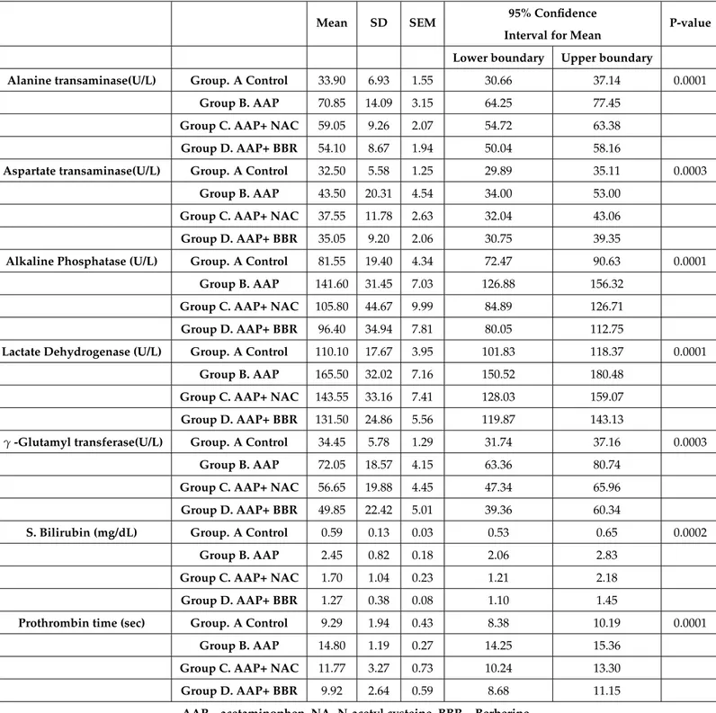
Alanine transaminase, aspartate transaminase, alkaline phos-phatase, lactate dehydrogenase, -glutamyl transferase, serum bilirubin and Prothrombin time (PT) are shown in Table 1. Es-cape special TeX symbols (Compress whitespace EsEs-cape special TeX symbols (Compress whitespace
Table 1Liver function tests of experimental rats (n=80)
Mean SD SEM 95% Confidence
Interval for Mean
P-value
Lower boundary Upper boundary
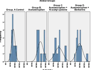
Alanine transaminase(U/L) Group. A Control 33.90 6.93 1.55 30.66 37.14 0.0001
Group B. AAP 70.85 14.09 3.15 64.25 77.45
Group C. AAP+ NAC 59.05 9.26 2.07 54.72 63.38
Group D. AAP+ BBR 54.10 8.67 1.94 50.04 58.16
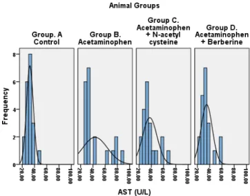
Aspartate transaminase(U/L) Group. A Control 32.50 5.58 1.25 29.89 35.11 0.0003
Group B. AAP 43.50 20.31 4.54 34.00 53.00
Group C. AAP+ NAC 37.55 11.78 2.63 32.04 43.06
Group D. AAP+ BBR 35.05 9.20 2.06 30.75 39.35
Alkaline Phosphatase (U/L) Group. A Control 81.55 19.40 4.34 72.47 90.63 0.0001
Group B. AAP 141.60 31.45 7.03 126.88 156.32
Group C. AAP+ NAC 105.80 44.67 9.99 84.89 126.71
Group D. AAP+ BBR 96.40 34.94 7.81 80.05 112.75
Lactate Dehydrogenase (U/L) Group. A Control 110.10 17.67 3.95 101.83 118.37 0.0001
Group B. AAP 165.50 32.02 7.16 150.52 180.48
Group C. AAP+ NAC 143.55 33.16 7.41 128.03 159.07
Group D. AAP+ BBR 131.50 24.86 5.56 119.87 143.13
γ-Glutamyl transferase(U/L) Group. A Control 34.45 5.78 1.29 31.74 37.16 0.0003
Group B. AAP 72.05 18.57 4.15 63.36 80.74
Group C. AAP+ NAC 56.65 19.88 4.45 47.34 65.96
Group D. AAP+ BBR 49.85 22.42 5.01 39.36 60.34
S. Bilirubin (mg/dL) Group. A Control 0.59 0.13 0.03 0.53 0.65 0.0002
Group B. AAP 2.45 0.82 0.18 2.06 2.83
Group C. AAP+ NAC 1.70 1.04 0.23 1.21 2.18
Group D. AAP+ BBR 1.27 0.38 0.08 1.10 1.45
Prothrombin time (sec) Group. A Control 9.29 1.94 0.43 8.38 10.19 0.0001
Group B. AAP 14.80 1.19 0.27 14.25 15.36
Group C. AAP+ NAC 11.77 3.27 0.73 10.24 13.30
Group D. AAP+ BBR 9.92 2.64 0.59 8.68 11.15
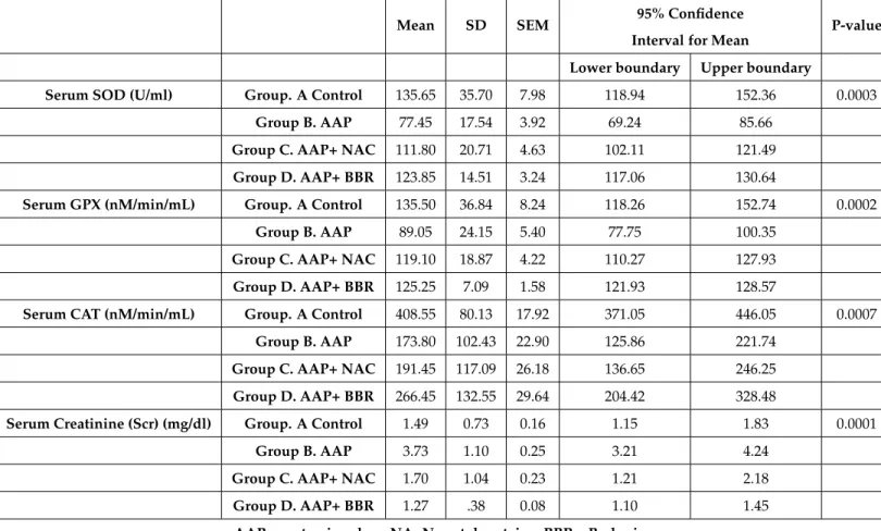
Table 2Serum superoxide dismutase, glutathione peroxidase, catalase and creatinine (n=80)
Mean SD SEM 95% Confidence
Interval for Mean
P-value
Lower boundary Upper boundary
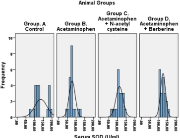
Serum SOD (U/ml) Group. A Control 135.65 35.70 7.98 118.94 152.36 0.0003
Group B. AAP 77.45 17.54 3.92 69.24 85.66
Group C. AAP+ NAC 111.80 20.71 4.63 102.11 121.49
Group D. AAP+ BBR 123.85 14.51 3.24 117.06 130.64
Serum GPX (nM/min/mL) Group. A Control 135.50 36.84 8.24 118.26 152.74 0.0002
Group B. AAP 89.05 24.15 5.40 77.75 100.35
Group C. AAP+ NAC 119.10 18.87 4.22 110.27 127.93
Group D. AAP+ BBR 125.25 7.09 1.58 121.93 128.57
Serum CAT (nM/min/mL) Group. A Control 408.55 80.13 17.92 371.05 446.05 0.0007
Group B. AAP 173.80 102.43 22.90 125.86 221.74
Group C. AAP+ NAC 191.45 117.09 26.18 136.65 246.25
Group D. AAP+ BBR 266.45 132.55 29.64 204.42 328.48
Serum Creatinine (Scr) (mg/dl) Group. A Control 1.49 0.73 0.16 1.15 1.83 0.0001
Group B. AAP 3.73 1.10 0.25 3.21 4.24
Group C. AAP+ NAC 1.70 1.04 0.23 1.21 2.18
Group D. AAP+ BBR 1.27 .38 0.08 1.10 1.45
Table 3Liver tissue anti oxidant and lipid peroxidant markers (n=80)
Mean SD SEM 95% Confidence Interval for Mean
P-value
Lower boundary Upper boundary
Tissue SOD (U/ml) Group. A Control 134.80 45.17 10.10 113.66 155.94 0.0003
Group B. AAP 86.40 6.10 1.36 83.55 89.25
Group C. AAP+ NAC 126.45 24.38 5.45 115.04 137.86
Group D. AAP+ BBR 131.20 31.76 7.10 116.34 146.06
Tissue GPX (nM/min/mg) Group. A Control 149.70 48.21 10.78 127.14 172.26 0.0002
Group B. AAP 82.80 21.72 4.86 72.63 92.97
Group C. AAP+ NAC 105.75 38.80 8.68 87.59 123.91
Group D. AAP+ BBR 118.55 29.76 6.66 104.62 132.48
Tissue CAT (nM/min/mg) Group. A Control 299.10 34.08 7.62 283.15 315.05 0.0007
Group B. AAP 116.70 61.70 13.80 87.82 145.58
Group C. AAP+ NAC 187.20 120.50 26.94 130.80 243.60
Group D. AAP+ BBR 239.20 125.37 28.03 180.53 297.87
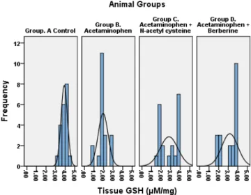
Tissue GSH (µM/mg) Group. A Control 3.91 0.34 0.08 3.75 4.06 0.0001
Group B. AAP 1.96 0.52 0.12 1.71 2.20
Group C. AAP+ NAC 2.82 0.93 0.21 2.39 3.26
Group D. AAP+ BBR 3.17 0.84 0.19 2.77 3.56
Tissue Malondialdehyde(µmol/gWTW) Group. A Control 3.05 1.45 0.32 2.37 3.72 0.0001
Group B. AAP 6.72 2.85 0.64 5.39 8.05
Group C. AAP+ NAC 5.10 1.72 0.38 4.30 5.91
Group D. AAP+ BBR 3.70 1.90 0.42 2.82 4.59
AAP – acetaminophen, NA- N-acetyl cysteine, BBR – Berberine
Figure 3:Serum Alkaline phosphatase
Figure 4:Serumγ-glutamyl transferase
Figure 6:Serum bilirubin
Figure 9:Serum Superoxide dismutase
Figure 10:Serum Glutathione peroxidase
Figure 11:Serum Catalase
Figure 12:Tissue superoxide dismutase levels
Figure 13:Tissue Glutathione peroxidase levels
Figure 15:Tissue Reduced Glutathione levels
Figure 16:Tissue Malondialdehyde
treated rats. The improvement of liver injury was significantly high in BBR group compared to other groups (p=0.0001).
Graphs 1-16 show the distribution of antioxidant enzymes, serum creatinine, and MDA. Acetaminophen-induced oxidative stress and lipid peroxidation revealed amelioration in the BBR and NAC-treated rats. Berberine response of ameliorating liver injury was superior to N-acetyl cysteine.
Discussion
Acetaminophen (AAP) induced liver toxicity was first reported in the United States in the mid-1980s, since then a rising trend has been reported from the World. AAP is one of the most commonly used drugs implicated in DILI. [1,3,4] A mortal-ity of 0.4% has been reported, approximating 300 deaths per year in the USA. [2] AAP-induced liver injury is mediated through production of reactive oxygen species (ROS). [1,25,26] AAP, when taken in megadoses, is metabolized into N-acetyl p-benzoquinoneimine (NAPQI). The NAPQI is a highly reac-tive agent which induces liver injury by free radical formation. [27,28] AAP induces oxidative stress and lipid peroxidation and depletes the liver of GSSH. [27-30] In the present study, the Berberine was evaluated for its free radical scavenging activity in comparison to a standard drug; the N-acetyl cysteine (NAC). In our current study the hepatotoxicity due to acetaminophen was confirmed by elevated levels of biochemical parameters; like ALT, AST, ALP,γ-GT, PT and serum bilirubin in the
ex-perimental group. Acetaminophen treated group B showed a relentless rise in liver aminotransferases. The NAC and BBR treated groups showed amelioration of cytoplasmic and mito-chondrial enzymes. The BBR treated animals showed more significant amelioration compared to NAC group (p = 0.0001). In the case of liver cell injury, the cytoplasmic and mitochondrial enzymes leak through the cell membrane, [31] and rise in the blood. [32] Treatment with NAC and BBR showed decreased levels of biochemical parameters; such as ALT, AST, ALP,γ-GT,
PT, and serum bilirubin. This may be due to free radical scaveng-ing activity of BBR and stabilization of cell membrane. Previous studies [30-34] supports the mechanisms. Serum ALP rises due to increased synthesis by the biliary canaliculi in response to biliary pressure because of cholestasis.[33,34] The present study reports free radical scavenging activity of BBR and NAC as evaluated by markers of oxidation and peroxidation. The BBR treated rats showed a statistically significant rise in serum and tissue SOD, GPX, CAT and GSSH and a decline in MDA. Antiox-idant and anti-lipid peroxAntiox-idant effect of BBR was superior to the NAC. Our findings are in agreement with the previous studies. [35,36] It is reported that the rise in MDA indicates failure of cellular antioxidant enzymes. [37] The BBR and NAC-treated rats showed a significant decrease in MDA; this suggests they possess antioxidant and anti peroxidant potential (Table 3). The BBR prevents hepatocyte cell injury by scavenging free radicals through an increase in cellular antioxidants – the SOD, GPX, CAT, and GSSH. The findings are in agreement with previous studies. [38,39] The berberine mitigates the oxidative injury by raising the SOD, GPX, CAT and GSSH which is visibly evident in the present study.
Conclusion
Berberine increases the antioxidant enzymes activity – the super-oxide dismutase, glutathione peroxidase, catalase and reduced glutathione, and decreases the malondialdehyde. The findings point towards free radical scavenging activity of Berberine.
Authors’ Statements
Competing Interests
The authors declare no conflict of interest.
ACKNOWLEDGMENT
We are thankful to staff of animal house of their help for comple-tion of this project.
Source of Support:
None
Conflict of Interest:
None
References
1. Yoon E, Babar A, Choudhary M, Kutner M, Pyrsopoulos N. Acetaminophen-Induced Hepatotoxicity: a Comprehensive Update. J Clin Translat Hepatol 2016; 4: 131–142.
2. Bunchorntavakul C, Reddy KR. Acetaminophen-related Hepatotoxicity. Clin Liver Dis 2013; 17: 587–607. doi: 10.1016/j.cld.2013.07.005.
3. Herndon CM, Dankenbring DM. Patient perception and knowledge of acetaminophen in a large family medicine service. J Pain Palliat Care Pharmacother 2014; 28:109–116. doi: 10.3109/15360288.2014.908993.
4. Clark R, Fisher JE, Sketris IS, Johnston GM. Population prevalence of high dose paracetamol in dispensed parac-etamol/opioid prescription combinations: an observational study. BMC Clin Pharmacol 2012; 12:11. doi: 10. 1186/1472-6904-12-11.
5. Cai Z, Lou Q, Wang F, Sun J, Fang H, Xi J, Ju L. N-acetylcysteine protects against liver injure induced by car-bon tetrachloride via activation of theNrf2/HO-1 pathway. Int J Clin Exp Pathol 2015; 8(7):8655-8662.
6. Priya S, Vijayalakshmi P, Vivekanandan P, Karthikeyan S. N-acetylcysteine attenuate di¬methylnitrosamine induced oxidative stress in rats. Eur J Pharmacol 2011; 654: 181-186.
7. Ye JF, Zhu H, Zhou ZF, Xiong RB, Wang XW, Su LX, Lou BD. Protective mechanism of an¬drographolide against car-bon tetrachloride-in¬duced acute liver injury in mice. Biol Pharm Bull 2011; 34: 1666-1670.
8. Ilavenil S, Kaleeswaran B, Ravikumar S. Protective effects of lycorine against carbon tetrachloride induced hepatotoxi-city in Swiss albino mice. Fundam Clin Pharmacol 2012; 26: 393-401.
9. Jiang W, Gao M, Sun S, Bi A, Xin Y, Han X, Wang L, Yin Z, Lou L. Protective effect of L-theanine on carbon tetrachloride-induced acute liver injury in mice. Biochem Biophys Res Commun 2012; 422: 344-350.
10. Xu M, Xiao Y, Yin J, Hou W, Yu X, Shen L, Liu F, Wei L, Jia W. Berberine Promotes Glucose Consumption Independently of AMP-Activated Protein Kinase Activation. PLoS ONE 2014; 9(7): e103702. doi:10.1371/journal.pone.0103702.
11. Yin J, Ye J, Jia W. Effects and mechanisms of berberine in diabetes treatment. Acta Pharmaceutica Sinica B 2012; 2: 327–334.
12. Yin J, Zhang H, Ye J. Traditional chinese medicine in treat-ment of metabolic syndrome. Endocr Metab Immune Dis-ord Drug Targets 2008; 8: 99–111.
13. Hu Y, Ehli EA, Kittelsrud J, Ronan PJ, Munger K, et al. Lipid-lowering effect of berberine in human subjects and rats. Phytomedicine 2012; 19: 861–867.
14. Xing LJ, Zhang L, Liu T, Hua YQ, Zheng PY, et al. Berberine reducing insulin resistance by up-regulating IRS-2 mRNA expression in nonalcoholic fatty liver disease (NAFLD) rat liver. Eur J Pharmacol 2011; 668: 467–471.
15. Li H, Dong B, Park SW, Lee HS, Chen W, et al. (2009) Hepa-tocyte nuclear factor 1alpha plays a critical role in PCSK9 gene transcription and regulation by the natural hypoc-holesterolemic compound berberine. J Biol Chem 2009; 284: 28885–28895.
16. Singh AB, Li H, Kan CF, Dong B, Nicolls MR, et al. The Critical Role of mRNA Destabilizing Protein Heteroge-neous Nuclear Ribonucleoprotein D in 39 Untranslated Region-Mediated Decay of Low-Density Lipoprotein Re-ceptor mRNA in Liver Tissue. Arterioscler Thromb Vasc Biol 2013; 284: 28885–28895.
17. Brusq JM, Ancellin N, Grondin P, Guillard R, Martin S, et al. Inhibition of lipid synthesis through activation of AMP kinase: an additional mechanism for the hypolipidemic effects of berberine. J Lipid Res 2006; 47: 1281–1288.
18. Bunchorntavakul C, Reddy KR. Acetaminophen-related hepatotoxicity. Clin Liver Dis 2013; 17:587-607.
19. Chen L, Guo W, Zhang S, Lu W, Liao S, Li Y. Berberine prevents high glucose-induced cell viability inhibition and apoptosis in podocytes. Int J Clin Exp Med 2016; 9(3):5942-5950.
20. Datta S, Dhar S, Nayak SS, Dinda SC. Hepatoprotective ac-tivity of Cyperus articulatus L against paracetamol induced hepatotoxicity in rats. J Chem Pharm Res 2013; 5:314-9.
21. Rajput MA, Khan RA, Qazi N, Feroz Z. Effect of Methanol Extract of Ajwain (Trachyspermum Ammi L) on Blood Co-agulation in Rats. J Liaq Univ Med Health Sci 2012; 11 (2): 105-108.
22. Girish C, Koner BC, Jayanthi S, Rao KR, Rajesh B, Pradhan SC. Hepatoprotective activity of six polyherbal formula-tions in paracetamol induced liver toxicity in mice. Indian J Med Res 2009; 129:569-78.
24. Galal RM, Zaki HF, Seif El-Nasr MM, Agha AM. Poten-tial protective effect of honey against paracetamol-induced hepatotoxicity. Arch Iran Med 2012; 15:674-80.
25. Biswas K, Kumar A, Babaria B, Prabhu K, Setty R. Hep-atoprotective effect of leaves of Peltophorumpterocarpum against paracetamol induced acute liver damage in rats ,” Journal of Basic and Clinical Pharmacy 2010; 1 (1): 10–15.
26. Setty SR, Quereshi AA, Swamy HMV. Hepatoprotective activity of Calotropis procera flowers against paracetamol-induced hepatic injury in rats. Fitoterapia 2007; 78 (7-8): 451–454.
27. Hanafy A, Aldawsari HM, Badr JM, Ibrahim AK, Abdel-Hady SES. Evaluation of Hepatoprotec-tive Activity of Adansonia digitata Extract on Acetaminophen-Induced Hepatotoxicity in Rats. Evid Bas Compl Alter Med 2016; Article ID 4579149: 1-7. http://dx.doi.org/10.1155/2016/4579149.
28. Bhawna S, Kumar SU. Hepatoprotective activity of some indigenous plants. Int‘l J PharmTech Res 2009; 1 (4): 1330–1334.
29. Hamza RZ, Al-Harbi MS. Amelioration of paracetamol hep-atotoxicity and oxidative stress on mice liver with silymarin and Nigella sativa extract supplements. Asian Pac J Trop Biomed 2015; 5(7): 521–531.
30. Yousef MI, Omar SA, El-Guendi MI, Abdelmegid LA. Poten-tial protective effects of quercetin and curcumin on parac-etamol induced histological changes, oxidative stress, im-paired liver and kidney functions and haematotoxicity in rat. Food Chem Toxicol 2010; 48: 3246-61.
31. Rajesh MG, Latha MS. Preliminary evaluation of the anti-hepatotoxic activity of Kamilari, a polyherbal formulation. J Ethnopharmacol 2004; 91 (1): 99–104, 2004.
32. Abraham P. Oxidative stress in paracetamol-induced patho-genesis: (I). Renal damage. Ind J Biochem Biophy 2005; 42 (1): 59–62.
33. Rabiul H, Subhasish M, Sinha S, Roy MG, Sinha D, Gupta S. Hepatoprotective activity of Clerodendron inerme against paracetamol induced hepatic injury in rats for pharmaceuti-cal product. Int‘l J Drug Develop Res 2011; 3 (1): 118–126.
34. Gini KC, Muraleedhara KG. Hepatoprotective effect of Spir-ulina lonar on paracetamol induced liver damage in rats. Asian J Exp Biol Sci 2010; 1: 614–623.
35. Dash DK, Yeligar VC, Nayak SS. Evaluation of hepatopro-tective and antioxidant activity of Ichnocarpus frutescens (Linn.) R.Br on paracetamol-induced hepatotoxicity in rats. Tropical J Pharma Res 2007; 6 (3): 755–765.
36. Biswas K, Kumar A, Babaria B, Prabhu K, Setty R. Hep-atoprotective effect of leaves of Peltophorumpterocarpum against paracetamol-induced acute liver damage in rats. J Basic Clin Pharmacy 2010; 1 (1):10–15.
37. Shao HB, Chu LY, Lu ZH, Kang CM.Primary antioxidant
38. Kalantari H, Khorsandi LS, Taherimobarekeh M. The protec-tive effect of the Curcuma longa extract on acetaminophen-induced hepatotoxicity in mice. Jundishapur J Natur Pharma Products 2007; 2 (1): 7–12.