Borrelia burgdorferi
Joppe W. R. Hovius1,2,3*, Maarten F. Bijlsma1,2, Gerritje J. W. van der Windt1,2, W. Joost Wiersinga1,2,3, Bastiaan J. D. Boukens4, Jeroen Coumou1, Anneke Oei5, Regina de Beer1,2, Alex F. de Vos1,2, Cornelis van ’t Veer1,2, Alje P. van Dam6, Penghua Wang7, Erol Fikrig7, Marcel M. Levi3, Joris J. T. H. Roelofs8, Tom van der Poll1,2,3
1Center for Experimental and Molecular Medicine (CEMM), Academic Medical Center, University of Amsterdam, AMC, Amsterdam, The Netherlands,2Center for Infection and Immunity Amsterdam (CINIMA), Academic Medical Center, University of Amsterdam, AMC, Amsterdam, The Netherlands,3Department of Medicine, Academic Medical Center, University of Amsterdam, AMC, Amsterdam, The Netherlands,4Heart Failure Research Center, Academic Medical Center, University of Amsterdam, AMC, Amsterdam, The Netherlands,5Department of Medical Microbiology, Academic Medical Center, University of Amsterdam, AMC, Amsterdam, The Netherlands,6Onze Lieve Vrouwe Gasthuis, Department of Medical Microbiology, Amsterdam, The Netherlands,7Yale University, School of Medicine, Section of Infectious Diseases, Department of Internal Medicine, New Haven, Connecticut, United States of America,8Department of Pathology, Academic Medical Center, University of Amsterdam, AMC, Amsterdam, The Netherlands
Abstract
The causative agent of Lyme borreliosis, the spirocheteBorrelia burgdorferi, has been shown to induce expression of the urokinase receptor (uPAR); however, the role of uPAR in the immune response againstBorreliahas never been investigated. uPAR not only acts as a proteinase receptor, but can also, dependently or independently of ligation to uPA, directly affect leukocyte function. We here demonstrate that uPAR is upregulated on murine and human leukocytes upon exposure toB. burgdorferi both in vitro as well as in vivo. Notably, B. burgdorferi-inoculated C57BL/6 uPAR knock-out mice harbored significantly higherBorrelianumbers compared to WT controls. This was associated with impaired phagocytotic capacity of B. burgdorferiby uPAR knock-out leukocytes in vitro.B. burgdorferinumbers in vivo, and phagocytotic capacity in vitro, were unaltered in uPA, tPA (low fibrinolytic activity) and PAI-1 (high fibrinolytic activity) knock-out mice compared to WT controls. Strikingly, in uPAR knock-out mice partially backcrossed to aB. burgdorferi susceptible C3H/HeN background, higherB. burgdorferinumbers were associated with more severe carditis and increased local TLR2 and IL-1bmRNA expression. In conclusion, inB. burgdorferiinfection, uPAR is required for phagocytosis and adequate eradication of the spirochete from the heart by a mechanism that is independent of binding of uPAR to uPA or its role in the fibrinolytic system.
Citation:Hovius JWR, Bijlsma MF, van der Windt GJW, Wiersinga WJ, Boukens BJD, et al. (2009) The Urokinase Receptor (uPAR) Facilitates Clearance ofBorrelia burgdorferi. PLoS Pathog 5(5): e1000447. doi:10.1371/journal.ppat.1000447
Editor:Linden Hu, Tufts University School of Medicine, United States of America ReceivedSeptember 25, 2008;AcceptedApril 25, 2009;PublishedMay 22, 2009
Copyright:ß2009 Hovius et al. This is an open-access article distributed under the terms of the Creative Commons Attribution License, which permits unrestricted use, distribution, and reproduction in any medium, provided the original author and source are credited.
Funding:JWRH is supported by ZonMw, the Netherlands organization for health research and development (http://www.zonmw.nl/en/). Grant number 92003370. The funders had no role in study design, data collection and analysis, decision to publish, or preparation of the manuscript.
Competing Interests:The authors have declared that no competing interests exist. * E-mail: j.w.hovius@amc.uva.nl
Introduction
Lyme borreliosis, an emerging tick-borne disease in both the New and Old world, is caused by spirochetes belonging to the Borrelia burgdorferisensu lato group and is predominantly transmit-ted byIxodesticks [1]. In the United StatesBorrelia burgdorferisensu stricto, from here on referred to as B. burgdorferi, is the only prevalentBorreliaspecies, whereas in Europe threeBorreliaspecies -B. burgdorferi, Borrelia gariniiandBorrelia afzelii – are able to cause Lyme borreliosis [2,3]. In humans, all three species frequently cause an erythematous cutaneous lesion,erythema migrans. In later stages of infection spirochetes can disseminate and cause disease that affects the joints, cardiac conduction system, central nervous system and the skin [4].
Borreliahas been shown to differentially express specific genes to inhibit, modulate or to bypass the host immune system [5] and to bind to host molecules in order to establish a persisting infection. In addition, B. burgdorferi can interact with the host fibrinolytic system [6]. B. burgdorferi abuses host plasminogen activators to
Importantly, uPAR has been shown to contribute to activation and mobilization of leukocytes in bacterial infections [14,15,19– 24]. To elucidate the role and function of uPAR in the development of Lyme borreliosis in vivo we infected wildtype (WT) and uPAR knock-out C57BL/6 mice withB. burgdorferisensu stricto and monitored B. burgdorferi numbers in multiple organs, histopathological changes of tibiotarsi and heart, and host immune responses. In addition, to investigate whether the observed phenotype in uPAR knock-out C57BL/6 mice was dependent on uPAR’s role in the fibrinolytic system or dependent on the interaction with uPA we also investigated the course of Lyme borreliosis in tPA, PAI-1 and uPA knock-out C57BL/6 mice. Moreover, we investigated the course ofBorreliainfection in uPAR knock-out mice partially backcrossed to a C3H/HeN genetic background to assess the role of uPAR in mice more susceptible for infection withB. burgdorferi.
Results
Borrelia burgdorferiupregulates uPAR expression in mice and humans
Previous reports have shown that uPAR is upregulated on both a monocytic cell line and primary monocytes upon activation with B. burgdorferi[12,13]. We here show that in vitro stimulation with different concentrations of viable B. burgdorferi resulted in significantly increased uPAR expression on both murine perito-neal macrophages and ex vivo generated – peripheral blood mononuclear cells-derived - human macrophages (Figure 1Aand Figure S1A). In addition, using murine and human whole blood we observed similar results for granulocytes and monocytes (Figure 1B and Figure S1B). By contrast, non-phagocytotic cells, i.e. T lymphocytes, did not upregulate uPAR upon ex vivo exposure toB. burgdorferi(Figure S1D). OtherBorreliaspecies, such asB. gariniistrain PBi andB. afzeliistrain pKo - both able to cause Lyme borreliosis - also induced enhanced uPAR expression on leukocytes (data not shown). To determine whether uPAR is upregulated in humans uponB. burgdorferiinfection, we quantified uPAR expression in transcutaneous skin biopsies fromB. burgdorferi PCR and culture confirmed positive erythema migrans patients and healthy controls. We could not detect uPAR expression in control patients, where as we could easily detect uPAR expression
in the diseased group (Figure 1C). Lastly, in WT C57BL/6 mice inoculated intraperitoneally with viableB. burgdorferifor 1 hour we observed a significant upregulation of uPAR on the surface of (F4/ 80 positive) macrophages (Figure S1C).
C57BL/6 uPAR knock-out mice exhibit increasedB.
burgdorferinumbers in vivo and impaired phagocytosis
ofB. burgdorferiin vitro
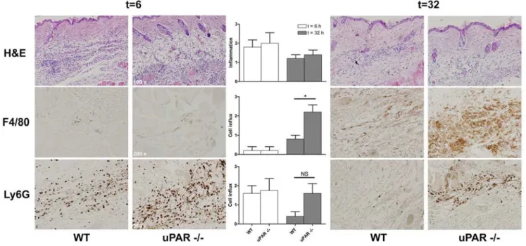
To assess the role of uPAR in the immune response againstB. burgdorferivivo, we infected C57BL/6 WT and uPAR knock-out mice withB. burgdorferiand sacrificed mice two and four weeks post infection. By quantitative PCR we assessedB. burgdorferinumbers in skin, bladder and tibiotarsi post mortem. C57BL/6 uPAR deficient mice harbored higherB. burgdorferinumbers compared to WT animals in all tissues examined. This was most pronounced, and statistically significant, four weeks post infection (Figure 2A). These data were underscored by the fact that two weeks post infection only 3/8 bladder tissue cultures were positive in WT mice versus 7/7 in uPAR knock-out mice (Chi-square p = 0,026). We did not determine B. burgdorferinumbers in cardiac tissue in these experiments since the heart were used in toto for histopathology. In line with higher systemicB. burgdorferinumbers in uPAR deficient mice a significant increase in total IgG against B. burgdorferiover time (Figure 2B), and significantly higher IgG1 antibody levels four week post infection, were observed (Figure 2C). We detected no differences in IgM and IgG2b subclass-levels four weeks post infection (data not shown). To obtain a first insight into the mechanism by which uPAR deficiency could impact pathogen burden after infection withB. burgdorferiwe stimulated leukocytes with viable spirochetes in vitro. We harvested peritoneal macrophages from C57BL/6 WT and uPAR knock-out mice, which we stimulated with viable B. burgdorferi(Cell:Borrelia= 1:50) for 16 hours. We demonstrate that Borreliainduced similar cytokine levels in WT and uPAR deficient macrophages (Figure 2D). We obtained comparable results when we stimulated whole blood in a similar fashion (data not shown). Next, because uPAR has been shown to play a crucial role in phagocytosis of Escherichia coli by neutrophils [19,21,22], we investigated whether WT and uPAR knock-out neutrophils and macrophages differed in their capacity to phagocytose B. burgdorferi. In these assays extracellular bacteria were quenched by addition of a quenching dye containing Trypan blue. We demonstrate that both uPAR knock-out neutrophils (in whole blood) and uPAR knock-out peritoneal macrophages were significantly less capable of phagocytosing B. burgdorferi, using either heat-killed FITC-labeled or viable CFSE-labeled B. burgdorferi (Figure 2E and F and Figure S2). Confocal microscopy confirmed labeled bacteria were localized intracellu-larly (Figure S3A and B). To distinguish between binding and phagocytosis we performed similar experiments, but at 4uC and without the addition of quenching solution. These experiments showed no difference in the capacity of WT and uPAR deficient leukocytes to bindB. burgdorferi(Figure 2G). In addition, binding experiments with recombinant human uPAR and viable B. burgdorferifailed to show direct binding of the spirochete to uPAR (data not shown). Since uPAR has been shown to be of importance in the migration of leukocytes, we also investigated whether there was impaired migration of leukocytes in B. burgdorferi-infected uPAR knock-out mice. We intradermally inoculated C57BL/6 WT and uPAR knock-out mice withB. burgdorferior controls and harvested skin at 0, 6 or 32 hours post infection. We did not observe influx of immune cells at t = 0 (data not shown). By H&E, Ly6G and F4/80 stainings on sagittal skin sections we did observe an evident influx of immune cells and inflammation at t = 6 hours,
Author Summary
Lyme borreliosis is caused by the spirochete Borrelia burgdorferi and is transmitted through ticks. Since its discovery approximately 30 years ago it has become the most important vector-borne disease in the Western world. The pathogenesis of this complex zoonosis is still not entirely understood. We here demonstrate that the urokinase receptor (uPAR) is upregulated in mice and humans upon exposure to B. burgdorferi in vitro and in vivo. Importantly, we describe the function of uPAR in the immune response against the spirochete; using uPAR knock-out mice, we show that uPAR plays an important role in phagocytosis ofB. burgdorferiby leukocytes both in vitro as well as in vivo. In addition, we show that the mechanism by which uPAR is involved in the phagocytosis of B. burgdorferi is independent of ligation to its natural ligand uPA or uPAR’s role in fibrinolysis. Our study contributes to the understanding of the pathogenesis of Lyme borreliosis and might contribute to the development of innovative novel treatment strategies for Lyme borrel-iosis.
however there were no differences between WT and uPAR knock-out mice (Figure 3). As has been shown by others [25], the predominant cells at this early time point were granulocytes (Figure 3). Importantly, these data show that the phenotype in uPAR knock-out mice is not explained by impaired influx of immune cells at the site of inoculation allowing for more dissemination of the spirochete. By contrast, later in the course of infection, at t = 32 hours, we observed a more pronounced influx of macrophages in uPAR knock-out mice compared to WT controls, which probably is explained by the increased Borrelia burden in uPAR knock-out mice (Figure 3). In conclusion, higher B. burgdorferi numbers in C57BL/6 uPAR knock-out mice compared to WT mice could be explained by a decreased phagocytotic capacity of uPAR deficient leukocytes observed
in vitro, but not by impaired migration of uPAR deficient leukocytes.
HigherB. burgdorferi numbers and impaired
phagocytotic capacity in C57BL/6 uPAR knock-out mice are independent of ligation of uPA to uPAR
Since uPAR has been suggested to affect function of leukocytes in both an uPA-dependent as well as an uPA-independent fashion we also assessed the course ofB. burgdorferiinfection in C57BL/6 uPA knock-out mice. Both 2 and 4 weeks post B. burgdorferi infection, C57BL/6 WT and uPA deficient mice displayed similar Borrelianumbers in all tissues examined as detected by quantitative PCR (Figure 4A). In addition, compared to WT controls, uPA
Figure 1.Borrelia burgdorferiinduces upregulation of the urokinase receptor on murine and human leukocytes in vitro and in vivo.
(A) ViableB. burgdorferiinduces uPAR expression on murine and human macrophages. Murine peritoneal macrophages, and ex vivo generated human macrophages, (16105) were stimulated with viableBorrelia burgdorferi(strain B31) for 16 hours (Cell:B. burgdorferi= 1:10 or 1:100). Cells were
harvested and analyzed for uPAR expression by FACS analysis. (B) ViableB. burgdorferiinduces uPAR expression on murine and human granulocytes and monocytes. Murine and human whole blood was incubated with viableB. burgdorferifor 16 hours. Erythrocytes were lysed and cells were co-stained for granulocytes or monocytes markers and uPAR and analyzed by FACS analysis. (C) Expression of uPAR is increased in skin biopsies from Lyme borreliosis patients. Total RNA was isolated from biopsies derived from culture and PCR confirmedB. burgdorferipositive erythema migrans lesions from Lyme borreliosis patients (n = 5) or healthy controls (n = 5) and subjected to quantitative uPAR andb-actin RT-PCR. We could not detect uPAR mRNA in healthy controls, for these samples the level of uPAR mRNA was set at the detection limit. Expression of uPAR mRNA was corrected for b-actin mRNA expression and depicted as a relative number. Expression of uPAR of one of the healthy controls was set at 1. Graphs in panels (A and B) are representative of at least three independent experiments and error bars represent the mean of triplicates within one experiment6SEM. Ap -value,0.05 was considered statistically significant. * indicatingp,0,05; **p,0,01 and ***p,0,001.
deficient neutrophils and peritoneal macrophages were equally capable of phagocytosing B. burgdorferi (Figure 4B). These data suggest that the phenotype observed in C57BL/6 uPAR knock-out mice was independent of ligation of uPA to uPAR.
HigherB. burgdorferinumbers and impaired
phagocytotic capacity in C57BL/6 uPAR knock-out mice are independent of uPAR’s role in the fibrinolytic system
Next, since uPAR has been shown to affect function of leukocytes through its role in the fibrinolytic system [14], we infected mice in which the activity of the fibrinolytic system was
either impaired, i.e. C57BL/6 tPA deficient mice, or enhanced, i.e. C57BL/6 PAI-1 knock-out mice. First we demonstrated thatB. burgdorferiinfection did not influence fibrinolytic activity in citrate plasma in either mouse strain, or WT controls, as measured by amidolytic plasminogen activator activity assays (Table 1). Next, we showed that, compared to C57BL/6 WT mice, both C57BL/6 tPA and as PAI-1 knock-out mice display normalBorrelianumbers in various tissues two weeks (Table 1) and four weeks (data not shown) post infection, as detected by quantitative PCR and tissue culture (data not shown). In line with these data, phagocytotic capacity of C57BL/6 tPA and PAI-1 deficient neutrophils was comparable to that of WT mice (Table 1). Importantly, uPAR
Figure 3. Leukocyte migration in uPAR knock-out mice in response toB. burgdorferiinfection in vivo.C57BL/6 WT and uPAR knock-out mice were intradermally injected with 16106B. burgdorferiin PBS in the midline of the neck and mice were sacrificed 6 or 32 hours post inoculation.
Skin was harvested, formalin fixed and imbedded in paraffin. Fivemm-thick sagittal skin sections were processed and H&E, Ly6G and F4/80 stained by routine histological techniques. Control animals injected with PBS alone did not display influx of leukocytes (data not shown). Slides were scored for influx of leukocytes by an independent pathologist who was blinded to the experimental design. Influx was semi-quantitatively scored on a scale from 0–3, with 0 being no, 1 mild, 2 moderate, and 3 being severe diffuse infiltration. Per group and time point 5 five mice were used, error bars represent SEM. Representative sections are depicted in the figure. Ap-value,0.05 was considered statistically significant. * indicatingp,0,05, i.e. p = 0,0259; NS, not significant, i.e. p = 0,1475.
doi:10.1371/journal.ppat.1000447.g003
Figure 2. The urokinase receptor (uPAR) is involved in clearance ofB. burgdorferi.(A) Urokinase receptor knock-out C57BL/6 mice display higher systemicB. burgdorferinumbers. WT and uPAR2/2mice were inoculated withB. burgdorferiand sacrificed two and four weeks post infection. DNA was extracted from the indicated tissues and subjected to quantitativeBorrelia flaband mouseb-actinPCR. In sham inoculated mice (2 to 3 per group) we did not detectB. burgdorferiDNA. Six to eight mice per group were used and bars represent the mean6SEM. (B and C) Urokinase receptor knock-out C57BL/6 mice develop more rigorous IgG responses. Sera from C57BL/6 WT and uPAR knock-out mice, 2 and 4 weeks postB. burgdorferi(B burg) or sham inoculation (SHAM) was used for whole cellB. burgdorferiELISA. Thus, we determined total IgG directed againstB. burgdorferi(B) and IgG subclasses, of which only IgG1 (C) is shown. (D) WT and uPAR2/2macrophages produce similar levels of pro-inflammatory cytokines when exposed to viableB. burgdorferiin vitro. Peritoneal macrophages were stimulated with control medium (medium) orB. burgdorferi (B burg) for 16 hours. The supernatant was analyzed for cytokine production using a mouse inflammation cytometric bead array. (E and F) Urokinase receptor deficient granulocytes and macrophages are incapable of adequately phagocytosingB. burgdorferi. Whole blood or peritoneal macrophages were incubated with CFSE-labeled viable or heat-killed FITC-labeledB. burgdorferiat 37uC or at 4uC as a control. Phagocytosis was stopped by transferring the tubes to ice and extracellular bacteria were quenched by addition of a quenching dye containing Trypan blue. When whole blood was used erythrocytes were lysed before cells were DAPI stained and subjected to fluorescent microscopy (E) or stained for Gr-1 (granulocytes) and subjected to FACS analysis (F; left panel). Peritoneal macrophages were directly subjected to FACS analysis (F; right panel). Phagocytosis was depicted as the phagocytosis index [64,65]: mean fluorescence intensity (MFI)6percentage (%) positive cells) at 37uC minus (MFI6% positive cells at 4uC). Six to eight mice per group were used, graphs represent the mean6SEM and are representative of three independent experiments. (G)B. burgdorferibinds equally well to WT and uPAR2/2macrophages. A similar experiment as described in (F) was performed, albeit at 4uC and without the addition of quenching dye to determine binding ofB. burgdorferito peritoneal macrophages. Binding is expressed as the binding index: % CFSE positive cells6MFI. Four to six mice per group were used and bars represent the mean6SEM. The experiment was repeated twice. Ap-value,0.05 was considered statistically significant. * indicatingp,0,05; **p,0,01 and ***p,0,001.
knock-out mice, regardless whether they were infected with B. burgdorferi, have comparable fibrinolytic activity to WT mice (data not shown). Together these data indicate that the impaired phagocytotic capacity of uPAR deficient mice, resulting in higher spirochete numbers upon B. burgdorferi infection in vivo, is not dependent on the role of uPAR in fibrinolysis.
The effect of uPAR deficiency on the development of Lyme borreliosis
We assessed carditis severity inB. burgdorferiinoculated C57BL/ 6 uPAR knock-out and WT mice two and four weeks post infection. Two weeks post infection, in hematoxylin and eosin (H&E) stained sagittal sections of mouse hearts, we found comparable carditis severity scores in C57BL/6 WT and uPAR knock-out mice (Figure S4AandB). The localization and severity of carditis in our experiments using C57BL/6 mice appeared to be similar to the localization and carditis severities reported by ourselves and others using the same, relatively resistant, mouse strain [26–29]. Sham inoculated mice did not develop carditis (data not shown). We were unable to reliably score carditis four weeks post infection, since, as observed by others, at this stage, carditis was characterized by an organizing rather than ongoing inflammation (Figure S4A) [26]. However, in 4/8 uPAR deficient mice and 0/8 WT mice a mild active carditis, characterized by the presence of small cellular infiltrates at the aortic root, could still be observed 4 weeks post inoculation (Chi-square p = 0,021) (data not shown). By contrast, in 5/8 of WT mice and only in 2/8 uPAR deficient mice we observed organized inflammatory infiltrates, characterized by sharply delineated foci (Figure S4A) of
mononuclear leukocytes situated in the atrial wall (Chi-square p = 0,0721). Together these findings suggest a difference with respect to the kinetics of the organization of carditis in C57BL/6 uPAR knock-out and WT mice. In line with the observed normal Borrelianumbers, in uPA, tPA and PAI-1 knock-out mice severity of carditis was comparable to that in WT mice (Figure S4Cand D). Together these data demonstrate that, despite higher B. burgdorferi numbers, C57BL/6 uPAR knock-out mice develop carditis with a similar severity, albeit for a prolonged period of time, compared to WT controls. Finally, although we observed ankle swelling in both WT and uPAR C57BL/6 knock-out mice during the course of infection, histological examination of H&E stained section of tibiotarsi did not reveal any signs of arthritis 2, 4 or 6 weeks post infection (data not shown).
The course ofB. burgdorferi infection in uPAR deficient mice on aB. burgdorferisusceptible genetic background
To further investigate the effect of uPAR deficiency on the development of Lyme borreliosis symptoms we generated uPAR deficient mice on a moreBorreliasusceptible genetic background. It is well-known that C57BL/6 mice are relatively resistant to B. burgdorferiand develop less severe symptoms after infection with the spirochete, and that C3H/HeN mice are more susceptible and develop more severe symptoms after infection withB. burgdorferi [30]. In addition, it has been described that F1 of WT C57BL/6 crossed with (x) C3H/HeN mice are intermediately sensitive toB. burgdorferiinfection [30]. Therefore we investigated the course of Lyme borreliosis in F2 of C57BL/66C3H/HeN uPAR knock-out
mice and WT littermate controls. We first showed that, similar to
Figure 4. The urokinase activator (uPA) is not involved in clearance of the spirochete.(A) Urokinase activator knock-out C57BL/6 mice display similar systemicB. burgdorferinumbers compared to WT controls. WT and uPA2/2mice were inoculated withB. burgdorferiand sacrificed two and four weeks post infection. DNA was extracted from the indicated tissues and subjected to quantitativeBorrelia flaband mouseb-actinPCR. Six to eight mice per group were used andB. burgdorferi numbers are depicted as described inFigure 2A. (B) Urokinase activator deficient granulocytes and macrophages (solid lines) are just as capable as WT controls (dotted lines) of phagocytosingB. burgdorferi. Phagocytosis assays were performed as described inFigure 2F. Six to eight mice per group were used, error bars represent SEM and the graphs are representative of two independent experiments. Ap-value,0,05 was considered statistically significant.
doi:10.1371/journal.ppat.1000447.g004
uPAR knock-out mice on a pure C57BL/6 background, these mice harbor higherBorrelianumbers in multiple tissues compared to WT littermate controls two weeks post infection (Figure 5A), indicating that the lack of uPAR in these mice also resulted in impaired phagocytosis and increased pathogen burden. Indeed, in in vitro phagocytosis assays, compared to WT littermate controls, C57BL/66C3H/HeN uPAR deficient neutrophils were
signifi-cantly less capable of phagocytosing B. burgdorferi (Figure 5B). Strikingly, compared to WT littermate controls (Figure 5C), C57BL/66C3H/HeN uPAR knock-out mice developed
signifi-cantly more severe carditis (Figure 5D), reflected by influx of greater numbers of leukocytes in more and larger parts of cardiac tissue two weeks post infection (Figure 5E). As has been shown by others the main cells involved in inflammation were macrophages, as determined by F4/80 immunostaining (Figure 5FandG). By multiplex ligation-dependent probe amplification (MLPA), we detected significantly increased levels of interleukin (IL)-1b, IL-1
Receptor Associated Kinase (IRAK)-3, and toll-like receptor (TLR)2 mRNA in hearts from uPAR knock-out mice compared to WT littermate controls two weeks post infection (Figure 5H), consistent with the observed higher B. burgdorferi numbers and more severe cardiac inflammation in uPAR knock-out mice. Since, uPAR has also been shown to enhance migration of leukocytes towards the site of infection for some, but not all bacteria, in these mice we performed in vitro migration assays with WT and uPAR deficient macrophages (Figure S5A). We observed impaired migration of uPAR deficient macrophages to C5a (Figure S5B), however not to supernatant from a cardiomyoblastic rodent cell line (Figure S5C), compared to migration of WT macrophages, which is in line with our in vivo observations in C57BL/6 mice, In this in vitro setting, whether or not this cell line was stimulated with viableB. burgdorferidid not affect migration of WT and uPAR deficient macrophages, which might be due to production of both stimulating and inhibitory chemotactic stimuli of these cardio-myoblastic cells upon exposure to B. burgdorferi, as has been recently shown for neutrophils [31]. WT and uPAR deficient C57BL/66C3H/HeN mice developed comparable ankle swelling
during the course ofB. burgdorferiinfection (Figure S5D), however despite the more susceptible phenotype of these mice compared to C57BL/6 mice, these mice did not develop any histological signs of arthritis, as determined by hematoxylin and eosin staining, but also Ly6G - a marker for granulocytes - immunostaining (data not shown). In line with these data, post mortem radiological examination of the hind limbs did not reveal any signs of arthritis (Figure S5E).
Discussion
Since its discovery approximately 30 years ago Lyme borreliosis has become the most important vector-borne disease in the Western world. We here demonstrate, to our knowledge for the first time, that uPAR plays an important role in the antibacterial innate immune response againstB. burgdorferi. We show that uPAR expression is upregulated in response toB. burgdorferi on human and murine leukocytes both in vitro, as well in vivo. Importantly, we describe the role of uPAR in the immune response againstB. burgdorferi. By using C57BL/6 WT and uPAR knock-out mice we show that uPAR plays an important role in phagocytosis of B. burgdorferi- a prerequisite for the eradication of the spirochete - by leukocytes. Moreover, experiments with C57BL/6 uPA, tPA and PAI-1 knock-out mice show that the mechanism by which uPAR is involved in the phagocytosis of B. burgdorferi is independent of ligation to uPA or uPAR’s role in fibrinolysis. Finally, we show that, in mice relatively susceptible toBorreliainfection - mice on a mixed C57BL/6 and C3H/HeN background - uPAR deficiency also impaired phagocytotic capacity in vitro, which was associated with higherB. burgdorferi numbers, more local inflammation and more severe carditis, compared to WT littermate control animals, further underscoring the in vivo relevance of our findings. Together these data demonstrate an important role for uPAR in the innate immune response against, and the clearance of, the causative agent of Lyme borreliosis.
Earlier studies documented that membrane bound uPAR and uPAR mRNA are upregulated in human peripheral blood-derived monocytes and the human monocyte-like cell line U937 upon exposure to viable and heat-killedB. burgdorferi[12,13]. We here show that viable B. burgdorferi induces upregulation uPAR (Figure 1 and Figure S1), not only on murine and human monocytes, but also on macrophages and granulocytes in vitro. Notably, uPAR expression in response toB. burgdorferiin vivo has never been investigated. We here show that in skin from Lyme Table 1.B. burgdorferiinfection in WT, tPA2/2and PAI-12/
2mice.
PA activity (in %)#
SHAM B. burgdorferi
WT 91,261,4 89,660,8
tPA2/2 10,760,9a 9,9
62,1c
PAI-12/2 132,764,6b 141,7 63,9d Pathogen numbers##
(Borrelia FlaB
copies/16106b-actincopies)
ankle
WT - 12926473
tPA2/2 - 6366366
PAI-12/2 - 5606149
skin
WT - 8726290
tPA2/2 - 7246301
PAI-12/2 - 14636488
bladder
WT - 5786173
tPA2/2 - 9676310
PAI-12/2 - 3676125
Phagocytosis index$(% pos * MFI)
WT ND 4101565826
tPA2/2 ND 3982863350
PAI-12/2 ND 4992862752
Note.C57BL/6 WT, tPA and PAI-1 knock-out mice (6–8 per group) were inoculated withB. burgdorferistrain B31 or sham and sacrificed two weeks later.
#Plasminogen activator (PA) activity was measured in citrate plasma using
amidolytic assays and expressed as a percentage.
##
B. burgdorferinumbers were determined by quantitative PCR and expressed as described inFigures 2and3.
$In addition, an in vitro phagocytosis assay was performed using naive mice
(n = 6–8 per group) as described inFigure 2. Whole blood was incubated with viable CFSE-labeledB. burgdorferifor 60 minutes at 37 or 4uC as a control and phagocytosis was depicted as the phagocytosis index as described inFigure 2.
a,cPA activity was significantly lower in tPA knock-out mice compared to WT
controls, regardless whether mice were inoculated withB. burgdorferior sham,p,0,0001.
b,dPA activity was significantly higher in PAI-1 knock-out mice compared to WT
controls, regardless whether mice were inoculated withB. burgdorferior sham,p,0,0001.
Results represent the mean6SEM. Non-parametric statistical tests were used to analyze the differences between the groups. Ap-value,0,05 was considered statistically significant.
Figure 5. The course of Lyme borreliosis in uPAR knock-out mice on aB. burgdorferisusceptible mixed C57BL/66C3H/HeN genetic
background.(A) Urokinase receptor deficient mice on the mixed genetic background also display higherB. burgdorferinumbers compared to WT littermate controls. C57BL/6 mice were backcrossed twice to a C3H/HeN background. We intercrossed F2 mice and used the homozygous and nullizygous offspring (F2 homozygous uPAR deficient C57BL/66C3H/HeN mice and WT littermate controls) for our experiments. Mice were inoculated
borreliosis patients with erythema migrans uPAR mRNA expression is significantly increased and could be readily detected by quantitative RT-PCR (Figure 1). Increased levels of uPAR are likely to be caused by influx of leukocytes to the site of the tick-bite. Indeed, in preliminary experiments in which we inoculated human skin ex vivo with viableB. burgdorferi- a model in which there is no influx of leukocytes [32] - we did not observe an increase in uPAR expression as determined by uPAR immunostaining on snap frozen sagittal skin sections (data not shown). Erythema migrans lesions are characterized by perivascular infiltrates in the dermis composed primarily of lymphocytes and macrophages [33]. We do not know which infiltrating cell type is responsible for the elevated uPAR levels, but based on our in vitro data we speculate that the macrophage is the most likely candidate. Indeed, macrophages from intraperitoneallyB. burgdorferi-inoculated WT C57BL/6 mice did upregulate uPAR expression, further indicating that Borrelia -phagocyte interaction in vivo results in induction of uPAR expression (Figure S1). Upregulation of uPAR appeared not to be specific forB. burgdorferisince, in our in vitro experiments, other bacteria, i.e.Klebsiella pneumoniaeand Burkholderia pseudomallei, also induce upregulation of uPAR to a similar extent (data not shown). To investigate the role of uPAR in the immune response against B. burgdorferi and the course of murine Lyme borreliosis we inoculated C57BL/6 WT and uPAR knock-out mice with B. burgdorferi. We demonstrate by quantitative PCR and culture that mice lacking uPAR display significantly increased B. burgdorferi numbers in all tissue examined, indicative of a more disseminated infection (Figure 2), although also in these mice there appeared to be clearance of B. burgdorferi, as suggested by lower numbers 4 weeks compared to 2 weeks post infection. The increased B. burgdorferi burden in uPAR deficient mice was underscored by a more abundant, putatively reactive, IgG response (Figure 2). The role of uPAR in leukocyte adhesion and migration, leading to recruitment of these cells to the site of infection, has been the topic of investigations for many years. Several in vivo studies show that migration of uPAR deficient leukocytes is impaired in response to, for example,Pseudomonas aeruginosum [22] andStreptococcus pneumo-niae[23]. In other studies, e.g. in E. coli-induced peritonitis [20] and pyelonephritis [21] uPAR deficiency did not affect leukocyte recruitment, indicating that the role of uPAR in migration of leukocytes is dependent on the pathogen, the site of infection and the disease model. Interestingly, in the mouse model for Lyme borreliosis uPAR is not crucially involved in migration of leukocytes to B. burgdorferiinfected tissues, as indicated in our in vivo migration experiments (Figure 3). Strikingly, the fact that we observed more macrophages 32 hours after injection with B. burgdorferiin uPAR knock-out skin compared to WT controls, but no differences in H&E staining, suggests that the quality of the inflammatory infiltrate is affected rather than the quantity; presumably due to higher B. burgdorferi numbers in the uPAR knock-out mice. Interestingly, recently it was shown that uPAR also facilitates phagocytosis of the gram-negative bacteriumE. coli by neutrophils [19,21]. We here show, by fluorescent microscopic
assays, and FACS-based phagocytosis assays, that both uPAR deficient granulocytes and macrophages are significantly less capable of phagocytosing viable spirochetes (Figure 2 and Figure S2andS3). Importantly, uPAR deficiency did not affect binding of the spirochete to the surface of leukocytes (Figure 2). In addition, in an in vitro killing assay uPAR appeared not to be involved in killing of the spirochete following phagocytosis (data not shown), indicating that uPAR is involved strictly in the process of internalization of B. burgdorferi by leukocytes. Others have previously shown that phagocytosis of spirochetes by immune cells can be crucial for adequate cytokine induction and leukocyte activation [34–36]. We did not observe defects in pro-inflamma-tory cytokine production in uPAR deficient leukocytes when stimulated in vitro with B. burgdorferi. In contrast to the studies described above our results describe more subtle differences in phagocytotic capacity between WT and uPAR deficient leuko-cytes; we demonstrate diminished, but not absent, phagocytosis in uPAR deficient macrophages compared to WT controls.
The role of uPAR in phagocytosis ofB. burgdorferiappeared to be independent of uPA and uPAR’s role in the fibrinolytic system, since in our phagocytosis assays uPA, tPA and PAI-1 knock-out mice all displayed normal phagocytotic capacity of the spirochete compared to WT mice (Figure 3andTable 1). In addition, in vivo experiments clearly show that when these mice were inoculated with B. burgdorferi and sacrificed two weeks post infection, normalB. burgdorferinumbers were detected (Figure 3 andTable 1). There are numerous in vitro studies reporting that B. burgdorferiinteracts with the fibrinolytic system (reviewed in [6]). Extrapolating these data to the in vivo situation, this interaction, mainly through binding to host derived plasminogen, was thought to enable the spirochete to penetrate tissues, the blood-brain barrier and migrate through the extracellular matrix [8,37–39]. Indeed, for the spirochetal causative agent of relapsing fever, using plasminogen knock-out mice, it has been clearly shown that plasminogen is required for dissemination of the spirochete to the heart and brain in vivo [40]. To our knowledge, forB. burgdorferi however, there is only one previously published study that describes the effect of diminished fibrinolytic activity on the course ofB. burgdorferiinfection in vivo [7]. In this study, in which plasminogen deficient mice were used, plasminogen was shown to be important for dissemination of the spirochete within the feeding tick. Strikingly, despite a short-lived spirochetemia, there were no differences inB. burgdorferinumbers in any of the tissues examined in plasminogen knock-out compared to WT mice at several time points post infection [7]. In line with these data, our results demonstrate that the fibrinolytic system per se does not affect the course ofB. burgdorferiinfection. Strikingly, we here show that one of the key players in the fibrinolytic system, uPAR, independently of ligation to uPA or its presumptive role in fibrinolysis, is importantly involved in the course of experimental murine Lyme borreliosis.
The fact that we show that the requirement of uPAR in phagocytosis ofB. burgdorferiis independent of uPA or uPAR’s role
group were used. (C, D and E) Peak carditis in these uPAR2/2mice (D) is more severe compared to carditis in WT littermate controls (C). Mice were inoculated with B. burgdorferi and sacrificed two weeks post infection. Pictures of hematoxylin and eosin stained sagittal sections depict representative sections. Carditis was scored as described inFigure 4within the same session (E). Six to eight mice per group were used. (F and G) The main cell involved in murine Lyme carditis is the macrophage. Representative pictures of F4/80 stained sagittal sections of hearts fromB. burgdorferiinfected uPAR deficient mice (G) and WT littermate controls (F). (H) More severe inflammation inB. burgdorferiinfected uPAR deficient animals (n = 7) compared to WT littermate controls (n = 7) as measured by multiplex ligation-dependent probe amplification (MPLA). MLPA was performed on RNA obtained from half of sagittally dissected hearts fromB. burgdorferior sham inoculated mice. Depicted are mRNA expression of TNF-a,CCL3,TLR2,CD14,IL1-b,IRAK3,ICAM1andTBP(housekeeping gene[66]). Other genes included in the assay wereIL6,IL10,INF-c,TFPI,F3,PROCR, SERPINE1P, PLAT, PLAUR, TLR4, TLR9 LY96, IRAK1, F2R, NFKB1a, NOS3,ITGA5,B2M,ITGAV,ITGAB3, TFRC, HIF1A, MMP2andHP. Bars represent the mean6SEM. Ap-value,0.05 was considered statistically significant. * indicatingp,0,05; **p,0,01.
in the fibrinolytic system suggests that the requirement of uPAR in internalization ofBorreliais dependent on interaction of uPAR with other cell surface molecules. Indeed, uPAR has been shown to facilitate various leukocyte functions, among which adhesion, migration and phagocytosis through interaction withab-integrins and other cell surface molecules, but also vitronectin [14,15]. This implies a role for uPAR as a signaling receptor. However, because uPAR is a glycosyl-phosphatidylinositol linked receptor and lacks a cytosolic domain it needs to form functional transmembrane units with other molecules, such as multiple ab-integrins, G-protein-coupled receptors, and caveolin in order to induce intracellular signaling events leading to cytoskeleton rearrangements and consequent cell movement [14,15]. Since both uPAR and B. burgdorferishare many molecules with which they can interact, for example ab-integrins and vitronectin, it will be challenging to identify the surface molecule with which uPAR associates to facilitate phagocytosis ofB. burgdorferi.
When we infected uPAR knock-out mice on a mixed C57BL/6 and C3H/HeN background withB. burgdorferithese mice exhibited higher B. burgdorferi numbers in cardiac tissue two weeks post infection compared to WT littermate controls, which was also associated with decreased phagocytosis ofB. burgdorferi. Strikingly, in these mice we observed a significantly increased influx of leukocytes, predominantly macrophages, at the atrioventricular junction and at the aortic root compared to WT littermate controls (Figure 5), further indicating that uPAR is not required for migration of leukocytes in response toB. burgdorferi, which was also underscored by the in vitro migration assays (Figure S5). Furthermore, our data indicate that, although the underlying mechanisms appeared to be the same, the consequences of uPAR deficiency for the course of murine Lyme borreliosis are dependent on the genetic background of the host. Others have shown that C57BL/6 and C3H/HeN mice harbor similar B. burgdorferi numbers after infection, but the severity of symptoms was more pronounced in C3H/HeN mice [30], indicating that the extent of the immune response that is mounted against the spirochete is dependent on the genetic background of the host. Indeed, we have demonstrated that uPAR deficiency inBorreliaresistant C57BL/6 mice leads to higherB. burgdorferiloads, but to comparable, albeit longer-lived active carditis compared to WT controls. By contrast, uPAR deficient mice on a more susceptible mixed C57BL/ 66C3H/HeN background also exhibited higher B. burgdorferi numbers, but more pronounced influx of leukocytes and more severe carditis. Local cytokines and chemokines induced by B. burgdorferiare thought to mediate Lyme carditis. A cytokine that has been implicated to be of paramount importance for local inflammation and migration of leukocytes is IL-1b[41–43]. Also inB. burgdorferiinfected mice and patients IL-1bhas been shown to be upregulated in heart or joints [44–47] Interestingly, by MLPA we found significantly higher levels of mRNA coding for IL-1b, IL-1 receptor associated kinase (IRAK)-3 (predominantly ex-pressed in macrophages) and TLR2 (the TLR preferentially recognizingB. burgdorferilipoproteins) in hearts fromB. burgdorferi infected uPAR knock-out mice on the mixed genetic background compared to WT littermate controls (Figure 5). Interestingly, in previous studies we showed that (human) peripheral blood-derived dendritic cells stimulated with the TLR2 ligand lipoteichoic acid (LTA) or viableB. burgdorferiproduced high levels of IL-1b [48]. One could argue against the use of F2 mice in our studies, however the fact that F1 WT C57BL/66WT C3H/HeN mice are
already intermediate susceptible toBorreliainfection [30], encour-aged us to perform our experiments with F2 mice.
Together, our data suggest that decreased phagocytosis of B. burgdorferiby uPAR deficient leukocytes (Figure 2,Figure S2and
Figure 5) resulted in higher local and systemic B. burgdorferi numbers, apparent in later stages post inoculation (Figure 2and 5). Early in infection increased Borrelia numbers might enhance local skin innate immune responses in uPAR knock-out mice resulting in significantly more influx of phagocytes, as shown by in vivo and in vitro migration assays (Figure 3andFigure S5). The increased influx of phagocytes might temporarily compensate for the impaired phagocytosis in uPAR deficient leukocytes and temporarily control dissemination ofBorrelia. However, as shown by our in vivo data, in uPAR knock-out mice,B. burgdorferiwill eventually manage to disseminate resulting in increased B. burgdorferinumbers in distant organs during later stages of infection compared to WT animals (Figure 2and5). In uPAR knock-out mice on the more susceptible genetic background, these higher Borrelia numbers were associated with an increased influx of leukocytes, as demonstrated by pathology of mouse hearts (Figure 5). Since, uPAR deficient leukocytes are as capable as WT leukocytes in producing pro-inflammatory cytokines upon exposure toB. burgdorferi(Figure 2), this could explain the increased inflammation and tissue damage observed in these mice compared to WT controls (Figure 5). Therefore, we postulate that in WT mice, upon B. burgdorferi infection, leukocytes upregulate uPAR (Figure 1 and Figure S1), which facilitates phagocytosis of the spirochete, reducing the number of disseminating spirochetes and thereby limiting the extent and severity of inflammation of distant sites, such as the heart. In conclusion, we here show that uPAR is importantly involved in the host defense againstB. burgdorferi in vivo by a mechanism that is independent of binding of uPAR to uPA or its role in the fibrinolytic system.
Materials and Methods
Mice, spirochetes and infection
Specific pathogen-free wildtype C57BL/6 mice were purchased from Harlan Sprague Dawley Inc. (Horst, The Netherlands) and uPAR knock-out C57BL/6 mice were purchased from Jackson Laboratories (Bar Harbor, ME) [49]. In addition C57BL/6 uPAR knockout mice were backcrossed twice to a C3H/HeN -purchased from Jackson Laboratories – background, generating F2 C57BL/66C3H/HeN heterozygous uPAR deficient mice. F2
mice were crossed among each other to generate homozygous C3H/HeN6C57BL/6 uPAR knock-out mice and WT littermate controls. uPA, tPA and PAI-1 knock-out mice were also purchased from Jackson Laboratories. All mice were bred in the animal facility of the Academic Medical Center (Amsterdam, The Netherlands). Age-and sex-matched animals were used in each experiment and the Animal Care and Use Committee of the University of Amsterdam approved all experiments. Six to eight-week old mice were infected by intradermal syringe inoculation with 16106B. burgdorferisensu stricto strain B31 clone 5A11 [50], that had previously been recovered from an experimentally infected mouse [29]. Spirochetes were cultured in BSK-II medium, enumerated and inoculated in the midline of the back or with BSK-II medium as a control (SHAM), as described previously [29,51]. Mice were sacrificed by bleeding from the inferior vena cava at the indicated time points, i.e. 2, 4 (or 6 weeks) post infection. Heparin or citrate plasma was stored at220uC for future use. Skin (inoculation site), urinary bladder, heart and tibiotarsi were saved for histopathological examination, culture or quantitative Polymerase Chain Reaction (q-PCR).
Q-PCR
DNA from murine tissues was obtained with the DNeasy KIT (Qiagen, Venlo, The Netherlands) as previously described [29].
Quantitative PCR detecting Borrelia flaB and mouse b-actin was performed, as described previously [29]. Standards consisted of dilutions of genomic DNA from B. burgdorferi or mouse ß-actin (252 bp) cloned into the PCR2.1-TOPO vector (Invitrogen, Breda, The Netherlands), as described previously [29,51]
Arthritis, paw swelling and radiological examination Histopathological changes in tibiotarsi were assessed as previously described [29,52]. We monitored ankle swelling of both tibiotarsal joints using a Mitutoyo pressure controlled microcaliper (Mitutoyo, Kanagawa, Japan). Measurements were performed several times throughout the course of the infection by the same observer blinded to the experimental design. Lastly, we performed post mortem radiological examination of formalin fixed right hind paws, as described previously [53].
Carditis
Fivemm-thick paraffin embedded sections of sagittally dissected hearts were processed and H&E stained by routine histological techniques. Carditis was scored on a scale from 0 to 3 by a pathologist blinded to the experimental design, essentially as previously described [27,28,51], with 0: no carditis; 1: mild carditis; 2: moderate carditis and 3: severe carditis. As described previously [26], 2 weeks post infection, carditis was characterized by disperse inflammation at the atrioventricular junction and aortic root, where as four weeks post infection, organizing inflammation was characterized by the presence of sharply delineated foci of.50 mononuclear cells in the atrial walls. An F4/80 immunostaining (BMA Biomedicals, Augst, Switzerland) was performed to detect influx of macrophages [54].
Multiplex ligation-dependent probe amplification MLPA was performed in essence as described before [55]. The genes that were analyzed are listed in the figure legend forFigure S3. Equal amounts of mRNA were included per reaction and all samples were tested in a single experiment using the same batch of reagents. The levels of mRNA for each gene were expressed as a normalized ratio of the peak area of the fluorescent intensity (in arbitrary units) and divided by the cumulative peak area of all genes in the assay, resulting in the relative abundances of mRNAs of the genes of interest [56].
Whole cellB. burgdorferiELISA
Borrelia burgdorferisensu stricto strain B31 specific total immuno-globulin (Ig)G and IgG subclasses were measured in heparin plasma from infected animals and controls by ELISA as described previously [29]. All measurements were performed in duplicate.
Amidolytic assays of PA activity
Plasminogen activator (PA) activity was measured as a measure for the activity of the fibrinolytic system using an amidolytic assay as described earlier [23,57]. Briefly, citrate plasma was incubated with S-2251 (Chromogenix, Mo¨lndal, Sweden), plasminogen and cyanogen bromide fragments of fibrinogen (Chromogenix, Mi-lano, Italy). Conversion of plasminogen to plasmin was assessed by subsequent conversion of the chromogenic substrate S-2251 and was detected with a spectrophotometer.
Stimulation assays
Whole blood and peritoneal macrophages from three naive uPAR knock-out or WT mice were harvested as described [58]. Briefly, 16105 adherent macrophages and heparinized whole
blood were stimulated in duplo in 96-well microtiter plates
(Greiner) with 16106 or 16107 viableB. burgdorferisuspended in
Roswell Park Memorial Institute (RPMI) 1640 medium or medium as a negative control for 16 h. Supernatants were collected and stored at 220uC until cytokine production was measured by CBA. For assessment of uPAR expression by fluorescence activated cell sorter (FACS), cells were harvested and stained with murine anti-CD87-Phycoerythrin (PE) (BD Pharmingen, Maarssen, The Netherlands). To assess uPAR expression on specific cells, cells were double-stained with anti-GR1-fluorescein isothiocyanate (FITC) (BD Pharmingen) (granu-locytes) or F4/80-allophycocyanin (APC) (BD Pharmingen) (monocytes and macrophages). In addition, in non-phagocytosing cells, i.e. CD4+
and CD8+
T cells - stained with anti-CD3-APC (BD Pharmingen) and anti-CD4-FITC or anti-CD8-PerCP respectively (BD Pharmingen) - we also assessed uPAR expression by FACS analysis. Similarly, uPAR expression on human cells derived from heparinized whole blood was analyzed with a human biotin-labeled antibody against uPAR (R&D Systems, Minneapo-lis, MN) in combination with streptavidin conjugated to PE; cells were triple-stained with also anti-CD15-APC (BD Pharmingen) (granulocytes) and anti-CD14-Cy-Chrome 5 (Cy5) (BD Pharmi-gen) (monocytes) (BD PharminPharmi-gen). Human macrophages were generated as described previously [59]. Briefly, human peripheral blood derived mononuclear cells were isolated from buffy coats by centrifugation over a Ficoll-Paque gradient. Subsequently, adher-ent monocytes were cultured in X-VIVO medium (BioWhittaker, Walkersville, MD) with 1% heat-inactivated autologous plasma to allow for differentiation to human monocyte-derived macrophages in 7 days. Antibodies were used in concentrations recommended by the manufacturer and FACS analysis was performed using the BD FACScalibur (BD Biosciences, Breda, The Netherlands). Endotoxin concentration in the B. burgdorferi culture media was approximately 1 IU/ml, as determined by a Cambrex QCL LAL assay (Cambrex). We established that the maximal amount of LPS that could have possibly contaminated the finalBorrelia prepara-tion used for the in vitro stimulaprepara-tions - after extensive washing and resuspension in different cell culture media - was insufficient to influence uPAR expression (data not shown). In a separate experiment viable B. burgdorferi (16108) were injected into the
peritoneal cavity of C57BL/6 WT or uPAR knock-out mice for one hour. Hereafter cells were harvested, stained for F4/80, and CD87 (uPAR) expression was measured by FACS analysis.
Phagocytosis assays
Phagocytosis assays were performed in essence as described before [60–62]. Viable B. burgdorferi were labeled with carboxy-fluorescein diacetate succinimidyl ester (CFSE, Invitrogen) as described by others [63] or heat-inactivated (30 min at 56uC) non-motile, but intact, B. burgdorferi were labeled with fluorescein isothiocyanate (FITC). Adhered peritoneal macrophages (derived from 6–8 mice per group) were incubated with CFSE-labeled B. burgdorferi(Cell:Borrelia= 1:50) in serum-free RPMI 1640 medium in 24-well microtiter plates (Greiner, Alphen a/d Rijn, The Netherlands) for 0, 15 and 60 minutes at 37uC. Phagocytosis was stopped by transferring the cells to 4uC. Extracellular signal ofB. burgdorferiwas eliminated by addition of a quenching solution for one minute - containing Trypan blue that absorbs the fluorescence emission of both FITC and CFSE (Orpegen, Groningen, The Netherlands; [62].) - and three washes with ice-cold PBS. For each sample and each time point 4uC controls were performed, however there was hardly any phagocytosis detectable under these conditions (data not shown). Cells were resuspended in FACS buffer (PBS supplemented with 0,5% bovine serum albumin (BSA), 0,01% NaN3 and 0,35 mM EDTA) followed by FACS analysis. At 37uC the majority of spirochetes was internalized as was determined by control experiments in which we did not add the quenching solution (data not shown). Similarly, to determine neutrophil phagocytosis capacity, 50ml of whole blood was incubated with 26106 viable CFSE-labeled B. burgdorferi for the indicated time, after which quenching solution was added for one minute and samples were washed twice with ice-cold FACS buffer. Thereafter cells were incubated with BD Lyse/Fix solution (BD Biosciences) and neutrophils were labeled using anti-Gr-1-PE (BD Pharmingen). Live cells were electronically gated and phagocytosis was determined using FACS. The phagocytosis index of each sample was calculated as previously, described: (mean fluorescence intensity (MFI)6percentage (%) positive cells) at 37uC minus
(MFI6% positive cells) at 4uC [64,65].
Migration assays
In vitro migration experiments with murine peritoneal macro-phages from WT and uPAR knock-out mice were performed essentially as described [64,65]. Prior to experimentation cells were labeled with CellTracker Green (Molecular Probes, Eugene, Or) in serum-free Dulbecco’s modified Eagle’s medium (DMEM). The dye was fixed by 1 h incubation in DMEM plus 10% FCS. Thereafter cells were washed and resuspended in serum-free medium and transferred to 3mM pore size HTS FluoroBlok Cell Culture Inserts (BD Falcon) which were inserted in fitting 24-well plates containing various attractants (B. burgdorferi, activated complement factor 5 (C5a)) also in DMEM serum-free medium. Fluorescence, representing the number of cells on the bottom side of the insert, was read every 2 min on a Series 4000 CytoFluor Multi-Well Plate Reader (Perseptive Biosystems, Framingham, MA). Raw fluorescence data were corrected for background fluorescence and no-attractants controls were subtracted at each measured time point to correct for random migration. Migration start points were set to zero. To mimic the in vivo situation more closely we also performed experiments with an embryonic rodent heart-derived cell line, H9c2 cells (CRL-1446, American Type Culture Collection, Queens Road, Teddington, UK). These cardiomyoblasts were maintained in DMEM with 10% foetal bovine serum (FBS). Prior to experimentation, cells were washed and resuspended in serum-free DMEM and incubated with viable Borrelia(Cell:Borrelia= 1:50) or medium as a control for 16 h. The supernatants were centrifuged for 5 minutes at 12006gto remove
cells and other particles, followed by centrifugation at 40006gfor
15 minute to remove the spirochetes. Supernatants were used undiluted or diluted (data not shown) as chemoattractants in the indicated experiments. All experiments were performed in duplo or in triplo and repeated three times. In addition, we also assessed migration of leukocytes in skin from C57BL/6 WT and uPAR deficient mice in response toB. burgdorferiin vivo (n = 5 per group). In these set of experiments we intradermally injected C57BL/6 WT mice with 16106B. burgdorferi in PBS in the midline of the neck and mice were sacrificed 0, 6 or 32 hours post inoculation. Control animals were injected with PBS. Skin was harvested, formalin fixed and imbedded in paraffin. Five mm-thick sagittal skin sections were processed and H&E, Ly6G and F4/80 stained by routine histological techniques [54]. The control animals did no display influx of leukocytes (data not shown). Slides were scored for influx of leukocytes by an independent pathologist who was blinded to the experimental design. Influx was semi-quantitatively scored on a scale from 0–3, with 0 being no, 1 mild, 2 moderate, and 3 being severe diffuse infiltration.
Statistical analysis
Differences between the groups were analyzed using the two-sided non-parametric Mann-Whitney U test (Graphpad Prism Software version 4.0, San Diego, CA). Where indicated a two-sided Chi-square indicated was applied. Data are presented as the mean6standard errors of the mean (SEM). Apvalue of,0.05 was considered significant, where * indicatedp,0,05, **p,0,01 and *** p,0,001. For ECG data statistical analysis was performed using a multivariate repeated measurements model (SPSS statistics software 17.0).
Supporting Information
Figure S1 Borrelia burgdorferiinduces upregulation of the urokinase receptor on leukocytes in vitro and in vivo. (A) ViableB. burgdorferi induces uPAR expression on ex vivo generated human macrophages. Cells were incubated with viableB. burgdorferifor 16 hours. Thereafter cells were stained with anti-CD87 (uPAR), electronically gated and analyzed by FACS analysis. Representative cytograms and histo-grams are shown. (B) ViableB. burgdorferiinduces uPAR expression on murine granulocytes. Whole blood was incubated with viable B. burgdorferifor 16 hours. Erythrocytes were lysed, cells were co-stained with anti-GR-1 and anti-CD87 (uPAR), electronically gated and analyzed by FACS analysis. Representative cytograms and histo-grams are shown. (C) ViableB. burgdorferi(16108) were injected into
the peritoneal cavity of C57BL/6 WT (n = 6) or uPAR knock-out (n = 4) mice for one hour. Hereafter cells were harvested, stained for F4/80, and CD87 (uPAR) expression was measured by FACS analysis. A p-value,0,05 was considered statistically significant. * indicatingp,0,05; **p,0,01. (D) In non-phagocytosing cells, i.e. CD4+and CD8+T cells - doublestained with anti-CD3-APC (BD
Pharmingen) and anti-CD4-FITC and anti-CD8-PerCP, respectively - we also assessed CD87 (uPAR) expression upon stimulation withB. burgdorferi by FACS analysis. Error bars represent the mean of triplicates within one experiment6SEM.
Found at: doi:10.1371/journal.ppat.1000447.s001 (0.81 MB JPG)
Figure S2 Impaired phagocytosis of B. burgdorferi by uPAR deficient leukocytes. (A and B) Representative cytograms (A) and histograms from phagocytosis assays ofB. burgdorferi by WT and uPAR deficient whole blood in time (B). Assays were performed as described inFigure 2. After the assays whole blood was lysed and stained with anti-GR-1 (granulocytes). Marker (M)1 encompasses positive cells.
Found at: doi:10.1371/journal.ppat.1000447.s002 (0.73 MB JPG)
Figure S3 Confocal microscopy ofB. burgdorferiphagocytosis. (A and B) Confocal microscopy confirmed thatB. burgdorferiin in vitro phagocytosis assays were localized intracellularly. Cells incubated with CFSE-labeled B. burgdorferi were subjected to confocal microscopy. Nuclei of cells were stained with DAPI. In Panel (A) we depicted the widest transversal section of a segmented nucleus of a granulocyte stained with DAPI and a CFSE-labeled B. burgdorferi spirochete. Superimposing the brightfield image con-firms the bacterium is localized intracellularly. Panel (B) shows another granulocyte andB. burgdorferi from different view points (left panel) and a stack movie (right panel) further verifying that we are assessing internalized bacteria in the in vitro phagocytosis assays. Note: The Figure S3 Powerpoint file should be saved in the same folder as the AVI file in order view the figure correctly. In addition, open the Powerpoint file in slideshow format.
Found at: doi:10.1371/journal.ppat.1000447.s003 (0.80 MB ZIP)
Figure S4 Carditis in WT, uPAR, uPA, tPA and PAI-1 knock-out mice. (A and B) Peak carditis in C57BL/6 uPAR2/2is of similar severity compared to WT controls, although active carditis persists longer in uPAR 2/2mice. WT and uPAR2/2mice were inoculated withB. burgdorferiand sacrificed two or four week post infection. Sagittal sections of formalin fixed and paraffin embedded hearts were H&E stained. The severity two weeks post infection was scored by a pathologist blinded to the experimental design on a scale of 0–3, with 0: no carditis; 1: mild carditis; 2: moderate carditis and 3: severe carditis. Sham inoculated mice did not develop carditis (data not shown). Pictures depict representa-tive sections. (C and D) Peak carditis in C57BL/6 uPA, tPA and PAI-1 knock-out mice is comparable to peak carditis in WT C57BL/6 mice infected withB. burgdorferi. Carditis was scored as described above. Six to eight mice per group were used and bars represent the mean6SEM. A p-value,0,05 was considered statistically significant.
Found at: doi:10.1371/journal.ppat.1000447.s004 (1.19 MB JPG)
Figure S5 Migration and arthritis in WT and uPAR knock-out mice on aB. burgdorferisusceptible genetic background. (A, B and C) Urokinase receptor deficient macrophages from mice on the mixed genetic background can migrate to cardiogenic stimuli just as well as macrophages from WT littermate controls.
Migration of CellTracker Green labeled WT or uPAR deficient macrophages towards several chemotactic stimuli was investigated in vitro (A). As chemotactic stimuli we used B. burgdorferi or activated complement factor 5 (C5a) (B) and supernatant from the cardiomyoblastic rodent cell line H9c2 stimulated with B. burgdorferior control medium for 16 hours prior to experimen-tation (C). All conditions were tested in duplo, in serum free DMEM medium without the addition of antibiotics, and migration was corrected for the no-attractant control. Graphs represent the mean of three independent experiments6SEM. The fluorescent signal in the lower chamber (indicative of migration) was measured in real time every two minutes (cycli). (D and E) Only edema, no arthritis inB. burgdorferiinfected uPAR knock-out mice (n = 7) and B. burgdorferi infected WT littermate controls (n = 8). Ankle swelling was measured using a microcaliper during the course of B. burgdorferi infection (D). In this particular experiment mice were monitored for three weeks. Post mortem, but before decalcification, radiological examination of the right hindlimb was performed (E). No differences between sham inoculated andB. burgdorferiinfected animals were observed. Ap -value,0,05 was considered statistically significant. * indicating p,0,05.
Found at: doi:10.1371/journal.ppat.1000447.s005 (0.47 MB JPG)
Acknowledgments
We would like to thank M.I. Cornelissen for the donation of the skin from healthy human individuals, J.B. Daalhuisen, M.S. ten Brink, E.P.M. van der Zanden, N. Claessen, D.W.M. Kruijswijk, J. Pater, W.A. van Dop and A.M. de Boer for excellent technical assistance, H.J.P.P. Eskes for performing radiological examinations, and finally L. Vermeulen for assistance with confocal microscopy.
Author Contributions
Conceived and designed the experiments: JWRH APvD EF TvdP. Performed the experiments: JWRH MFB GJWvdW WJW BJDB JC AO RdB AFdV PW MML JJTHR. Analyzed the data: JWRH BJDB CvV APvD PW EF MML JJTHR TvdP. Contributed reagents/materials/ analysis tools: AO AFdV CvV EF MML JJTHR TvdP. Wrote the paper: JWRH MFB TvdP.
References
1. Burgdorfer W, Barbour AG, Hayes SF, Benach JL, Grunwaldt E, et al. (1982) Lyme disease-a tick-borne spirochetosis? Science 216: 1317–1319.
2. Steere AC (1989) Lyme disease. N Engl J Med 321: 586–596.
3. van Dam AP, Kuiper H, Vos K, Widjojokusumo A, de Jongh BM, et al. (1993) Different genospecies ofBorrelia burgdorferiare associated with distinct clinical manifestations of Lyme borreliosis. Clin Infect Dis 17: 708–717.
4. Steere AC (2001) Lyme disease. N Engl J Med 345: 115–125.
5. Hovius JW, van Dam AP, Fikrig E (2007) Tick-host-pathogen interactions in Lyme borreliosis. Trends Parasitol 23: 434–438.
6. Bergmann S, Hammerschmidt S (2007) Fibrinolysis and host response in bacterial infections. Thromb Haemost 98: 512–520.
7. Coleman JL, Gebbia JA, Piesman J, Degen JL, Bugge TH, et al. (1997) Plasminogen is required for efficient dissemination ofB. burgdorferiin ticks and for enhancement of spirochetemia in mice. Cell 89: 1111–1119.
8. Klempner MS, Noring R, Epstein MP, McCloud B, Hu R, et al. (1995) Binding of human plasminogen and urokinase-type plasminogen activator to the Lyme disease spirochete, Borrelia burgdorferi. J Infect Dis 171: 1258– 1265.
9. Klempner MS, Noring R, Epstein MP, McCloud B, Rogers RA (1996) Binding of human urokinase type plasminogen activator and plasminogen toBorrelia species. J Infect Dis 174: 97–104.
10. Haile WB, Coleman JL, Benach JL (2006) Reciprocal upregulation of urokinase plasminogen activator and its inhibitor, PAI-2, by Borrelia burgdorferi affects bacterial penetration and host-inflammatory response. Cell Microbiol 8: 1349–1360.
11. Perides G, Noring R, Klempner MS (1996) Inhibition ofBorrelia burgdorferi-bound fibrinolytic enzymes by alpha2-antiplasmin, PAI-1 and PAI-2. Biochem Biophys Res Commun 219: 690–695.
12. Coleman JL, Gebbia JA, Benach JL (2001)Borrelia burgdorferiand other bacterial products induce expression and release of the urokinase receptor (CD87). J Immunol 166: 473–480.
13. Coleman JL, Benach JL (2003) The urokinase receptor can be induced by Borrelia burgdorferithrough receptors of the innate immune system. Infect Immun 71: 5556–5564.
14. Mondino A, Blasi F (2004) uPA and uPAR in fibrinolysis, immunity and pathology. Trends Immunol 25: 450–455.
15. Blasi F, Carmeliet P (2002) uPAR: a versatile signalling orchestrator. Nat Rev Mol Cell Biol 3: 932–943.
16. Furlan F, Orlando S, Laudanna C, Resnati M, Basso V, et al. (2004) The soluble D2D3(88-274) fragment of the urokinase receptor inhibits monocyte chemotaxis and integrin-dependent cell adhesion. J Cell Sci 117: 2909–2916.
17. Gyetko MR, Todd RF III, Wilkinson CC, Sitrin RG (1994) The urokinase receptor is required for human monocyte chemotaxis in vitro. J Clin Invest 93: 1380–1387.
18. Gyetko MR, Sitrin RG, Fuller JA, Todd RF III, Petty H, et al. (1995) Function of the urokinase receptor (CD87) in neutrophil chemotaxis. J Leukoc Biol 58: 533–538. 19. Gyetko MR, Aizenberg D, Mayo-Bond L (2004) Urokinase-deficient and urokinase receptor-deficient mice have impaired neutrophil antimicrobial activation in vitro. J Leukoc Biol 76: 648–656.
20. Renckens R, Roelofs JJ, Florquin S, van der Poll T (2006) Urokinase-type plasminogen activator receptor plays a role in neutrophil migration during lipopolysaccharide-induced peritoneal inflammation but not duringEscherichia coli-induced peritonitis. J Infect Dis 193: 522–530.