against
Brucella abortus
Infections
Jerod A. Skyberg, Theresa Thornburg, MaryClare Rollins, Eduardo Huarte, Mark A. Jutila, David W. Pascual*
Department of Immunology and Infectious Diseases, Montana State University, Bozeman, Montana, United States of America
Abstract
cdT cells have been postulated to act as a first line of defense against infectious agents, particularly intracellular pathogens, representing an important link between the innate and adaptive immune responses. HumancdT cells expand in the blood of brucellosis patients and are active againstBrucella in vitro. However, the role ofcdT cellsin vivoduring experimental brucellosis has not been studied. Here we report TCRd2/2mice are more susceptible toB. abortusinfection than C57BL/6 mice at one week post-infection as measured by splenic colonization and splenomegaly. An increase in TCRcdcells was observed in the spleens ofB. abortus-infected C57BL/6 mice, which peaked at two weeks post-infection and occurred concomitantly with diminished brucellae.cdT cells were the major source of IL-17 following infection and also produced IFN-c. Depletion ofcdT cells from C57BL/6, IL-17Ra2/2, and GMCSF2/2mice enhanced susceptibility toB. abortusinfection although this susceptibility was unaltered in the mutant mice; however, whencdT cells were depleted from IFN-c2/2mice, enhanced susceptibility was observed. Neutralization ofcdT cells in the absence of TNF-adid not further impair immunity. In the absence of TNF-aorcdT cells,B. abortus-infected mice showed enhanced IFN-c, suggesting that they augmented production to compensate for the loss of cd T cells and/or TNF-a. While the protective role ofcd T cells was TNF-a -dependent,cdT cells were not the major source of TNF-aand activation ofcdT cells followingB. abortusinfection was
TNF-a-independent. Additionally, bovine TCRcdcells were found to respond rapidly toB. abortusinfection upon co-culture with autologous macrophages and could impair the intramacrophage replication of B. abortus via IFN-c. Collectively, these results demonstratecdT cells are important for early protection toB. abortusinfections.
Citation:Skyberg JA, Thornburg T, Rollins M, Huarte E, Jutila MA, et al. (2011) Murine and BovinecdT Cells Enhance Innate Immunity againstBrucella abortus
Infections. PLoS ONE 6(7): e21978. doi:10.1371/journal.pone.0021978
Editor:Hiranmoy Das, Ohio State University Medical Center, United States of America
ReceivedFebruary 12, 2011;AcceptedJune 14, 2011;PublishedJuly 12, 2011
Copyright:ß2011 Skyberg et al. This is an open-access article distributed under the terms of the Creative Commons Attribution License, which permits unrestricted use, distribution, and reproduction in any medium, provided the original author and source are credited.
Funding:This work is supported by grants from USDA 2007-01612, USDA 2009-34397-20133, Montana Agricultural Station, U.S. Department of Agriculture Formula Funds, National Institutes of Health Grant P20 RR020185, NIH contract HHSN266200400009N01-AI, P01 AT004986, and an equipment grant from the M.J. Murdock Charitable Trust. The funders had no role in study design, data collection and analysis, decision to publish, or preparation of the manuscript.
Competing Interests:The authors have declared that no competing interests exist.
* E-mail: dpascual@montana.edu
Introduction
Brucellosis is a widespread and economically important agricul-tural and human disease in many regions of the world. The etiologic agent of brucellosis in cattle is Brucella abortus, a Gram-negative, facultative, intracellular pathogen [1]. The pathological manifesta-tions of brucellosis are diverse, depending upon host, and in humans include arthritis, endocarditis, and meningitis, while animal brucellosis is characterized by spontaneous abortions [2]. Although use of theB. abortusvaccine strains S19 and RB51 reduces disease incidence and preventsB. abortus-induced abortions, these vaccines are less than ideal because of their limited efficacy and potential to cause disease in humans [3,4]. Consequently, the need exists for improved animal protective measures that will combine safety and efficacy to all species at risk, including domestic herds and wildlife [5]. While much emphasis has been placed on identifying new vaccine candidates, relatively little attention has been paid to the effective coordination of the innate immune response by the host. Moreover, the need exists to determine protective innate immune responses against brucellosis to lessen the progression of infection and to allow a protective, adaptive immune response to develop [6]. cdT cells constitute a small percentage of circulating T cells in adult humans and in mice, but comprise a majority of the
intraepithelial lymphocyte population in the gut and in other epithelial mucosa [7]. Moreover,cdT cells are a major subset in adult ruminants and make up a majority (up to 70%) of circulating lymphocytes in neonatal calves [8]. cd T cells are the first to develop and are recruited and expand in response to various infections in humans, rodents, and other animals [7].
Studies conducted with TCRd2/2mice demonstratecdT cells can play a crucial role in protection from bacterial infection [9–12], and it has been proposed that mice lackingcdT cells have an altered or impaired response to Gram-negative bacteremia [10]. Different mechanisms have been proposed for the protection conferred bycdT cells in murine models. Mooreet al.[10] postulates decreased levels of IFN-c and TNF-a observed in Klebsiella-infected TCRd2/2 mice results in enhanced susceptibility to infection. However, in studies with Nocardia-infected mice, it is shown that TCRd2/2 mice are unable to effectively recruit inflammatory cells, leading to increased susceptibility to infection (and death) [9].cdT cells have also been shown to be a major producer of IL-17 in response to infection with intracellular bacteria [13,14].
pathogens, includingBrucella[16]. Also,B. suisis shown to produce soluble factors that can directly activate Vc9d2 T cells to produce IFN-c and TNF-a [17]. In vitro, Vc9d2 T cells exhibit strong cytolytic activity againstBrucella-infected cells and are able to impair intracellular growth of B. suis in autologous macrophages [17]. Inhibition of bacterial multiplication is partially attributed to IFN-c and TNF-aproduction by Vc9d2 T cells, while granule exocytosis is a mechanism used by Vc9d2 T cells to reduce the intracellular numbers ofB. suisby lysing infected macrophages [15]. In addition, Vc9d2 T cells are shown to play a part in innate immunity to
Brucellaby releasing LL-37 as an antimicrobial defense mechanism [18]. While the effects of human Vc9d2 T cells againstBrucellahave been studiedin vitro, this subset ofcdT cells is absent in nonhuman primates [15]. In addition, the role of cd T cells in response to
Brucellahas not been studiedin vivoin an animal model [6]. Here we demonstratecdT cells can be protective againstB. abortusinfection in both murine and bovine models.
Results
TCRd2/2mice have impaired innate immunity to B.
abortusinfection
To assess the role ofcdT cells in protection against brucellosis, TCRd2/2and wild-type (wt) C57BL/6 mice were infected withB.
abortus, and at select time points (1, 3, 7, 14, 21, and 28 days post-infection), the extent of splenic colonization was determined
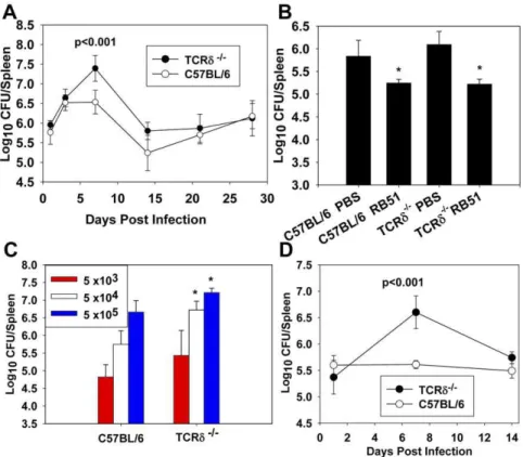
(Figure 1A). At one week post-infection, TCRd2/2 spleens contained,ten-fold moreBrucellathan spleens from wt mice, and
by two weeks post-infection, no significant differences in coloniza-tion were observed between TCRd2/2and wt mice. To determine if the protective role ofcdT cells was limited to innate immunity, wt and TCRd2/2mice were immunized with RB51 and eight weeks later challenged withB. abortus2308. Four weeks after challenge, vaccination efficacy was assessed. RB51 was found to be equally protective in wt and TCRd2/2mice (Figure 1B), indicating the protective function ofcdT cells againstB. abortusmust be limited to early infection and most likely to innate immunity. In an effort to determine if the enhanced susceptibility of TCRd2/2 mice to infection was dose-dependent, both wt and TCRd2/2mice were infected with varying doses, 56103, 56104, or 56105CFUs ofB. abortus, and splenic colonization was assessed one week post-infection (Figure 1C). TCRd2/2mice infected with either 56104
or 56105CFUs ofB. abortuswere more susceptible to infection than wt
mice (Figure 1C). The kinetics of infection and susceptibility following infection with 56104CFUs are similar (Figure 1D) to that found with 56105CFUs ofB. abortus(Figure 1A).
SpleniccdT cells proliferate in response toB. abortus infection
To determine if cd T cells are induced subsequent B. abortus
infection, C57BL/6 mice were infected with 56104CFUs ofB. abortus, and the relative proportions of TCRcd+
, TCRab+
, CD4+
,
Figure 1.cdT cells are required for innate, but not acquired, immunity toB. abortusinfection. A.TCRd2/2and C57BL/6 mice were infected with 56105CFUs ofB. abortus2308 i.p and splenic colonization was determined 1, 3, 7, 14, 21, and 28 days post-infection. The mean
and CD8+
T cells were determined at varying time points post-infection when compared to splenic T cells of naı¨ve mice. Approximately a 3-fold increase in the proportion ofcdT cells was observed in the spleens ofB. abortus-infected C57BL/6 mice, which peaked at two weeks post-infection and occurred concomitantly with diminished brucellae (Table 1; Figure 1A and D). As the total number of splenic mononuclear cells increased dramatically during infection, the increase in the proportion of cd T cells observed was due to an increase in the absolute number ofcdT cells, rather than a reduction in the number of other lymphocyte subsets. The proportion of CD4+
T cells also appeared to decrease by day 28 following infection, which could be due to an increase in the proportion of B cells, a phenomenon that has been observed in other murine models in which mice are infected with intracellular bacteria, such asMycobacterium[19].
cdT cells fromB. abortus infected mice produce IL-17 and IFN-c
SincecdT cells are enhanced byB. abortusinfection, we queried which cytokines are produced. cd T cells (.95% purity) were purified from C57BL/6 mice infected 14 days earlier. This time point was selected since it was the peak of splenic cd T cell induction. For comparison, total T cells (neutralized ofcdT cells) were also assayed for cytokine production in parallel with the purified cd T cells. Both T cell subsets were stimulated with ionomycin and PMA for three days (Figure 2). cd T cells were found to be the major producer of IL-17 during infection and were also found to produce IFN-c. IL-17 production by purifiedcdT cells fromB. abortus-infected mice was also found to be induced by TCRcd stimulation (data not shown). Minimal IL-17 was produced by the enriched TCRab cell fraction; however, this fraction was found to contain elevated levels of IFN-cand IL-6 relative tocdT cells. IL-4, IL-10, and TNF-awere not detected in any cell culture supernatants.
cdT cells do not require IL-17Ra, IFN-c, or GM-CSF to confer protection against systemicB. abortusinfection
Since cd T cells were found to be the main source of IL-17 during infection, we sought to determine if the protection conferred by cd T cells was IL-17-dependent. Wt and IL-17 receptor deficient (IL-17Ra2/2) mice were neutralized ofcd T cells via neutralizing mAb (control mice received normal hamster IgG) and were then infected withB. abortus. Neutralization ofcdT cells enhanced the susceptibility of both wt and IL-17Ra2/2mice (Figure 3A and B), and IL-17 receptor deficiency did not impact splenomegaly (Figure 3A), nor tissue colonization by B. abortus
(Figure 3B) at one week post-infection relative to infected wt mice. Thus, these data show that the protective effect by cd T cells during infection is independent of IL-17. Intracellular cytokine staining revealed that whilecdT cells were the main source of IL-17 during infection, the proportion of cells producing IL-IL-17 was actually diminished byB. abortusinfection (Figure S1A), which may explain while IL-17Ra was dispensable for protection.
IFN-cis required for immunity to experimental brucellosis [20– 22] and also is produced bycd T cells fromB. abortus-infected mice. Therefore, wt and IFN-c2/2 mice (B6 background) were neutralized of cd T cells via neutralizing mAb (control mice received normal hamster IgG) and were then infected with B. abortus(Figure 3C and D). Deficiency of either IFN-corcdT cells enhanced susceptibility to colonization by,10-fold at one-week
post-infection, while mice deficient in both IFN-candcd T cells possessed.300-fold more viableB. abortusin their spleens than did wt mice (Figure 3D). IFN-c2/2 mice neutralized of cd T cells prior to infection with B. abortus displayed ruffled fur and a hunched posture (data not shown), while no clinical symptoms of disease were observed in any other treatment group. Thus, the protection conferred bycdT cells was independent of IFN-c. In addition, cd T cell deficiency in wt mice resulted in enhanced splenomegaly and brucellar colonization, while splenomegaly in IFN-c2/2mice was similar to that observed wt mice (Figure 3C and D). GM-CSF has been found by others to be required for protection to intracellular bacterial pathogens [23,24]; however, GM-CSF was not important for protection againstB. abortusand was dispensable for cd T cell-mediated protection against colonization and splenomegaly at one week post-infection (Figure 3E and F).
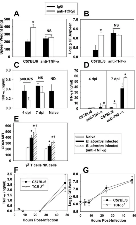
cdT cell-mediated protection against B. abortus infection is dependent on TNF-a
While cd T cells were found not to be a major producer of TNF-a (Figure 2), others have shown TNF-a is required for protection toB. abortus[25]. Thus, mice were neutralized ofcdT cells, TNF-a, or both, prior to infection with B. abortus. Neutralization of either TNF-a or cd T cells from wt mice enhanced susceptibility toB. abortus colonization one week after infection (Figure 4A and B). However, in vivo neutralization of TNF-a did not exacerbate susceptibility to colonization or splenomegaly in mice neutralized of cd T cells (Figure 4B), indicating TNF-a is required forcd T cell-mediated protection againstB. abortus. Depletion ofcdT cells did not appear to affect TNF-aproduction by splenocytes fromB. abortus-infected mice at 4 or 7 days post-infection (Figure 4C), and stimulation of
Table 1.Percentages of splenic T cell subsets and splenic weights followingB. abortusinfectiona.
Uninfected 3 days PI 7 days PI 14 days PI 21 days PI 28 days PI
TCRd+
0.52 (0.056) 0.73 (0.136) 0.50 (0.054) 1.84 (0.13)* 1.31 (0.031)* 0.80 (0.070)*
TCRb+ 37.50 (1.75) 34.73 (0.76) 38.05 (1.57) 38.80 (1.82) 26.33 (3.12) 27.29 (3.03)*
CD4+ 27.20 (1.76) 22.23 (1.41) 24.14 (1.53) 27.40 (3.19) 16.67 (3.33) 16.77 (1.39)*
CD8+
10.17 (1.48) 9.34 (0.66) 11.40 (0.83) 9.50 (1.53) 10.77 (0.84) 7.85(1.36)
Lymphocytes (6106) 77.3 (0.98) ND 194.1 (3.53)* 357.5 (6.52)* ND ND
Spleen weight (mg) 75.3 (6.4) 86.0 (2.1) 170.0 (14.1)* 284 (37.3)* 275.3 (26.6)* 280.0 (27.7)*
aSpleen cells fromBrucella-infected and naı¨ve C57BL/6 mice were stained for FACS analysis using conventional methods. Total splenic lymphocyte number and spleen
weights are shown. At least three mice were used per time point, while the 14 day time point represented six mice from two independent experiments. Standard error is shown in parentheses. ND = not determined.
splenocytes with plate-bound anti-cdTCR mAb did not enhance TNF-aproduction (data not shown). Also, TNF-aproduction by splenocytes from naı¨ve and B. abortus-infected mice was similar (Figure 4C), and intracellular TNF-a levels in splenocytes from bothB. abortus-infected and naı¨ve mice were similar as measured by flow cytometry (data not shown). However, increased IFN-c production by splenocytes from B. abortus-infected mice was observed in mice depleted of TNF-aand/orcdT cells (Figure 4D), suggesting possible compensation by IFN-c. Others have shown that TNF-apreferentially activatescdT cells as measured by up-regulation of surface CD69 expression [26]. While B. abortus
infection did lead to activation of cdT cells (and NK cells to a lesser extent), this effect was independent of TNF-aat 4 and 7 days post-infection, as mice neutralized of TNF-ain vivostill displayed enhanced CD69 expression by theircdT cells and, in fact, more so than mice simply subjected toB. abortusinfection (Figure 4E and data not shown). In addition, it has been shown that macrophages from TCRd2/2mice display impaired TNF-a production when stimulatedin vitrowith LPS [27]. However, no differences in the ability of wt or TCRd2/2 peritoneal macrophages to produce TNF-aor control brucellae infection was evident (Figure 4F and G).
BovinecdT cells can impair the intramacrophage growth ofB. abortusin autologous macrophages
In order to corroborate our findings with murinecdT cells in a bovine model, a co-culture system was used in which bovine macrophages were infected withB. abortus, and then fresh media containing either media only, or media including autologouscdT cells were added to the macrophage-containing wells. Intracellular bacterial burden was then determined at several time points post-infection. We found that at 5 days post-infection, bovinecdT cells could augment the clearance of B. abortus in autologous macrophages. This effect varied depending on the donor of leukocytes, ascdT cells from Calf#1 were protective (Figure 5A); however,cdT cells from Calf#2 (Figure 5C) and Calf#3 were
not as effective (Figure 5E). Protection appeared to correlate with IFN-cproduction, and the addition ofcd T cells to macrophage containing wells from all donors resulted in an increase in IFN-c concentration (Figure 5B, D, and F); however, the additioncdT cells from Calf #1 resulted in the greatest increase in IFN-c production (Figure 5B). Neutralization of IFN-c in vitro also abrogated the protective effectcdT cells from Calf#1, indicating an IFN-c-dependent mechanism. To assess whether cd T cells could protect against B. abortus in an in vivo adoptive transfer model, Rag-12/2 mice were depleted of NK cells and reconsti-tuted with bovine macrophages, macrophages plus autologouscd T cells, or macrophages plus autologous CD4+T cells prior to
infection withB. abortus(leukocytes for adoptive transfer were all derived from Calf #1). When mice were sacrificed seven days later, adoptive transfer of macrophages withcd T cells, but not transfer of macrophages only, or macrophages with CD4+
T cells, resulted in a significant reduction in splenic colonization by brucellae (Figure 5G).
BovinecdT cells respond rapidly to B. abortusinfection and alter the transcriptional profile of autologous macrophages
To determine the transcriptional responses of protective bovine cdT cells during infection at several time points, non-adherentcd T cells were aspirated from macrophage-containing wells, and RNA was extracted. Following aspiration of cd T cells, the remaining (adherent) macrophages were washed repeatedly to remove any remaining non-adherent cells. This process allowed determination of the transcriptional responses by both cell populations via RT-PCR. After 5 h of co-culture with autologous
B. abortus-infected bovine macrophages, cd T cell mRNA transcripts for IL-8, IL-1b, GM-CSF, MIP-1a, and CD25 were up-regulated (Figure 6A). A slight up-regulation of IL-17 after 5 and 24 h of co-culture was also observed incdT cells (data not shown). Later in infection, particularly after 72 h of co-culture,cd T cells produced markedly stronger granzyme B, RANTES, IFN-c, and CD36 transcripts (Figure 6A). To corroborate the transcriptional analysis at the protein level, surface expression for CD25 (IL-2Ra) bycdT cells was assayed via flow cytometry. Co-culture of cd T cells with B. abortus-infected macrophages enhanced CD25 expression bycdT cells within 5 h, and by 72 h, the percentage ofcdT cells expressing CD25 had increased.15 fold (Figure 7). The presence of cd T cells also altered the transcriptional profile of macrophages in the co-culture. Bovine macrophages produced elevated IL-6 mRNAs following infection with B. abortus; however, this response was accelerated and amplified bycdT cells (Figure 6B).cdT cells also enhanced the expression of IL-8 and IL-23p19 by macrophages, particularly, early in infection. IL-8 was also detected within 5 h ofcdT cells/ macrophage culture, but was not detected in wells containing macrophages only (Figure S2). iNOS mRNA expression was only detected at 5 h post-infection, a response unaltered by the presence ofcd T cells. Significant changes in mRNA levels for IL-4, IL-10, IL-12p35, IL-12p40, TNF-a, FoxP3, and TGF-b mRNA fromcdT cells or macrophages were not observed under the conditions tested (data not shown).
Discussion
With over 500,000 new human cases a year [28], brucellosis is the most common zoonotic infection in the world [29]. The pathological manifestations of brucellosis are diverse and include arthritis, endocarditis, and meningitis in humans, while animal brucellosis is characterized by spontaneous abortion [2]. While Figure 2.cdT cells are the primary source of IL-17 duringB.
abortusinfection.C57BL/6 mice were infected i.p. with 56104CFUs of B. abortus2308, and two weeks latercdT cells (.95% purity) and an enriched TCRab(,55% CD4+, 25% CD8+) cell fraction were isolated from the spleens of infected mice. Cells were stimulated with 500 ng/ml ionomycin and 50 ng/ml PMA for three days, and cell-free supernatants from triplicate wells were assayed for cytokine production via ELISA. The mean6SD is shown; * P,0.05 versus the enriched TCRabcells. Results are representative of two independent experiments.
much emphasis has been placed on identifying new vaccine candidates for human and animal brucellosis, relatively little attention has been paid to effective coordination of the innate immune response by the host. cd T cells represent a small percentage of circulating T cells in adult humans and mice, [7]; however, cd T cells are a major lymphocyte subset in adult ruminants and make up a majority (up to 70%) of circulating lymphocytes in neonatal calves [8].cdT cells play an active role in the regulation and resolution of pathogen induced immune responses [30]; however, different subsets ofcdT cells may have different functions. Transcriptional analyses of bovinecd T cells suggest that while CD82cdT cells are activated, proliferative, and inflammatory, the CD8+
subset suppressed genes are consistent with quiescence trafficking to the mucosa and immune suppression [7]. While human Vc9d2 T cells exhibit strong cytolytic activity againstBrucella-infected cells and are able to impair intracellular growth ofB. suisin autologous macrophages [17], this subset ofcd T cells is absent in nonhuman primates, and the role ofcdT cells in anin vivomodel of brucellosis has not been determined.
Here we report mice deficient incdT cells had impaired innate immunity toB. abortus. The protective role of cd T cells during infection appeared to be temporal, as TCRd2/2mice were more susceptible to B. abortus colonization at 7 days post-infection; however, the absence of cd T cells did not impair immunity, particularly, at time points after 2 wks post-infection. Vaccination of mice with RB51 before challenge with wild-typeB. abortus2308 also resulted in reduced colonization in wt and TCRd2/2mice, suggesting that the protective function ofcdT cells may be limited to innate immunity. In addition, the temporal protection conferred bycdT cells correlates with the theory thatcdT cells may be a link between innate and adaptive immunity [31]. Studies in which cdT cells are protective against the organismListeria monocytogenes
[32] have found the magnitude and timing of the cd T cell response are related to the infectious dose of bacteria used, withcd T cell expansion being quicker and of a larger magnitude in affected organs following a higher dose of bacteria [33]. Therefore, as our initial studies used a high dose ofB. abortus(56105CFUs), we queried whether the protection mediated bycdT cells in our Figure 3.cdT cells do not require IL-17Ra, IFN-c, or GM-CSF to mediate protection toB. abortusinfection. A.–F.C57BL/6 orA.andB. IL-17Ra2/2,C.andD.IFN-c2/2, andE.andF.GM-CSF2/2mice (5–6 per group) were treated with anti-TCRcdmAb or hamster IgG on day -1 and day 3 post-infection with 56104CFUs ofB. abortus2308. Mice were sacrificed seven days after infection.A.,C., andE.for splenic weights andB.,D., andF. for tissue colonization were determined; *P,0.05 versus mice of the same genotype treated with hamster IgG;{
P,0.05 versus anti-TCRcd-treated C57BL/6 mice.
Figure 4.cdT cells require TNF-ato protect againstB. abortusinfection.C57BL/6 mice treated with anti-TCRcdmAb or hamster IgG on day -1 and day 3 post-infection with 56104CFUs ofB. abortus2308. Some mice were also neutralized of their TNF-aon days -1 and 3. Seven days after infection,A.splenic weights andB.extent of brucellae colonization were determined. The mean6SEM of 10 mice/group is depicted; *P,0.05 versus hamster IgG-treated C57BL/6 mice. Results are from two independent experiments.C.andD.Splenocytes (56106/ml) from 4 or 7 dayB. abortus-infected mice depleted or not ofcdT cells and/or TNF-aor splenocytes from naı¨ve C57BL/6 mice were left unstimulated and cultured for 3 days at 37uC/5%CO2; supernatants were harvested forC.TNF-a- orD.IFN-c- specific ELISA. The mean6SD of triplicate wells is shown. The results from 7 dpi are representative of two independent experiments. NS = not significant. ND = cytokine production from uninfected mice neutralized ofcd
T cells was not determined. * P,0.05 as compared to mice not depleted ofcdT cells within the same TNF-atreatment group at the same time point. {
P,0.05 as compared to C57BL/6 mice treated with IgG only at the same time point.E.The median fluorescence intensity (MFI) of CD69 expression by splenic NK andcdT cells as measured by flow cytometry is shown for naı¨ve C57BL/6 andB. abortus-infected (after 4 days of infection with 56104CFUs of strain 2308) C57BL/6 mice. Data depict the mean
6SD from 5 mice/group; *P,0.05 versus naı¨ve mice and{
P,0.05 versusB. abortus -infected mice not neutralized of their TNF-a.F.andG.Peritoneal macrophages from C57BL/6 and TCRd2/2mice were infected withB. abortus(30 bacteria:1 macrophage), andF.TNF-alevels in supernatants andG.intracellular colonization were measured. Data represent the mean6SD of triplicate wells/group.
model was dose-dependent by infecting wt and TCRd2/2 mice with three doses ofB. abortus (56103, 56104, and 56105CFUs). Protection against colonization conferred bycdT cells at 7 days post-infection appeared to be similar, regardless of the dose of infection, and the difference in the kinetics of clearance of B. abortusbetween wt and TCRd2/2mice was also similar, regardless of whether 56104or 56105CFUs were used as an infectious dose. Notably, regardless of whether TCRd2/2 mice or wt mice neutralized of cd T cells via mAb (clone UC7-13D5) treatment was used, either model showed similar impairment of immunity to
B. abortus, indicating that the effect ofcdT cells is not due to a developmental defect in TCRd2/2 mice. A recent study has shown that treatment of mice with mAb against the TCRcd(such as UC7-13D5) may not actually deplete cells, but rather block TCRcd signaling [34], which according to the results presented here would indicate the protective effect of murine cd T cells againstB. abortusrequires TCRcd-specific signaling.
To determine ifcdT cells expand in response to infection, the lymphocyte composition of spleens from B. abortus-infected C57BL/6 mice was characterized at several time points post-infection. At two weeks post-infection, the proportion ofcdT cells amongst lymphocytes in the spleens from infected mice increased nearly three-fold. The total number of lymphocytes in spleens increased following B. abortus infection, indicating the enhanced percentage ofcdT cells was due to an increase in the total number ofcdT cells rather than a reduction of other T cell subsets. The finding thatcdT cells expanded or were recruited to the spleens of
infected mice lent further support to the importance ofcdT cells in resolving infection byBrucella. The observed spike in spleniccd T cells occurring after the peak ofBrucellainfection was similar to what has been found in mice infected withNocardia asteroides or
Mycobacterium bovisBCG [9,35].
Different mechanisms have been proposed for the protection conferred by cd T cells in murine models. Klebsiella-infected TCRd2/2mice are found to have decreased levels of IFN-cand TNF-a, which has been postulated to result in enhanced susceptibility to infection [10].cd T cells are the main producer of IL-17 in naı¨ve mice [14], and IL-17 production bycd T cells has been shown to be augmented in response to infection [11,13,14]. In addition, the protection conferred bycd T cells against infection withL. monocytogenes,E. coli, andF. tularensisLVS has been shown to be dependent on IL-17 [11,13,36]. Therefore, thecdT cell cytokine profile was assessed following infection with
B. abortusto ascertain the mechanism by which these cells confer protection. Similar to what others have found,cdT cells were the main producer of IL-17 in infected mice. Interestingly, IL-17Ra2/2 mice were not impaired in their ability to controlB. abortusinfection, and IL-17Ra signaling was not required forcd T cell-mediated protection. Intracellular cytokine staining revealed that whilecdT cells were the major source of IL-17 in both naı¨ve andB. abortus -infected mice, IL-17 production was not enhanced by Brucella
infected mice, production of IFN-c by an enriched TCRab cell fraction was much more robust. B. abortus-induced IFN-c production by CD4+
T cells was confirmed by intracellular cytokine staining (Figure S1A–B). Subsequent secretion analysis ofcdT cells sorted from naı¨ve andB. abortusinfected TCRa2/2 mice (which have a higher proportion of cd T cells) revealed that B. abortus
infection may actually suppress IL-17 and IFN-cproduction bycd T cells (Figure S1B). However, it is important to consider the function ofcdT cells may be different in TCRa2/2mice than in wt mice, and we must not over-interpret these results. The protective effects of IFN-candcdT cells againstB. abortuswere found to be independent of each other and, in fact, could be compensatory, as depletion ofcdT cells from IFN-c2/2mice resulted in a greater impairment of immunity toB. abortusthan did depletion ofcdT cells from wt mice. Indeed, depletion of TNF-aand/orcdT cells from
B. abortus-infected mice was found to enhance IFN-cproduction by splenocytes. This finding further suggested the host may enhance IFN-cproduction to compensate for the loss of protection conferred by TNF-aand/orcdT cells and may also indicate the protective effects of IFN-cin C57BL/6 mice may be independent of TNF-a and/orcdT cells.
cdT cells were not found to be a major producer of TNF-a, and neutralization ofcd T cells fromB. abortus-infected mice did not significantly reduce TNF-aproduction. Others have shown that macrophages from TCRd2/2 mice display impaired TNF-a production uponin vitrostimulation [27], yet no differences in the ability of peritoneal macrophages from either wt or TCRd2/2 mice to control brucellae infection or produce TNF-a were observed. However, TNF-awas required forcdT cells to confer protection againstB. abortusinfection. While it has been shown that TNF-a preferentially activates cd T cells as measured by
up-regulation of surface CD69 expression [26], TNF-aneutralization had no effect upon CD69 expression bycdT cells or NK cells in
B. abortus-infected mice. TNF-ais also known to enhance T cell cytotoxicity [37]; thus, future studies will investigate whethercdT cells mediate protection against B. abortusinfection via TNF-a -dependent cytotoxic effects. Interestingly, B. abortus infection enhanced CD69 expression more robustly incd T cells than in NK cells, which may help explain work by others indicating that NK cells are dispensable for innate immunity toB. abortus[38].
In order to corroborate our murine findings using bovine lymphocytes, a co-culture system was adopted and purified bovine cd T cells were cultured with autologous B. abortus-infected macrophages. BovinecdT cells could impair the intramacrophage replication ofB. abortusby co-cultured macrophages, but this effect varied depending on the donor of cd T cells, which was not entirely surprising, since cattle exhibit wide variability in their immune response to infection [39]. While the addition ofcd T cells from all donors to autologousB. abortus-infected macrophages resulted in enhanced IFN-c production in cell culture superna-tants,cdT cells only conferred significant protection when high levels (.40 ng/ml) of IFN-c were produced. Neutralization of IFN-cin vitroabrogated the protective effect of bovinecdT cells in our co-culture system, further demonstrating the requirement of IFN-cfor bovinecdT cell-mediated protection. IFN-chas been shown to enhance the resistance of bovine macrophages to bacterial infection by inducing apoptosis [40]; therefore, future studies will assess whether cd T cells and IFN-c affect bovine macrophage apoptosis duringB. abortusinfection.
Transcriptional analyses of bovine co-cultured with Brucella -infected macrophages suggestcdT cells follow a ‘‘priming’’ model of activation [41]. Early in infection (,5 h), IL-8, MIP-1a(CCL3),
GM-CSF, IL-1b, IL-17, and CD25 mRNAs were upregulated by cdT cells. At later time points after infection (,72 h),cdT cells
enhanced expression of granzyme B, RANTES, and IFN-c mRNAs. This biphasic immune response indicates thatcdT cells initially respond to infection by expressing signals that lead to direct enhancement of macrophage function, along with an enhanced responsiveness of the cd T cells to further secondary signals, such as antigen or cytokines, while later in infection,cdT cells express signals indicative of an ‘‘effector’’ state [41]. In addition, the presence of cd T cells seemed to alter the early mRNA profile of infected macrophages as increased levels of IL-8, IL-6, and IL23p19 were observed in infected macrophages co-cultured withcdT cells relative to infected macrophages alone. These cytokines are all considered part of the Th17 response [42], suggesting that the presence ofcdT cells may shift the subsequent immune response to infection. TNF-amRNA was not found to be induced in bovine cells under the conditions tested, which is not surprising as others have found that intravenous or subcutaneous vaccination of cattle withB. abortusRB51 does not increase serum TNF-alevels [43].
The need for model systems to analyze the immune system and pathogenesis of disease in cattle is great since studies in cattle are difficult owing to the limited availability of genetically similar animals and the high costs involved in animal purchase and housing [44].In vivobovine studies with virulentBrucellaspecies are particularly difficult to perform due to the limited availability of large animal BSL-3 facilities. Therefore, to determine if bovinecd T cells that were protective in our co-culture system could protect in anin vivomodel, Rag-12/2mice were depleted of NK cells and reconstituted with bovine macrophages alone, macrophages plus autologouscd T cells, or macrophages plus autologous CD4+T
cells prior to infection withB. abortus. When mice were sacrificed seven days later, adoptive transfer of macrophages withcdT cells, Figure 6. Transcriptional response of bovine cd T cells to
infection. A.Total RNA was extracted from restingcdT cells, orcdT cells co-cultured with infected autologous macrophages for 5, 24, or 72 h. B. RNA was also isolated from mock- or B. abortus-infected macrophages cultured with or withoutcdT cells after removal ofcdT cells and repeated washing of adherent macrophages. cDNA was synthesized, and RT-PCR was conducted for several immune-related genes and b-actin Results are representative of two independent experiments with Calf#1.
but not macrophages only nor macrophages with CD4+
T cells, resulted in a significant reduction in splenic colonization by B. abortus. These findings further demonstrate bovinecdT cells can protect againstB. abortusinfection, and reconstitution of Rag-12/2 mice with bovine cells is a viable method, which can be used to assay the role of bovine leukocytes during infection.
In this study, we showed that both murine and bovinecdT cells rapidly responded and could provide protection against, infection with B. abortus. While murine and bovine cd T cells utilized different mechanisms of protection to confer protection againstB. abortus,cdT cells from both species were found to produce IFN-c, enhance surface expression of activation markers, and help coordinate the host cytokine response followingB. abortusinfection. Characterization of the transcriptional profile of bovinecdT cells subsequent to infection revealed a biphasic immune response. Initially, bovine cd T cells expressed signals leading to direct enhancement of macrophage function along with enhancedcdT cells responsiveness to further secondary signals, while later in infectioncdT cells expressed signals indicative of an effector state. Collectively, these findings demonstratecdT cells are important for controlling B. abortus infection and provide insight into the
response of both murine and bovine cd T cells in response to infection. In addition, these results indicate immunotherapeutic strategies that targetcdT cells could be viable means to augment innate immunity against brucellosis.
Materials and Methods
Ethics Statement
All animal care and procedures were in accordance with institutional policies for animal health and well-being, and approved by MSU Institutional Animal Care and Use Committee under protocol 38.
Mice
Breeder pairs of cd T cell-deficient (TCRd2/2), TCRab -deficient (TCRa2/2), T and B cell-deficient (Rag-12/2), and GM-CSF (GM-GM-CSF2/2) deficient mice on a C57BL/6 background were obtained from The Jackson Laboratory (Bar Harbor, ME), and breeder pairs IL-17Ra2/2mice on B6 background were a gift from Amgen (Seattle, WA); mice were bred and maintained at the Montana State University Animal Resource Center (Bozeman, Figure 7. BovinecdT cells upregulate surface CD25 expression following infection of autologous macrophages withB. abortus. Surface CD25 expression was measured on naı¨vecdT cells along withcdT cells co-cultured withB. abortus-infected autologous macrophages for 5, 24, or 72 h. Data depict the mean6S.D. of triplicate measurements/group.
MT). C57BL/6 and IFN-c2/2 mice (B6 background; Jackson Laboratory) were used at 7 to 11 weeks of age. All mice were maintained at Montana State University Animal Resource Center under pathogen-free conditions in individually ventilated cages under HEPA-filtered barrier conditions and were fed sterile food and waterad libitum. In some experiments,cdT cells and/or TNF-awere neutralizedin vivovia intraperitoneal (i.p.) administration of 0.5 mg of anti-cdTCR mAb (clone UC7-13D5; BioXcell) and/or 0.5 mg of anti-TNF-amAb (clone XT3.11; BioXcell) on days -1 and 3 post-infection. For challenge studies with B. abortus strain 2308, mice were maintained under similar isolation conditions, in our institutional ABSL-3 facilities. All animal care and procedures were in accordance with institutional policies for animal health and well-being.
Bacterial Strains and Growth Conditions
B. abortus strain 2308 and the B. abortus vaccine strain RB51 were obtained from the National Veterinary Services Laboratory, USDA (Ames, IA). Bacteria were grown under aerobic conditions in potato infusion agar for 72 h (Difco Laboratories) at 37uC and 5% CO2. For inoculation, a colony was chosen and incubated
overnight at 37uC with shaking in Brucella broth (Difco); the bacterial suspension was adjusted spectrophotometrically to an optical density at 600 nm corresponding to desired inoculum concentration. All experiments with live brucellae were performed in our institutional biosafety level 3 facilities.
Tissue Colony Counts
At selected time points post-infection, spleens and livers were harvested for CFU determinations. Organs were dounce homog-enized, and serial 10-fold dilutions in triplicate of homogenates in sterile water were grown onBrucellaagar (BA). After incubation for 3 to 5 days at 37uC with 5% CO2, Brucella colonies were
enumerated, and the number of CFU per tissue was calculated from the dilutions.
FACS Analysis
Murine and bovine lymphocytes were stained for FACS analysis using conventional methods [45,46]. Following the removal of erythrocytes, whole murine spleen cells were stained with fluorochrome-conjugated or biotinylated mAbs (Becton Dickinson or eBioscience): anti-CD4 (clone L34T4), anti-CD8 (clone 853-6.7), anti-TCRb chain (clone H57-597), anti-B220 (clone RA3-6B2), NK1.1 (clone PK136), CD69 (clone H1.2F3), anti-CD3 (clone 17A2), or anti-TCRd chain (clone GL3); cells were then fixed with 2% paraformaldehyde. Bovine lymphocytes were stained with anti-TCRd chain (clone GD3.8) and anti-CD25 (clone LCTB2A), as described [47]. Stained lymphocytes were analyzed using a FACSCalibur, FACSCanto, or LSRII flow cytometer (BD Biosciences) and analyzed using FlowJo software (Tree Star). For intracellular staining, cells were isolated as described above and stimulated overnight with ionomycin (500 ng/ml)/phorbol 12-myristate 13-acetate (PMA; 50 ng/ml). For the last 3 h of culture, brefeldin A (10mg/ml) was added to the
cultures. Cells were then stained for cell surface markers as described above and fixed in paraformaldehyde. Cells were then permeabilized with 0.2% saponin prior to intracellular staining for IL-17A (clone TC11-18H10) and IFN-c (clone XMG1.2). For determination of total lymphocytes, splenic mononuclear cells were isolated over a Lympholyte M gradient (Cedarlane Labs), and viable cells counted via trypan blue exclusion. The proportion of lymphocytes was confirmed via flow cytometry by gating on forward and side scatter.
Isolation and Infection of Murine Peritoneal Macrophages Peritoneal macrophages were isolated, as previously described [48,49]. Briefly, mice were given a single i.p. injection of 1.0 ml of expired thioglycolate medium (Difco), and 3 days later, the peritoneum of each mouse was washed with RPMI 1640 (Gibco BRL-Life Technologies [Life Technologies] containing 2% fetal calf serum (Life Technologies) without antibiotics. Peritoneal cells were washed twice in the same medium without antibiotics and allowed to adhere overnight to 24 well microtiter plates. Macrophages (16106/ml) were infected with B. abortus (30 bacteria:1 macrophage) for 1 hour at 37uC/5% CO2.
Macro-phages were then washed with PBS, fresh complete medium (CM): RPMI 1640 medium supplemented with 1 mM sodium pyruvate, 1 mM nonessential amino acids, penicillin/streptomycin (10 U/ ml), and 10% FBS (Atlanta Biologicals) containing 50mg/ml gentamicin (Sigma-Aldrich) were added, and cells were incubated for 30 min at 37uC/5% CO2. After washing twice, fresh CM
containing gentamicin (2.5mg/ml) was added, and cells were incubated 37C/5% CO2. At various time points post-infection, the
wells were washed three times, macrophages were lysed with sterile water, and intracellular bacterial burden was determined by serial dilution of macrophage lysates on BA.
Cytokine ELISAs
Spleens were aseptically removed from C57BL/6 mice at various time points after challenge withB. abortus2308. For whole cell cultures, murine splenocytes (56106
cells/ml) were cultured following the removal of erythrocytes at for 72 h in CM. To culture purified populations, mononuclear cells were prepared from a Lympholyte M (Cedarlane Labs) gradient, andcdT cells along with an enriched T cell fraction were purified using acdT cell isolation kit (Miltenyi Biotec), according to the manufacturer’s instructions. Cells were cultured at 56105cells/ml in CM alone or in the presence of ionomycin (500 ng/ml)/PMA (50 ng/ml) or plate bound anti-TCRcdmAb (UC7-13D5; 100mg/ml) for 72 h
at 37uC/5%CO2. Supernatants were also harvested from murine
peritoneal macrophages and bovine macrophages (in some instances co-cultured with autologous cd T cells), which were infected withB. abortus. All supernatants were filtered through a 0.4mm filter and stored at –80uC. Capture ELISA for IL-4, IL-6, IL-10, IL-17, IFN-c, and TNF-a was used to quantify cytokine levels, as previously described [45,46,49].
Generation of Bovine Monocyte-Derived Macrophages Whole blood was collected from 6–10 month old Holstein calves into sodium heparin tubes (BD Biosciences). Leukocytes were separated from whole blood using Histopaque-1077 (Sigma-Aldrich) for bovine cells, as previously described [47]. Mono-cyte-derived macrophages were generated via modification of a previous protocol [50]. PBMCs (16107/ml) were cultured in CM without antibiotics containing 12.5% autologous serum. Cells were allowed to adhere to tissue culture flasks for 2 h at 37uC/5%CO2
when the flasks were agitated. The cells were then cultured at 37uC/5%CO2for 72 h when flasks were agitated again, and the
supernatant was aspirated and fresh CM without antibiotics containing 12.5% autologous serum was added. Adherent cells were cultured for an additional 72 h at 37uC/5%CO2, when the
Montana State University Institutional Animal Care and Use Committee (Bozeman, MT).
Purification of Bovine T Cell Subsets
Bovine PBMCs were isolated as described above and then incubated with biotinylated mAbs against CD4+ T cells (clone
CC30) [51] or the TCRcd(clone GD3.8) [52]. Washed cells were incubated with streptavidin microbeads (Miltenyi). Cells were washed again and positively sorted using magnetic LS columns (Miltenyi) according to manufacturer’s instructions. The resulting purity was greater than 95%. Purified T cells were allowed to rest overnight and were resuspended in fresh CM the following day immediately prior to being added to wells containingB. abortus-infected macrophages.
Infection of Bovine Macrophage and cdT Cell Co-Cultures
Bovine macrophages (prepared as described above) were infected withB. abortusin a manner similar to that described previously [5]. Macrophages (26105/ml) were infected withB. abortus (30 bacter-ia:macrophage) for 1 hour at 37uC/5% CO2. Macrophages were
then washed with PBS, fresh CM containing 50mg/ml gentamicin
(Sigma-Aldrich) was added, and cells were incubated for 30 min at 37uC/5% CO2. After washing twice, as described above, fresh CM
containing gentamicin (2.5mg/ml) with or without autologous CD4+
cells or cd T cells (26106/ml prepared as described above,) was added, and cells were incubated 37uC/5% CO2. A neutralizing
anti-bovine IFN-cmAb (clone CC302, Serotec, 50 ng/ml: [53]) was also added to some wells. At various time points post-infection, the wells were washed three times, macrophages were lysed with sterile water, and intracellular bacterial burden was determined by serial dilutions of macrophage lysates on BA. The nonadherent T cells were aspirated from the co-culture, centrifuged, and stored in Tri Reagent (Sigma-Aldrich) for RNA extraction. In some instances, after the aspiration of T cells (or CM only) and three washes with PBS, adherent macrophages were lysed in Tri Reagent for RNA extraction. Supernatants were also harvested and 0.4mm- filtered and stored at -80uC to determine IFN-clevels using mAbs from ABD Serotec: the capture mAb was clone CC330, and the detecting mAb was clone CC302. IL-8 concentrations were also measured using the anti-human CXCL-8/IL-8 DuoSet Kit from R&D Systems, which cross-reacts with bovine IL-8.
Reconstitution of Rag-12/2Mice with Bovine Leukocytes
Rag-12/2mice were depleted of NK cells via i.p. administra-tion of 0.5 mg of an anti-NK1.1 mAb (clone PK136, BioXCell) on days -2 and day 3 post-infection. One day before infection, PBS, macrophages (56105/mouse) only or macrophages plus T cells (16107/mouse) were adoptively transferred into mice intraperito-neally. Macrophages and T cells were purified, as described above. One day later, mice were infected i.p. with 16104B. abortus2308. Splenic colonization was determined seven days later.
RNA Extraction and RT-PCR Analysis
RNA was extracted from cells in Tri Reagent according to manufacturer’s guidelines. RNA was then further purified via extraction with an RNeasy Mini Kit (Qiagen). cDNA was
generated using the Superscript III First Strand Synthesis System (Invitrogen). Primers for immune-related genes along withb-actin (endogenous control) were designed using the PrimerQuest application from IDTDNA.com and purchased from IDT. Amplicons were visualized under UV illumination on a 2% agarose gel containing GelRed (Biotium).
Statistical Analysis
The Student t test was used to evaluate the differences in colonization, splenic weights, cytokine production, intracellular bacterial burden, and lymphocyte populations when two groups were compared. When more than two groups were compared, an ANOVA followed by Tukey’s test was used. Apvalue of,0.05 was considered significant.
Supporting Information
Figure S1 B. abortus infection does not induce IL-17 or IFN-cproduction bycdT cells. A.Splenocytes from naı¨ve or
B. abortus-infected mice (7 dpi) were stimulated overnight with PMA/Ionomycin and brefeldin A was added for the last 3 h of culture. Following surface staining, cells were permeabilized and stained for intracellular IL-17 or IFN-c. Top panel, the proportion of IL-17 producingcdT cells was determined following gating on lymphocytes. Second panel from top, cells were gated on CD4+
(CD3+
) T cells and assayed for IL-17 production. Third panel from top, cells were gated oncd T cells (CD3+/TCRcd+) and
assayed for IFN-cproduction. Bottom panel, cells were gated on CD4+
(CD3+
) T cells and assayed for IFN-cproduction. Depicted is the mean6 SD of 5 mice/group and is representative of two independent experiments.B.cdT cells were sorted from naı¨ve or
B. abortus-infected (7 dpi) mice and stimulated for 72 h with PMA/ Ionomycin. Cytokine levels in supernatant were determined by ELISA. Depicted is the mean6SD of triplicate wells. *P,0.05 versus cytokine production bycdT cells from naı¨ve mice. (TIF)
Figure S2 IL-8 production is augmented by cd T cells when co-cultured with bovine macrophage during infection withB. abortus.IL-8 concentrations were measured
by ELISA in supernatants from B. abortus-infected bovine macrophages cultured with or without autologous T cells at various time points after infection. Data depict the mean6S.D. of triplicate measurements/group. *P,0.05 versus wells containing macrophages only.
(TIF)
Acknowledgments
We thank Amgen Inc. for providing us breeder pairs of IL-17ra2/2mice
for our studies. In addition, we thank Ms. Nancy Kommers for her assistance in preparing this manuscript.
Author Contributions
Conceived and designed the experiments: JAS DWP. Performed the experiments: JAS TT MCR EH. Analyzed the data: JAS MAJ DWP. Contributed reagents/materials/analysis tools: JAS MAJ. Wrote the paper: JAS DWP.
References
1. Edmonds M, Booth N, Hagius S, Walker J, Enright F, et al. (2000) Attenuation and immunogenicity of aBrucella abortus htrA cycLdouble mutant in cattle. Vet Microbiol 76: 81–90.
2. Cardoso PG, Macedo GC, Azevedo V, Oliveira SC (2006) Brucella spp noncanonical LPS: structure, biosynthesis, and interaction with host immune system. Microb Cell Fact 5: 13.
3. Yang X, Hudson M, Walters N, Bargatze RF, Pascual DW (2005) Selection of protective epitopes forBrucella melitensisby DNA vaccination. Infect Immun 73: 7297–7303.
5. Yang X, Becker T, Walters N, Pascual DW (2006) Deletion ofznuAvirulence factor attenuates Brucella abortus and confers protection against wild-type challenge. Infect Immun 74: 3874–3879.
6. Ko J, Splitter GA (2003) Molecular host-pathogen interaction in brucellosis: current understanding and future approaches to vaccine development for mice and humans. Clin Microbiol Rev 16: 65–78.
7. Hedges JF, Graff JC, Jutila MA (2003) Transcriptional profiling ofcdT cells. J Immunol 171: 4959–4964.
8. Hein WR, Mackay CR (1991) Prominence ofcdT cells in the ruminant immune system. Immunol Today 12: 30–34.
9. King DP, Hyde DM, Jackson KA, Novosad DM, Ellis TN, et al. (1999) Cutting edge: protective response to pulmonary injury requirescd T lymphocytes. J Immunol 162: 5033–5036.
10. Moore TA, Moore BB, Newstead MW, Standiford TJ (2000)cdT cells are critical for survival and early proinflammatory cytokine gene expression during murine Klebsiella pneumonia. J Immunol 165: 2643–2650.
11. Shibata K, Yamada H, Hara H, Kishihara K, Yoshikai Y (2007) Resident Vd1+
cdT cells control early infiltration of neutrophils afterEscherichia coliinfection via IL-17 production. J Immunol 178: 4466–4472.
12. Takano M, Nishimura H, Kimura Y, Mokuno Y, Washizu J, et al. (1998) Protective roles ofcdT cells and IL-15 inEscherichia coliinfection in mice. Infect Immun 66: 3270–3278.
13. Lin Y, Ritchea S, Logar A, Slight S, Messmer M, et al. (2009) Interleukin-17 is required for T helper 1 cell immunity and host resistance to the intracellular pathogen Francisella tularensis. Immunity 31: 799–810.
14. Lockhart E, Green AM, Flynn JL (2006) IL-17 production is dominated bycdT cells rather than CD4 T cells duringMycobacterium tuberculosisinfection. J Immunol 177: 4662–4669.
15. Oliaro J, Dudal S, Liautard J, Andrault JB, Liautard JP, et al. (2005) Vc9Vd2 T cells use a combination of mechanisms to limit the spread of the pathogenic bacteriaBrucella. J Leukoc Biol 77: 652–660.
16. Bertotto A, Gerli R, Spinozzi F, Muscat C, Scalise F, et al. (1993) Lymphocytes bearing thecdT cell receptor in acuteBrucella melitensisinfection. Eur J Immunol 23: 1177–1180.
17. Ottones F, Dornand J, Naroeni A, Liautard JP, Favero J (2000) Vc9Vd2 T cells impair intracellular multiplication of Brucella suis in autologous monocytes through soluble factor release and contact-dependent cytotoxic effect. J Immunol 165: 7133–7139.
18. Dudal S, Turriere C, Bessoles S, Fontes P, Sanchez F, et al. (2006) Release of LL-37 by activated human Vc9Vd2 T cells: a microbicidal weapon against
Brucella suis. J Immunol 177: 5533–5539.
19. Ladel CH, Daugelat S, Kaufmann SH (1995) Immune response toMycobacterium bovisbacille Calmette Guerin infection in major histocompatibility complex class I- and II-deficient knock-out mice: contribution of CD4 and CD8 T cells to acquired resistance. Eur J Immunol 25: 377–384.
20. Baldwin CL (2002) Immune response overview. Vet Microbiol 90: 365–366. 21. Murphy EA, Sathiyaseelan J, Parent MA, Zou B, Baldwin CL (2001) IFN-cis
crucial for surviving aBrucella abortusinfection in both resistant C57BL/6 and susceptible BALB/c mice. Immunology 103: 511–518.
22. Jiang X, Baldwin CL (1993) Effects of cytokines on intracellular growth of
Brucella abortus. Infect Immun 61: 124–134.
23. LeVine AM, Reed JA, Kurak KE, Cianciolo E, Whitsett JA (1999) GM-CSF-deficient mice are susceptible to pulmonary group B streptococcal infection. J Clin Invest 103: 563–569.
24. Zhan Y, Lieschke GJ, Grail D, Dunn AR, Cheers C (1998) Essential roles for GM-CSF and G-CSF in the sustained hematopoietic response of Listeria monocytogenes-infected mice. Blood 91: 863–869.
25. Murphy EA, Parent M, Sathiyaseelan J, Jiang X, Baldwin CL (2001) Immune control ofBrucella abortus2308 infections in BALB/c mice. FEMS Immunol Med Microbiol 32: 85–88.
26. Lahn M, Kalataradi H, Mittelstadt P, Pflum E, Vollmer M, et al. (1998) Early preferential stimulation ofcdT cells by TNF-a. J Immunol 160: 5221–5230. 27. Nishimura H, Emoto M, Hiromatsu K, Yamamoto S, Matsuura K, et al. (1995)
The role ofcdT cells in priming macrophages to produce TNF-a. Eur J Immunol 25: 1465–1468.
28. Colmenero JD, Reguera JM, Fernandez-Nebro A, Cabrera-Franquelo F (1991) Osteoarticular complications of brucellosis. Ann Rheum Dis 50: 23–26. 29. Pappas G, Papadimitriou P, Akritidis N, Christou L, Tsianos EV (2006) The
new global map of human brucellosis. Lancet Infect Dis 6: 91–99.
30. Tramonti D, Andrew EM, Rhodes K, Newton DJ, Carding SR (2006) Evidence for the opposing roles of differentcdT cell subsets in macrophage homeostasis. Eur J Immunol 36: 1729–1738.
31. Holtmeier W, Kabelitz D (2005)cdT cells link innate and adaptive immune responses. Chem Immunol Allergy 86: 151–183.
32. Hiromatsu K, Yoshikai Y, Matsuzaki G, Ohga S, Muramori K, et al. (1992) A protective role ofcdT cells in primary infection withListeria monocytogenesin mice. J Exp Med 175: 49–56.
33. Belles C, Kuhl AK, Donoghue AJ, Sano Y, O’Brien RL, et al. (1996) Bias in the
cdT cells response toListeria monocytogenes. Vd6.3+cells are a major component of thecdT cell response toListeria monocytogenes. J Immunol 156: 4280–4289. 34. Koenecke C, Chennupati V, Schmitz S, Malissen B, Forster R, et al. (2009) In
vivo application of mAb directed against thecd TCR does not deplete but generates ‘‘invisible’’cdT cells. Eur J Immunol 39: 372–379.
35. Muller D, Pakpreo P, Filla J, Pederson K, Cigel F, Malkovska V (1995) Increased cd T lymphocyte response to Mycobacterium bovis BCG in major histocompatibility complex class I-deficient mice. Infect Immun 63: 2361–2366. 36. Hamada S, Umemura M, Shiono T, Tanaka K, Yahagi A, et al. (2008) IL-17A produced bycdT cells plays a critical role in innate immunity againstListeria monocytogenesinfection in the liver. J Immunol 181: 3456–3463.
37. Ranges GE, Figari IS, Espevik T, Palladino MA, Jr. (1987) Inhibition of cytotoxic T cell development by transforming growth factor beta and reversal by recombinant tumor necrosis factor alpha. J Exp Med 166: 991–998. 38. Fernandes DM, Benson R, Baldwin CL (1995) Lack of a role for natural killer
cells in early control ofBrucella abortus2308 infections in mice. Infect Immun 63: 4029–4033.
39. Morris CA (2007) A review of genetic resistance to disease inBos tauruscattle. Vet J 174: 481–491.
40. Denis M, Wedlock DN, Buddle BM (2005) IFN-cenhances bovine macrophage responsiveness toMycobacterium bovis: Impact on bacterial replication, cytokine release and macrophage apoptosis. Immunol Cell Biol 83: 643–650. 41. Jutila MA, Holderness J, Graff JC, Hedges JF (2008) Antigen-independent
priming: a transitional response of bovinecdT cells to infection. Anim Health Res Rev 9: 47–57.
42. McKenzie BS, Kastelein RA, Cua DJ (2006) Understanding the IL-23-IL-17 immune pathway. Trends Immunol 27: 17–23.
43. Palmer MV, Elsasser TH, Cheville NF (1998) TNF-ain pregnant cattle after intravenous or subcutaneous vaccination with Brucella abortus strain RB51. Am J Vet Res 59: 153–156.
44. Smith RA, Kreeger JM, Alvarez AJ, Goin JC, Davis WC, et al. (1999) Role of CD8+
and WC-1+cdT cells in resistance toMycobacterium bovisinfection in the SCID-bo mouse. J Leukoc Biol 65: 28–34.
45. Ochoa-Repa´raz J, Riccardi C, Rynda A, Jun S, Callis G, Pascual DW (2007) Regulatory T cell vaccination without autoantigen protects against experimental autoimmune encephalomyelitis. J Immunol 178: 1791–1799.
46. Pascual DW, White MD, Larson T, Walters N (2001) Impaired mucosal immunity in L-selectin-deficient mice orally immunized with aSalmonellavaccine vector. J Immunol 167: 407–415.
47. Holderness J, Jackiw L, Kimmel E, Kerns H, Radke M, et al. (2007) Select plant tannins induce IL-2Raup-regulation and augment cell division incdT cells. J Immunol 179: 6468–6478.
48. Ochoa-Repa´raz J, Sentissi J, Trunkle T, Riccardi C, Pascual DW (2007) AttenuatedCoxiella burnetiiphase II causes a febrile response in IFN-cknockout and Toll-like receptor 2 knockout mice and protects against reinfection. Infect Immun 75: 5845–5858.
49. Pascual DW, Trunkle T, Sura J (2002) FimbriatedSalmonella entericaserovar typhimurium abates initial inflammatory responses by macrophages. Infect Immun 70: 4273–4281.
50. Campbell GA, Adams LG (1992) The long-term culture of bovine monocyte-derived macrophages and their use in the study of intracellular proliferation of
Brucella abortus. Vet Immunol Immunopathol 34: 291–305.
51. Naessens J, Howard CJ, Hopkins J (1997) Nomenclature and characterization of leukocyte differentiation antigens in ruminants. Immunol Today 18: 365–368. 52. Wilson E, Walcheck B, Davis WC, Jutila MA (1998) Preferential tissue
localization of bovine cdT cells subsets defined by anti-T cell receptor for antigen antibodies. Immunol Lett 64: 39–44.
53. Leite F, Atapattu D, Kuckleburg C, Schultz R, Czuprynski CJ (2005) Incubation of bovine PMNs with conditioned medium from BHV-1 infected peripheral blood mononuclear cells increases their susceptibility toMannheimia haemolytica