Analytical Sensitivity Comparison between
Singleplex Real-Time PCR and a Multiplex
PCR Platform for Detecting Respiratory
Viruses
Jayme Parker1,2, Nisha Fowler1, Mary Louise Walmsley1, Terri Schmidt1, Jason Scharrer1, James Kowaleski1, Teresa Grimes1, Shanann Hoyos2, Jack Chen1,2*
1Department of Health and Social Services, Division of Public Health, Alaska State Public Health Virology Laboratory, Fairbanks, Alaska, United States of America,2Department of Biology and Wildlife, Institute of Arctic Biology, University of Alaska Fairbanks, Fairbanks, Alaska, United States of America
Abstract
Multiplex PCR methods are attractive to clinical laboratories wanting to broaden their detec-tion of respiratory viral pathogens in clinical specimens. However, multiplexed assays must be well optimized to retain or improve upon the analytic sensitivity of their singleplex coun-terparts. In this experiment, the lower limit of detection (LOD) of singleplex real-time PCR assays targeting respiratory viruses is compared to an equivalent panel on a multiplex PCR platform, the GenMark eSensor RVP. LODs were measured for each singleplex real-time PCR assay and expressed as the lowest copy number detected 95–100% of the time,
depending on the assay. The GenMark eSensor RVP LODs were obtained by converting the TCID50/mL concentrations reported in the package insert to copies/μL using qPCR.
Analytical sensitivity between the two methods varied from 1.2–1280.8 copies/μL (0.08–
3.11 log differences) for all 12 assays compared. Assays targeting influenza A/H3N2, influ-enza A/H1N1pdm09, influinflu-enza B, and human parainfluinflu-enza 1 and 2 were most comparable (1.2–8.4 copies/μL,<1 log difference). Largest differences in LOD were demonstrated for
assays targeting adenovirus group E, respiratory syncytial virus subtype A, and a generic assay for all influenza A viruses regardless of subtype (319.4–1280.8 copies/μL, 2.50–3.11
log difference). The multiplex PCR platform, the GenMark eSensor RVP, demonstrated improved analytical sensitivity for detecting influenza A/H3 viruses, influenza B virus, human parainfluenza virus 2, and human rhinovirus (1.6–94.8 copies/μL, 0.20–1.98 logs).
Broader detection of influenza A/H3 viruses was demonstrated by the GenMark eSensor RVP. The relationship between TCID50/mL concentrations and the corresponding copy
number related to various ATCC cultures is also reported. OPEN ACCESS
Citation:Parker J, Fowler N, Walmsley ML, Schmidt T, Scharrer J, Kowaleski J, et al. (2015) Analytical Sensitivity Comparison between Singleplex Real-Time PCR and a Multiplex PCR Platform for Detecting Respiratory Viruses. PLoS ONE 10(11): e0143164. doi:10.1371/journal.pone.0143164
Editor:Lars Kaderali, University Medicine Greifswald, GERMANY
Received:February 25, 2015
Accepted:November 2, 2015
Published:November 16, 2015
Copyright:© 2015 Parker et al. This is an open access article distributed under the terms of the
Creative Commons Attribution License, which permits unrestricted use, distribution, and reproduction in any medium, provided the original author and source are credited.
Data Availability Statement:All relevant data is within the paper.
Introduction
Multiplex PCR methods, those that target more than one pathogen in a single test, benefit diag-nostics in a clinical laboratory due to their ability to detect and rule-out many related patho-gens in the same amount of time. New and improved workflow designs make it possible for laboratories with varied molecular technical ability to implement multiplex PCR platforms.
The Respiratory Viral Panel (RVP) manufactured by GenMark Diagnostics, Inc. is a multi-plex PCR panel that detects the amplification of various viral gene fragments electrochemically. Nucleic acids from targeted viral pathogens are amplified using a multiplex PCR reaction fol-lowed by denaturation of the double stranded molecules into single oligonucleotide strands using exonuclease. Once the amplicons are in a single-stranded state, they are hybridized to a complementary virus-specific signal probe tagged with ferrocene, a reducing agent. This hybridized molecule is then exposed to another sequence-specific probe which is bound to a solid phase, a gold electrode. Upon application of a low voltage current, the hybridized mole-cule bound to this solid phase brings the ferrocene in close proximity to the gold electrode where reversible electron transfer can occur and the resulting current can be measured. Viral pathogenic nucleic acid can be detected with confidence when measurements are at or exceed 3 nanoamps (nA) on the GenMark XT-8 instrument. The GenMark eSensor RVP has been shown to be highly comparable to other multiplex PCR platforms as well as singleplex real-time PCR in terms of diagnostic sensitivity and specificity[1,2], which measures the level of correlation between two methods. In this experiment, the primary interest is the analytical sen-sitivity of the PCR assays, or the minimum detectable concentration of the target. The Gen-Mark eSensor RVP LODs as determined by the manufacturer are compared to singleplex real-time PCR assay LODs determined by our laboratory and expressed as lowest copy number reli-ably detected 95–100% of the time.
Limit of detections for FDA-approved clinical assays, including those described in the Gen-Mark eSensor RVP package insert, are commonly expressed as 50% tissue culture infectious dose per milliliter, or TCID50/mL. Although this is a standard practice, other quantification
methods such as real-time PCR are also reliable and may be able to more precisely describe quantities of viral particles with or without TCID50/mL calculations as a reference[3–6]. Since
the LODs for the GenMark RVP assays are expressed exclusively as TCID50/mL
concentra-tions, these values needed to be converted to copy number perμL in order to meet our goals of
comparing analytical sensitivity as lowest copy number. The LODs of each GenMark RVP assay were not re-established in our laboratory. Instead, manufacturer established TCID50/mL
values were converted to copy number using quantitative real-time PCR (qPCR). Performing this conversion also provided an opportunity to view the relationship between TCID50/mL and
copy number and relate this information to various virus-infected ATCC cell cultures. The respiratory assays evaluated in this experiment target the following virus species: influ-enza A virus (InfA/H3N2 and InfA/H1N1pdm09), influinflu-enza B virus (InfB), human respiratory syncytial virus (RSV), human parainfluenza virus (hPIV 1, 2, and 3), human adenovirus (Adeno), and human rhinovirus (hRV). The multiplex GenMark eSensor RVP assays were able to further distinguish human adenoviruses as belonging to subgenera C or E and respiratory syncytial viruses as belonging to subgroup A or B, unlike the singleplex real-time PCR assays that were designed to detect human adenovirus and respiratory syncytial virus universally across all subgroups. A generic influenza A virus assay, one that targets a conserved region of all influenza A viruses regardless of strain, was also evaluated.
Methods and Materials
Clinical specimens
Clinical specimens used in this study were de-identified. The University of Alaska Fairbanks Institutional Review Board (IRB) has determined that the proposed research qualifies for exemption from the requirements of 45 CFR 46 (Approval number: 667418–1).
Preparation of standard materials
Specific plasmids were created for each real-time PCR assay by ligating single copies of the diagnostic amplicon onto vectors (pCR 2.1 or pCR4, Invitrogen) and amplifying via TOPO cloning (Invitrogen). TransformantE.colicompetent cells were extracted using a phenol/chlo-roform mixture and the presence of viral-specific inserts was verified by sequencing (Elim Bio-pharmaceuticals, Inc.).
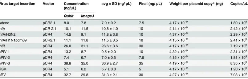
Plasmid concentrations were calculated by performing two quantification methods: 1) fluo-rometry specific to double stranded DNA (Qubit 2.0, dsDNA br Assay Kit, Invitrogen) and 2) pixel intensity measurements using the ImageJ application[7]. Using ImageJ, the pixel intensity of linearized plasmid DNA gel bands could be interpolated into a standard curve consisting of 1KB ladder dilutions (New England Biolaboratories) to predict quantities of unknown bands on the gel. Plasmid DNA was linearized using restriction enzyme NcoI (New England Biolabs) prior to gel electrophoresis. These quantification strategies were chosen to focus on the DNA of interest and to help exclude possible quantification pitfalls of over or underestimating DNA concentrations. Used in combination, these methods accounted for contaminating RNA (fluo-rometry specific for DNA only) as well as contaminating DNA as seen as different sized bands on the gel which could be excluded by only measuring the pixel intensity of gel bands of expected size (~4KB).
Differences between the two quantification methods ranged from 0.2 to 5.4 ng/μL (average
2.6 ng/μL ± 1.8). Final concentrations were calculated by rounding the average of the two
methods to the nearest 2.5ng/μL. The weight of each plasmid was calculated using Geneious
(v.8.1.3), using the known sequence of the vector in addition to the confirmed sequence of the insert. Final copy numbers (perμL) were calculated by dividing the plasmid weights (ng/copy)
into the concentrations of each plasmid (ng/μL). Results of the quantification methods and
downstream calculations are shown inTable 1.
Determination of singleplex real-time LOD
Plasmid DNA was serially diluted to produce eight (8) test concentrations ranging between 1 copies/μL and 1250 copies/μL, depending on the assay. This narrow range was chosen to
iden-tify the lowest potential copy number able to be detected repeatedly, but keep it above theoreti-cal limitations of real time PCR,<3 copies (0.6 copies/uL when using 5uL per reaction)[8]. Seven (7) replicates were tested at each concentration. This process was repeated twice, once using nuclease-free water as the diluent background for the plasmids to assess basic analytical sensitivity and once using total nucleic acid extract (TNA) as background for the plasmids to simulate real clinical matrices. TNA was isolated from clinical specimens using the easyMAG total nucleic acid automated extractor (Biomerieux). A total of 200μL of the clinical specimen
was extracted and final eluate volumes were 60μL. TNA from clinical specimens were screened
by PCR, and only those that demonstrated the absence of target DNA or RNA were qualified to be pooled as clinical background diluent.
reagents and all other assays were performed using Ambion AgPath ID reagents. For assays using the Invitrogen reagents, the following PCR thermal cycling profile was used; 50°C hold for 30 minutes, 95°C hold for 2 minutes, and 45 cycles of 95°C for 15 seconds then 55°C for 30 seconds. For assays using the Ambion reagents, the following PCR thermal cycling profile was used; 45°C hold for 10 minutes, 95°C for 10 minutes, and 45 cycles of 95°C for 15 seconds then 55°C for 1 minute. Reactions were tested using ABI 7500Dx thermal cyclers (Life
Technologies).
Negative controls consisted of no template control replicates (NTC, n = 3) and diluent blank replicates, made up of water or TNA diluent (n = 7) to assess contamination. Positive reactions were defined as those amplification curves that produced cycle threshold (Ct) values at or below 40 cycles. The LOD was chosen as the concentration that demonstrated a percent-age of positivity over all replicates at a particular dilution. The percentpercent-age of positivity was cho-sen using those that were set by the manufacturer for each matching GenMark RVP assay. All but three assays were set by the manufacturer below 100% positivity (InfA/H1N1pdm09, RSVA, and hRV assays only); therefore, the LOD for these particular singleplex assays were estimated using probit analysis to match these probabilities for comparison purposes[11]. Final LODs were expressed as a concentration, copies/μL (Table 2).
Conversion of TCID50/mL concentrations to copies/
μ
L
Cell cultures with known TCID50/mL quantities of target viruses (ATCC) were used to estimate
the LOD for the GenMark RVP assay. Cultures were stored in liquid nitrogen until they were extracted using the easyMAG total nucleic acid automated extractor (Biomerieux). A total of 200μL of the TCID50/mL culture was extracted and final eluate volumes were 60μL. Purified
nucleic acid was stored at -80°C until tested by quantitative real time PCR (qPCR).
Using quantified plasmids containing inserts specific to each assay, ten-fold dilutions were prepared covering 101to 106copies/5μL. Each dilution was tested in triplicate to create a
stan-dard curve. All qPCR assays utilized a sequence-specific hydrolysis probe with the exception of the H3 due to sequence incompatibilities with the ATCC strain being analyzed (seeresults). In this case, a SYBR Green assay (GoTaq, Promega) with new primers were designed to target this specific strain of Influenza A/H3. Alongside the standard curve, dilutions of the isolated nucleic Table 1. Plasmid concentrations and copy number determination.
Virus target insertion Vector Concentration (ng/μL)
avg±SD (ng/μL) Final (ng/μL) Weight per plasmid copy*(ng) Copies/μL
Qubit ImageJ
Adeno pCR2.1 8.0 7.8 7.9±0.2 7.5 4.17 x 10⁻⁹ 1.80 x 10⁹
InfA pCR 2.1 10.1 11.5 10.8±1.0 10 4.14 x 10⁻⁹ 2.42 x 10⁹
InfA/H3N2 pCR4 14.5 9.1 11.8±3.8 10 4.37 x 10⁻⁹ 2.29 x 10⁹
InfA/H1N1pdm09 pCR2.1 11.1 11.8 11.5±0.5 10 4.15 x 10⁻⁹ 2.41 x 10⁹
InfB pCR4 26.0 31.1 28.6±3.6 30 4.17 x 10⁻⁹ 7.19 x 10⁹
hPIV-1 pCR4 13.2 8.7 9.5±2.0 10 4.32 x 10⁻⁹ 2.31 x 10⁹
hPIV-2 pCR4 7.4 6.7 7.0±0.5 7.5 4.15 x 10⁻⁹ 1.81 x 10⁹
hPIV-3 pCR4 38.8 35.0 36.9±2.7 35 4.19 x 10⁻⁹ 8.35 x 10⁹
RSV pCR4 5.1 6.2 5.7±0.8 5 4.17 x 10⁻⁹ 1.20 x 10⁹
hRV pCR4 32.7 29.8 31.3±2.1 30 4.27 x 10⁻⁹ 7.03 x 10⁹
*Weight/copy was calculated using Geneious (v.8.1.3) which considers the exact sequence of the plasmid.
acid derived from the ATCC cultures were tested in triplicate at dilutions that would include reported GenMark eSensor RVP LOD TCID50/mL values. As with the singleplex real-time
PCR assays, reactions were tested on ABI 7500Dx thermal cyclers (Life Technologies) and stan-dard curves and associated unknown quantities were calculated using ABI 7500 v2.3 software. The copy number equivalents for each GenMark eSensor RVP assay’s LOD is shown in
Table 2. The relationship between copy number and TCID50/mL for each ATCC culture tested
is shown inTable 3.
Results
Ten singleplex real-time PCR assays were compared in terms of analytical sensitivity to twelve multiplex assays on the GenMark eSensor RVP. This difference stems from the fact that the singleplex real-time PCR assays are not designed to distinguish between different subgenera of human adenovirus or different subtypes of respiratory syncytial viruses (RSV), while the Gen-Mark eSensor RVP differentiates between human adenovirus C and E as well as RSV subtype A and B. Thus, two additional assays were evaluated for the GenMark eSensor RVP. Analytical sensitivity was expressed as lowest copies/μL concentration for all assays.
The Genmark eSensor RVP capable of distinguishing between different subgenera of adeno-viruses (C vs. E) demonstrated less analytical sensitivity than the generic singleplex real-time PCR assay targeting all adenoviruses, differing by 108.8 copies/μL (2.04 log difference), and
388.8 copies/μL (2.59 log difference), respectively. The difference in sensitivity may be due to
slight variations in the targeted priming region. The singleplex real-time PCR assays use prim-ers designed to anneal highly conserved sequences within the hexon-coding region in order to target all adenoviruses, whereas the GenMark eSensor RVP assays use subgenera-specific hexon primers to make possible the distinction between adenovirus subgenera C and E. Upper respiratory tract infections associated with adenovirus C viruses infect more than 80% of the Table 2. LOD comparison summary.
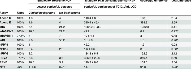
Singleplex Real-time PCR Multiplex PCR GenMark eSensor RVP copies/μL difference Log Difference
Lowest copies/μL detected copies/μL equivalent of TCID50/mL LOD
Assay %pos Clinical background No Background
Adeno C 100% 1.6 4 110.4±8 108.8 2.04
Adeno E 100% 1.6 4 390.4±45.4 388.8 2.59
InfA 100% 5.4 21.2 1286.2±23.2 1280.8 3.11
InfA/H3N2 100% 10.6 21.2 <2.2 8.4 0.92*
InfA/H1N1 97.5% 7 7 10±4.4 3 0.48
InfB 100% 2.6 53.2 1±2.8 1.6 0.20*
hPIV-1 100% 1 1 <2.2 1.2 0.08
hPIV-2 100% 5.4 2.2 1.6±0.6 3.8 0.58*
hPIV-3 100% 2.2 1 134.8±8.4 132.6 2.12
RSVA 97.5% 6.8 3.6 326.2±22.8 319.4 2.50
RSVB 100% 10.6 5.2 120.2±8.6 109.6 2.04
hRV 95% 111.8 82.4 <17 94.8 1.98*
*lower LOD demonstrated for the multiplex assay; 5μL used in each reaction. Adenovirus and RSV assays were not differentiated with the singleplex
real-time PCR assay, although RSV assays were calculated differently based on %pos to be compared. The TCID50/mL concentration for InfA/H3, HPIV 1,
and hRV exceeded the detection limit on the qPCR assay. Copy number difference was calculated by subtracting the lowest copies/μL detected with
clinical background on the singleplex assays from the average copies/μL equivalent converted from TCID50/mL.
population early in life[12]; however, infections with the adenovirus E (serotype 4) can prove to be more severe and even fatal for people living in close quarters, such as military recruits [13]. In terms of surveillance, differentiation of virus subgenera within a population may be clinically useful, regardless of lost sensitivity.
Similarly, the singleplex real-time PCR assay generically targeting respiratory syncytial viruses also demonstrated better sensitivity than the GenMark eSensor RVP assays which are capable of distinguishing between subtypes A and B (319.4 copies/μL, 2.50 log difference and
109.6 copies/μL, 2.04 log difference, respectively). Respiratory syncytial viruses in subtype A
are thought to be more prevalent and virulent than those in subtype B[14]. Subtyping respira-tory syncytial virus may be clinically beneficial when surveilling populations that experience high hospitalization rates associated with the virus, such as Native Americans living in south-west United States and Alaska[15].
Analytical sensitivity of assays targeting the current circulating strains of influenza A viruses in the human population, H3N2 and H1N1pdm09, were highly comparable between the sin-gleplex real-time PCR and multiplex GenMark eSensor RVP assays (8.4 copies/μL, 0.92 log
dif-ference and 3 copies/μL, 0.48 log difference, respectively). Comparing the LOD between the
influenza H3N2 assays proved to be the most challenging. When converting TCID50/mL
con-centrations to copies/μL using qPCR, it was determined that this particular culture contained
an uncommon virus, an Aichi strain (A/Aichi/2/35) circa 1968 (ATCC) and therefore could not be amplified using the singleplex real-time PCR assay, which is designed to detect current influenza A/H3N2 virus strains. However, it was repeatedly detected using the GenMark eSen-sor RVP. This finding suggests that the eSeneSen-sor RVP is capable of detecting a broader range of Influenza A/H3N2 strains while maintaining a comparable analytic sensitivity to that of its sin-gleplex real-time PCR counterpart.
The greatest difference measured between analytic sensitivities was seen with the generic influenza A assay showing a 3.11 log difference in LOD (1280.8 copies/μL difference). Because
the LOD for the generic influenza A assay is much higher than the subtype assays (as described above) for the multiplex GenMark eSensor RVP, difficulty in result interpretation from speci-mens with low influenza A virus titers is likely, since subtypes (H3N2 or H1N1pdm09) have a lower LOD than the generic influenza A assay (e.g. + H3N2,—influenza A). The performance Table 3. Relationship between TCID50/mL concentrations and copy number.
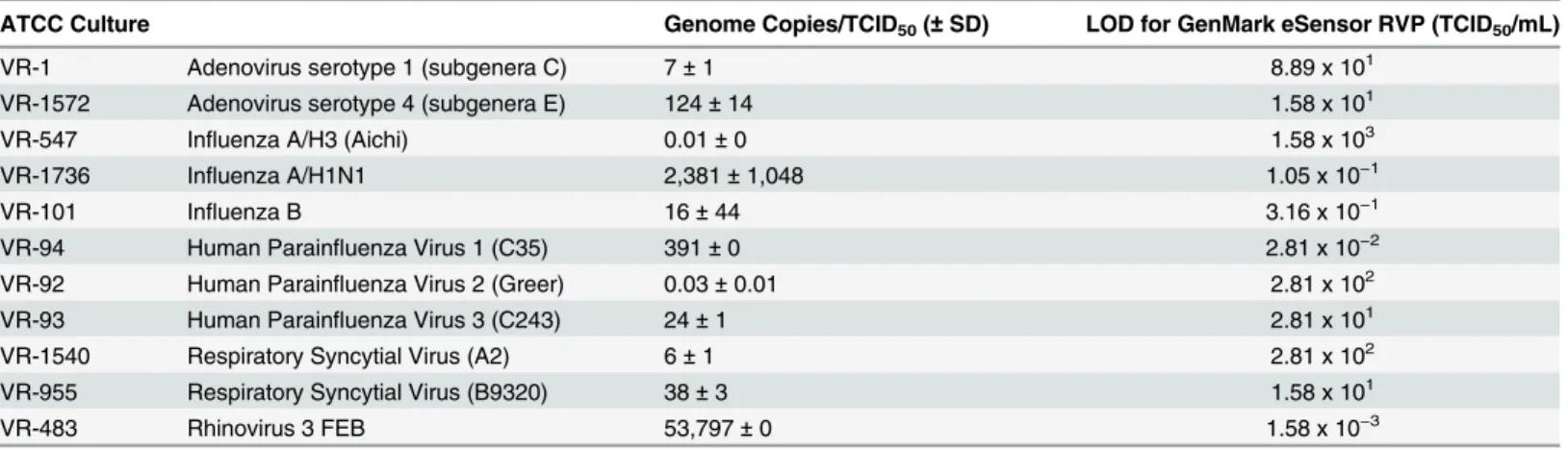
ATCC Culture Genome Copies/TCID50(±SD) LOD for GenMark eSensor RVP (TCID50/mL)
VR-1 Adenovirus serotype 1 (subgenera C) 7±1 8.89 x 101
VR-1572 Adenovirus serotype 4 (subgenera E) 124±14 1.58 x 101
VR-547 Influenza A/H3 (Aichi) 0.01±0 1.58 x 10³
VR-1736 Influenza A/H1N1 2,381±1,048 1.05 x 10⁻¹
VR-101 Influenza B 16±44 3.16 x 10⁻¹
VR-94 Human Parainfluenza Virus 1 (C35) 391±0 2.81 x 10⁻²
VR-92 Human Parainfluenza Virus 2 (Greer) 0.03±0.01 2.81 x 10²
VR-93 Human Parainfluenza Virus 3 (C243) 24±1 2.81 x 101
VR-1540 Respiratory Syncytial Virus (A2) 6±1 2.81 x 10²
VR-955 Respiratory Syncytial Virus (B9320) 38±3 1.58 x 101
VR-483 Rhinovirus 3 FEB 53,797±0 1.58 x 10⁻³
SD = standard deviation, SD could not be calculated for VR-547, VR-94, and VR-483 since the TCID50/mL concentration exceeded the detection limit on
the qPCR assay.
of the generic influenza A assay is an important surveillance tool for tracking genetic changes among influenza A viruses. For instance, specimens demonstrating positivity for influenza A using this generic, highly conserved matrix-coding region may not subtype using the H3N2 or H1N1pdm09 assays, which may indicate that the virus is novel and worthy of alerting public health authorities. In contrast, the influenza B assays were shown to be highly comparable between the singleplex and multiplex assays, with a difference of only 1.6 copies/μL (0.20 log
difference).
Human parainfluenza 1 assays were highly comparable (1.2 copies/μL, 0.08 log difference).
Human parainfluenza 2 assays demonstrated improved sensitivity on the multiplex GenMark eSensor assay (3.8 copies/μL, 0.58 log difference). Human parainfluenza 3 assays demonstrated
the largest difference in analytical sensitivity among the human parainfluenza serotypes, dem-onstrating a 2.12 log improvement in detectability when using the singleplex real-time PCR assay (132.6 copies/μL difference).
Five of the twelve GenMark eSensor RVP assays matched (<1 log difference in copies/μL) the LOD of the real-time singleplex PCR assay targets in this study (Table 2). These include influenza A/H3N2, influenza A/H1N1pdm09, influenza B, and human parainfluenza 1 and 2. Six of the twelve assays compared showed greater sensitivity using the real-time singleplex assays. These include the adenovirus assays (C & E), influenza A, human parainfluenza 3, and RSV (A & B). The GenMark eSensor human rhinovirus assay demonstrated the biggest differ-ence in terms of improved detection when compared to its singleplex counterpart (94.8 copies/
μL, 1.98 log difference, 95% positivity).
The number of genome copies per TCID50/mL value was highly variable ranging from 0.01
to 53,797 (Table 3). LODs set at higher TCID50/mL concentrations (102–103) corresponded to
stock cultures with lower copy numbers (0.01 to 6 copies). LODs set at in the mid-range TCID50/mL concentrations (101to 10−1) corresponded to stock cultures with variable copy
numbers per TCID50/mL (7–2,381 copies). LODs set at lower TCID50/mL concentrations
(10−2
–10−3) corresponded to stock cultures with somewhat higher copy numbers per TCID
50/
mL (391–53,797 copies).
Conclusion
Multiplex PCR applications benefit diagnostics in a clinical laboratory due to their ability to detect and rule-out many related pathogens in a single reaction, reducing tech-time by more than 3 hours for a panel of 10 viruses[1]. However, multiplex PCR platforms continue to carry higher overall costs. Analytic sensitivity, or the lowest possible concentration necessary to pro-duce a reliable result, is an important parameter to consider when replacing singleplex real-time PCR assays with multiplex PCR platforms evolving from newer, more expensive technolo-gies. This experiment aims at finding a method in which to compare LODs of various assays using copy number as the unit of expression.
Choosing a 2.5 log difference to express considerable loss in sensitivity, the multiplex PCR strategy in combination with the GenMark eSensor technology demonstrates a considerable loss in sensitivity for three of the twelve assays assessed. Two of the assays were adenovirus E and respiratory syncytial virus subtype A. Although sensitivity is reduced, further characteriza-tion of viruses in clinical specimens may be of greater clinical importance, especially when par-ticular subtypes are known to be more virulent in the population as is the case with adenovirus serotype 4 (subgenera E) and respiratory syncytial virus subtype A in particular populations.
rule out novel influenza. Better analytic sensitivity was achieved using singleplex real-time PCR, which indicates that influenza A can be detected in clinical specimens even at low titers using this method. Specimens collected from patients that are suspected to have influenza infections that test negative on the GenMark eSensor RVP may need to be tested by more sen-sitive methods to rule out cases of novel influenza.
Expressing LOD in units that can be comparable across methodologies can prove to be diffi-cult experimentally. TCID50/mL measurements can vary depending on how these cultures are
handled in the laboratory in regards to preserving the concentration of infectious virus parti-cles for purposes of experimentation and quantity comparisons. Molecular detection strategies used in clinical laboratories are non-discriminating when identifying infectious or non-infec-tious viruses. PCR methodologies used to detect viral targets in clinical specimens do not pro-vide information regarding the viability of the virus and, therefore, every detection may not point to a causative agent of disease. Other complicating factors to consider when interpreting PCR results are that patients can be asymptomatic carriers or may be exhibiting evidence of a past infections. Viral copy numbers provide an estimate of the number of virus particles in a given volume, but in our experiment, they did not correlate well with the number of infectious particles. To test the analytical sensitivity of a PCR-based methodology, it is important to understand that the intent of the assay is to detect any genome copy targeted by the designed primers, whether these be from infectious or non-infectious virus particles.
Acknowledgments
This research was supported by the Alaska Department of Health and Social Services, Division of Public Health, Section of laboratories. Much of the plasmid development and sequencing was supported in part by the University of Alaska Fairbanks. The ATCC cultures were pur-chased by GenMark Diagnostics, Incorporated in an effort to be consistent with the particular strains used in the FDA validation testing. We would like to thank the staff at the Alaska State Virology Laboratory for all of their help with carrying out testing for this project.
Author Contributions
Conceived and designed the experiments: JP JC. Performed the experiments: JP NF MLW TS JS JK TG SH. Analyzed the data: JP JC. Wrote the paper: JP JC.
References
1. Pierce VM, Hodinka RL (2012) Comparison of the GenMark Diagnostics eSensor Respiratory Viral Panel to Real-Time PCR for Detection of Respiratory Viruses in Children. Journal of Clinical Microbiol-ogy 50: 3458–3465. doi:10.1128/JCM.01384-12PMID:22875893
2. Popowitch EB, O'Neill SS, Miller MB (2013) Comparison of the Biofire FilmArray RP, Genmark eSensor RVP, Luminex xTAG RVPv1, and Luminex xTAG RVP Fast Multiplex Assays for Detection of Respira-tory Viruses. Journal of Clinical Microbiology 51: 1528–1533. doi:10.1128/JCM.03368-12PMID: 23486707
3. Jonsson N, Gullberg M, Lindberg AM (2009) Real-time polymerase chain reaction as a rapid and effi-cient alternative to estimation of picornavirus titers by tissue culture infectious dose 50% or plaque forming units. Microbiology and Immunology 53: 149–154. doi:10.1111/j.1348-0421.2009.00107.x PMID:19302525
4. Gustafsson RKL, Engdahl EE, Fogdell-Hahn A (2012) Development and validation of a Q-PCR based TCID(50) method for human herpesvirus 6. Virology Journal 9: 311–311. doi: 10.1186/1743-422X-9-311PMID:23249654
6. Iwami S, Holder BP, Beauchemin C, Morita S, Tada T, et al. (2012) Quantification system for the viral dynamics of a highly pathogenic simian/human immunodeficiency virus based on an in vitro experiment and a mathematical model. Retrovirology 9: 18. doi:10.1186/1742-4690-9-18PMID:22364292 7. Rasband WS ImageJ, U. S. National Institutes of Health, Bethesda, Maryland, USA,http://imagej.nih.
gov/ij/, 1997–2014.
8. Bustin SA, Benes V, Garson JA, Hellemans J, Huggett J, et al. (2009) The MIQE Guidelines: Minimum Information for Publication of Quantitative Real-Time PCR Experiments. Clinical Chemistry 55: 611– 622. doi:10.1373/clinchem.2008.112797PMID:19246619
9. Weinberg GA, Schnabel KC, Erdman DD, Prill MM, Iwane MK, et al. (2013) Field evaluation of TaqMan Array Card (TAC) for the simultaneous detection of multiple respiratory viruses in children with acute respiratory infection. J Clin Virol 57: 254–260. doi:10.1016/j.jcv.2013.03.016PMID:23608639 10. Wangchuk S, Thapa B, Zangmo S, Jarman RG, Bhoomiboonchoo P, et al. (2013) Influenza
surveil-lance from November 2008 to 2011; including pandemic influenza A(H1N1)pdm09 in Bhutan. Influenza and Other Respiratory Viruses 7: 426–430. doi:10.1111/j.1750-2659.2012.00409.xPMID:22813389 11. Sloan LM (2007) Real-time PCR in clinical microbiology: verification, validation, and contamination
con-trol. Clinical Microbiology Newsletter 29: 87–95.
12. Garnett CT, Erdman D, Xu W, Gooding LR (2002) Prevalence and Quantitation of Species C Adenovi-rus DNA in Human Mucosal Lymphocytes. Journal of Virology 76: 10608–10616. PMID:12368303 13. Robert NP, Joyce AC, Craig TM, Joel CG (2012) Adenovirus-associated Deaths in US Military during
Postvaccination Period, 1999–2010. Emerging Infectious Disease journal 18: 507.
14. Walsh EE, McConnochie KM, Long CE, Hall CB (1997) Severity of Respiratory Syncytial Virus Infection Is Related to Virus Strain. Journal of Infectious Diseases 175: 814–820. PMID:9086135