Peri-implant evaluation of osseointegrated implants
subjected to orthodontic forces: results after three years
of functional loading
Bruna de Rezende Marins1, Suy Ellen Pramiu2, Mauro Carlos Agner Busato3, Luiz Carlos Marchi3, Adriane Yaeko Togashi4
Objective: The objective of this study was to clinically and radiographically assess the peri-implant conditions of implants used as orthodontic anchorage. Methods: Two groups were studied: 1) a test group in which osseointegrated implants were used as orthodontic anchorage, with the application of 200-cN force; and 2) a control group in which implants were not subjected to orthodontic force, but supported a screw-retained prosthesis. Clinical evaluations were performed three, six and nine months after prosthesis installation and 1- and 3-year follow-up examinations. Intraoral periapical radiographs were obtained 30 days after surgical implant placement, at the time of prosthesis installation, and one, two and three years thereafter. The results were compared by Kruskal-Wallis test. Results: There was no statistically significant difference in clinical probing depth (p = 0.1078) or mesial and distal crestal bone resorption (p = 0.1832) during the study period. After three years of follow-up, the mean prob-ing depth was 2.21 mm for the control group and 2.39 mm for the test group. The implants of the control group showed a mean distance between the bone crest and implant shoulder of 2.39 mm, whereas the implants used as orthodontic anchorage showed a mean distance of 2.58 mm at the distal site. Conclusion: Results suggest that the use of stable intraoral orthodontic anchorage did not compromise the health of peri-implant tissues or the longevity of the implant.
Keywords: Bones. Dental implants. Orthodontic appliances. Osseointegration.
1 Graduate student in Oral and Maxillofacial Surgery, Universidade Estadual do
Oeste do Paraná (UNIOESTE), School of Dentistry, Cascavel, Paraná, Brazil.
2 Undergraduate student, Universidade Estadual do Oeste do Paraná
(UNIOESTE), School of Dentistry, Cascavel, Paraná, Brazil.
3 Professor, Universidade Estadual do Oeste do Paraná (UNIOESTE),
Department of Orthodontics, School of Dentistry, Cascavel, Paraná, Brazil.
4 Professor of Periodontology and Oral Implantology, Universidade Estadual
do Oeste do Paraná (UNIOESTE), Department of Implantology, School of Dentistry, Cascavel, Paraná, Brazil.
Submitted: October 05, 2015 - Revised and accepted: January 08, 2016
DOI: http://dx.doi.org/10.1590/2177-6709.21.2.073-080.oar
How to cite this article: Marins BR, Pramiu SE, Busato MCA, Marchi LC,
Togashi AY. Peri-implant evaluation of osseointegrated implants subjected to orthodontic forces: results after three years of functional loading. Dental Press J Orthod. 2016 Mar-Apr;21(2):73-80.
DOI: http://dx.doi.org/10.1590/2177-6709.21.2.073-080.oar
» The authors report no commercial, proprietary or financial interest in the products or companies described in this article.
Contact address: Adriane Yaeko Togashi
Rua Universitária, 2069 – Jd. Universitário, Cascavel/PR – Brasil CEP: 85.814-110 – E-mail: [email protected]
Introdução: o objetivo do presente estudo foi avaliar, clínica e radiograficamente, as condições peri-implantares de implantes usados como ancoragem ortodôntica. Métodos: dois grupos foram estudados: 1) Grupo Teste – no qual os implantes os-seointegráveis foram utilizados como ancoragem ortodôntica, com aplicação de uma força de 200cN; e 2) Grupo Controle – no qual os implantes não foram submetidos à ancoragem ortodôntica, apenas serviram de suporte para fixação de prótese implantossuportada. Avaliações clínicas foram realizadas aos 3, 6 e 9 meses após a instalação das próteses, e após 1 e 3 anos de acompanhamento. Radiografias periapicais intrabucais foram obtidas 30 dias após a colocação do implante, no momento da instalação da prótese e após 1, 2 e 3 anos de acompanhamento. Os resultados foram comparados pelo teste de Kruskal-Wallis. Resultados: não houve diferenças quanto à profundidade clínica de sondagem (p = 0,1078) e a reabsorção das cristas ósseas mesial e distal (p = 0,1832) durante o período avaliado. Após três anos de acompanhamento, a média de profundidade de sonda-gem foi de 2,21mm para o Grupo Controle e de 2,39mm para o Grupo Teste. Os implantes do Grupo Controle apresentaram distância média de 2.39mm entre a crista óssea e o ombro do implante, enquanto os implantes usados como ancoragem orto-dôntica mostraram distância média de 2,58mm na distal. Conclusão: esses resultados sugerem que o uso de uma ancoragem intrabucal estável não compromete a saúde dos tecidos peri-implantares ou a longevidade do implante.
INTRODUCTION
Osseointegrated titanium implants were initially used as abutment for prosthetic reconstruction in fully edentulous patients in order to increase masticatory function.1,2,3 The implants were later used extensively to replace missing teeth in partially edentulous patients, allowing for preservation of the remaining dental struc-tures.1,4,5,6 Other indications have been proposed for osseointegrated implants, such as orthodontic or max-illofacial anchorage,7-11 since decayed or missing teeth can impair orthodontic treatment due to absence of ap-propriate dental anchorage for orthodontic movement. The advantage of osseointegrated implants for orth-odontic anchorage is the absolute immobility of the im-plant, as the periodontal ligament is inexistent, allowing for the application of controlled orthodontic forces without bone resorption. This phenomenon is known as “absolute anchorage.”12 Thus, the implant will irst function as intra-oral orthodontic anchorage and later as prosthetic anchor-age, providing stability, biocompatibility and comfort.13
Since several studies have demonstrated that anchor-age of orthodontic forces on implants seems to be a good alternative in partially edentulous patients who re-quire orthodontic treatment,7,14-18 the present study pro-posed to assess the long-term peri-implant behavior of implants subjected to orthodontic anchorage. Thus, the objective of this study was to clinically and radiographi-cally assess implants used as orthodontic anchorage, as well as the success rate of such implants over a period of three years ater the installation of prostheses over them.
MATERIAL AND METHODS
Patient selection and study design
A prospective clinical study using titanium implants as orthodontic anchorage was conducted. The pa-tients were recruited from the Undergraduate and Postgraduate clinics of the Department of Dental Im-plantology, School of Dentistry, Universidade Estadual do Oeste do Paraná (UNIOESTE), Brazil. The study was approved by the Ethics Committee of the same uni-versity (Process 301/2008-CEP) and were asked to sign a free informed consent form ater receiving detailed in-formation about the study.
Ater patient selection according to inclusion and ex-clusion criteria, the sample was randomly divided into two groups: 1) test group in which osseointegrated implants were used as orthodontic anchorage (n = 26 implants);
and 2) control group in which the implants were only used as support for prostheses (n = 24 implants).
Criteria for inclusion in the study were: 18 years of age or older (mean patient age was 41 years, with a range of 35 to 56 years old); willingness to cooperate with the require-ments of the study; no systemic health condition; good oral hygiene; good periodontal health; suicient alveolar bone volume at the implant recipient site (width: ≥ 6 mm and height: ≥ 8 mm) exclusively for the study group; and type I-III bone quality. Exclusion criteria were: pregnancy or breast-feeding; smoking and use of alcohol or drugs; previous reconstruction at the implant recipient site; insuf-icient alveolar bone volume at the implant recipient site (width: < 6 and height: < 8 mm); presence of residual roots at the recipient site; type IV bone quality; keratinized mu-cosa < 2 mm at the implant recipient site; stomatological and periodontal diseases; and clinical signs of temporo-mandibular dysfunction and bruxism.
The implants were installed according to the num-ber of missing teeth and bone availability in the poste-rior region of the mandible, which required prosthetic rehabilitation and dental movements.
Surgical procedures
Titanium implants were placed under local anesthesia by a dental surgeon and within a single intervention. The surgical procedure consisted of an incision in the alveo-lar ridge crest for preservation of the keratinized mucosa. Subsequently, lingual and buccal mucoperiosteal laps were carefully elevated from the top of the alveolar crest. The implants were placed supracrestally, according to the protocol of the system, and primary stability was always achieved. The mucoperiosteal laps were repositioned for healing by irst intention. Ater one week, the sutures were removed and postoperative control was performed. The patient was advised to properly clean the treated area.
The surgical phase of implant installation consisted of the use of self-tapping external hexagon titanium implants (Dentolex Comércio e Indústria de Materiais Odontológicos, São Paulo, Brazil), installed according to Branemark’s surgical protocol.19 Implants with a di-ameter of 3.75 or 4.0 mm and 8, 10 and 11.5 mm in length were used according to bone availability.
Clinical sequence
Four months ater implant placement, the period cor-responding to osseointegration, reopening and placement of healing abutments were performed. Subsequently, the implant was transferred and the crowns were screwed in place with a torque of 45 Nm. Molds of each patient were taken and each case was planned by an implantodontist and two orthodontists participating in the study.
Occlusion of the provisional acrylic resin screw-retained restorations was established with contact in maximum in-tercuspation and no contact in excursive movements. In the test group, provisional screw-retained restorations received the following orthodontic accessories: TMA wire cantile-ver (0.018 x 0.025-in, Morelli, Sorocaba, Brazil) and NiTi spring (0.25 mm diameter – Morelli, Sorocaba, Brazil). The maximum force applied to the implants was 200 cN. Orthodontic force was used in order to correct minor dental movements, such as molar uprighting and incisor relation-ship, and to improve the occlusal relationship with the ob-jective of obtaining prosthetic space for implant placement. Orthodontic treatment period varied between 9 and 12 months. At the end of orthodontic treatment, provisional restorations were replaced with deinitive prostheses.
Clinical evaluation
Clinical evaluations were performed three, six and nine months ater prosthesis installation. The 1- and 3-year follow-up examinations included the following parameters: 1) modiied plaque index (mPlI)20 for all implants; 2) modi-ied bleeding index (mBlI)20 for all implants; 3) pocket prob-ing depth (PPD): distance between the gprob-ingival margin and pocket depth in millimeters;21,22 and 4) keratinized mucosa width (KMW): distance between the keratinized gingival-mucosa junction and the free gingival margin in millime-ters for implants. All measurements were performed at six aspects of each implant site by means of a Hu-Friedy PCP-UNC probe (Hu-Friedy, Chicago, IL, USA) .
Radiographic evaluation
Intraoral periapical radiographs were obtained before implant placement and 30 days ater surgery for implant placement at the time of prosthesis installation and one, two and three years thereater (Fig 1). The paralleling technique was used. Radiographs were taken with the aid of a digital radiographic sensor (Kavo-Kerr), an individual acrylic positioning device, and an exposure time of 0.4 sec-onds. For the evaluation of changes in peri-implant crestal
bone height, a single examiner measured the linear dis-tance (in mm) from the implant shoulder to the most cor-onary part of the mesial and distal bone crest,23 using the Image Tool analysis program (UTHSCSA, Texas, USA).
Crestal bone measurements were made on the peri-apical radiographs obtained for the 8-mm, 10-mm and 11.5-mm implants. The length of the implant repre-sents the reference to compensate for radiographic dis-tortion. Subsequently, crestal bone measurements were obtained on the mesial and distal side for all implants at the pre-established intervals.24
Follow-up and maintenance
Periodic visits were held for maintenance and rein-forcement of oral hygiene instructions at 3-month inter-vals during the irst year ater prosthesis installation, and at 6-month intervals during the subsequent two years.
Clinical parameters were evaluated at three, six and nine months and one and three years after pros-thesis installation. Radiographic analysis was per-formed 30 days after surgery for implant placement at the time of prosthesis installation and one, two and three years thereafter.
Success criteria established for the present study followed those of Karoussis et al;21 i.e., absence of mobility, absence of subjective complaints (pain, for-eign body sensation, and/or paresthesia), no probing depth of 5 mm or more and positive modified sulcus bleeding index (mSBl), absence of continuous radio-lucency around the implant, and an annual vertical bone loss not exceeding 0.2 mm after the first year since installation.
The results of the clinical parameters as well as bone crestal distance mesially and distally were compared by
Kruskal-Wallis test. A p-value < 0.05 was considered to indicate statistical signiicance, and all calculations were performed by means of GraphPad InStat and GraphPad Prisma 5 sotware (GraphPad Sotware Inc, USA).
RESULTS
Clinical, radiographic and peri-implant parameters showed that the biological response of gingival tissue and bone structure surrounding the implant subjected to orthodontic anchorage was similar to control. Peri-implant health was maintained for approximately one year of anchorage on the implant and over a period of three years of follow-up.
The mean bone crest/shoulder distance of the im-plant during a period of 30 days ater imim-plant instal-lation, at the time of prosthesis instalinstal-lation, and one, two and three years thereater revealed similar bone
Bone crest/shoulder distance of the implant (mm) mean ±SD
Group
Periods
p
30 days 0 1 year 2 years 3 years
mc dc mc dc mc dc mc dc mc dc
Control 2,13
(0.72)
2.33 (0.79)
2.09 (0.74)
2.27 (0.84)
2.15 (0.52)
2.47 (0.78)
2.22 (0.64)
2.53 (0.71)
2.14 (0.63)
2.39
(0.79) 0.1832
(NS)
Test 2.17
(0.74)
2.45
(1.02)
2.08
(0.59)
2.28
(1.03)
2.22
(0.68)
2.06
(0.96)
2.32
(1.02)
2.41
(1.11)
2.36
(0,92)
2.58
(1.19)
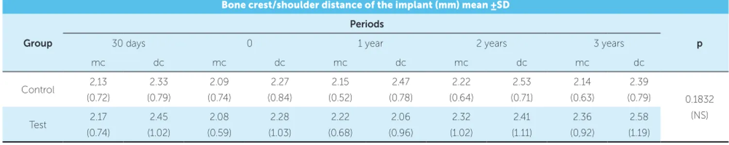
Table 1 - Mean values of the bone crest/shoulder distance of the implant during a period of 30 days after implant installation, at the time of prosthesis installation and one, two and three years thereafter. Crestal bone measurements were obtained on the mesial (mc) and distal (dc) side for all implants at the pre-established time points.
Data are expressed as mean (SD). Kruskal-Wallis test, * p < 0.05; NS = non significant (p > 0.05). Crestal bone measurements: mesial (mc) and distal (dc) sides. Figure 2 - Comparison of the bone crest-implant shoulder distance of the implant during a period of 30 days after implant installation, at the time of prosthesis installation and after one, two and three years. Crestal bone mea-surements were obtained on the mesial (mc) and distal (dc) side for all im-plants, on groups Control (C) and Test (T), at the pre-established time points.
Bone Crest - Implant Shoulder distance (BC-ISD)
Mean BC-ISD (mm)
30d 3 4
1 2
0
0 1y 2y 3y
Periods
Table 2 - Comparison of pocket probing depth (PPD) measurements of the implant at three, six and nine months and at one and three years after prosthesis installation.
Pocket probing depth (mm)
Group Periods p
3 m 6 m 9 m 1 y 3 y
Control 2.30 (0.54) 2.10 (0.41) 2.39 (0.56) 2.25 (0.27) 2.21 (0.47)
p = 0.1078 (NS)
Test 2.57 (0.40) 2.39 (0.29) 2.24 (0.78) 2.51 (0.64) 2.39 (0.45)
Data are expressed as mean (SD); Kruskal-Wallis test, *: p < 0.05; NS = non significant (p > 0.05).
Figure 3 - Comparison of pocket probing depth (PPD) measurements of im-plant at 3, 6 and 9 months and at 1 and 3 years after prosthesis installation, on Control (C) and Test (T) groups.
remodeling of the implant crests for the test and con-trol groups, with no statistically signiicant diference between groups, since the time of implant place-ment, during the application of orthodontic force and throughout the study period (Fig 2 and Table 1). However, the mean bone crest/implant shoulder dis-tance was 2.58 ± 1.19 mm on the distal surface for the test group and 2.39 ± 0.79 mm for the control group ater three years of follow-up.
There was no significant difference in pock-et probing depth bpock-etween groups throughout the study period (Fig 3 and Table 2). The mean prob-ing depth was 2.57 ± 0,40 mm and 2.39 ± 0.45 mm three months and three years after prosthesis instal-lation, respectively, for implants of the test group, and 2.30 ± 0.54 mm and 2.21 ± 0.47 mm for the con-trol group, showing that mean probing depth was unchanged throughout the study period.
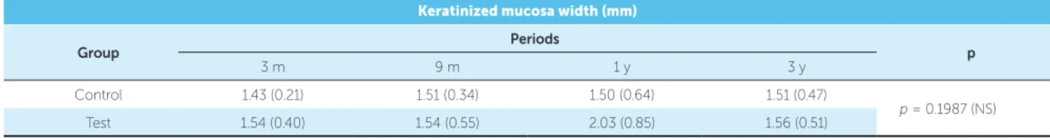
Keratinized mucosa width (KMW) did not differ significantly between groups during the study, with mean values of 1.43 ± 0.21 mm for the control group and 1.54 ± 0.40 mm for the test group, three months after prosthesis installation, and of 1.51 ± 0.47 mm and 1.56 ± 0.51 mm, respectively, after three years of follow-up (Fig 4 and Table 3). Thus, keratinized mu-cosa width remained stable and in sufficient quantity to protect the implant and the health of peri-implant tissue, providing better safety regarding the mainte-nance of peri-implant health.
Table 3 - Comparison of keratinized mucosa width (KMW) measurements of the implant at three and nine months and at one and three years after prosthesis installation.
Keratinized mucosa width (mm)
Group Periods p
3 m 9 m 1 y 3 y
Control 1.43 (0.21) 1.51 (0.34) 1.50 (0.64) 1.51 (0.47)
p = 0.1987 (NS)
Test 1.54 (0.40) 1.54 (0.55) 2.03 (0.85) 1.56 (0.51)
Data are expressed as mean (SD); Kruskal-Wallis test, *: p < 0.05; NS = non significant (p > 0.05).
Figure 4 - Comparison of keratinized mucosa width (KMW) measurements of the implant at 3 and 9 months and at 1 and 3 years after prosthesis installa-tion, on Control (C) and Test (T) groups.
Pocket Probing Depth (PPD)
Keratinized Mucosa Width (KMW)
Mean PPD (mm)
Mean KM
W (mm)
3m
3m 0.0 1.0 2.0
0.5 1.5 2.5
NS
NS C
C
C
C C
C
C
C C T
T
T
T T
T
T
T T 9m
9m
6m 1y
1y
3y
3y 3
1 2
0
Periods
Table 4 - Comparison of the modified bleeding index (mBlI) for all implants at 3, 6 and 9 months and at 1 and 3 years after prosthesis installation.
Table 5 - Comparison of the modified plaque index (mPlI) for all implants at 3, 6 and 9 months and at 1 and 3 years after prosthesis installation.
Modified bleeding index
Group Periods p
3 m 6 m 9 m 1 y 3 y
Control 0.13 (0.29) 0.15 (0.29) 0.06 (0.15) 0.01 (0.05) 0.01 (0.05)
0.017
Test 0.24 (0.40) 0.13 (0.12) 0.12 (0.35) 0.03 (0.10) 0.0 (0.0)
Modified plaque index
Group Periods p
3 m 6 m 9 m 1 y 3 y
Control 0.01 (0.05) 0.0 (0.0) 0.09 (0.30) 0.09 (0.30) 0.0 (0.0)
p < 0.0001
Test 0.01 (0.04) 0.01 (0.04) 0.46 (0.49)a 0.46 (0.60)b 0.08 (0.27)
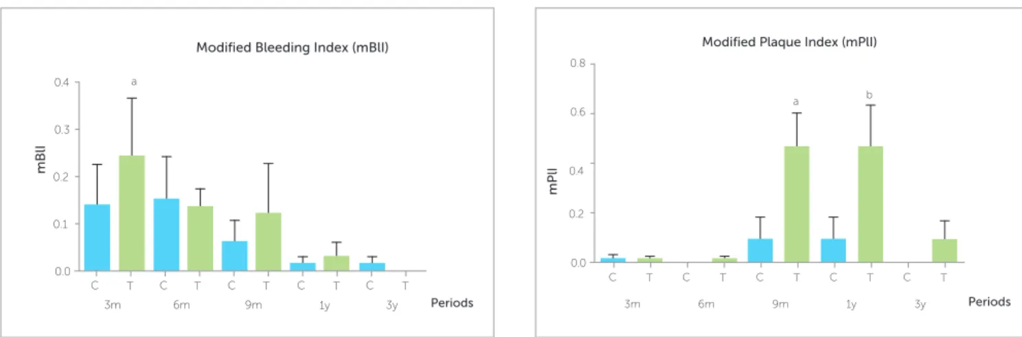
Data are expressed as mean (SD). Kruskal-Wallis test: * = p < 0.05; superscript a = p < 0.05, for T9m group versus C6m and C3y groups; superscript b = p < 0.05, for T1y group versus C3m, T3m, C6m, T6m and C3y groups; NS = non significant: p > 0.05.
Data are expressed as mean (SD); Kruskal-Wallis test: * = p < 0.05; superscript a = p < 0.05, for T3m group versus T3y group; NS = non significant (p > 0.05).
Figure 6 - Comparison of the modified plaque index (mPlI) for all implants at 3, 6 and 9 months and at 1 and 3 years after prosthesis installation. Figure 5 - Comparison of the modified bleeding index (mBlI) for all implants
at 3, 6 and 9 months and at 1 and 3 years after prosthesis installation.
The mean mBlI values did not differ signifi-cantly between groups, with both groups main-taining healthy peri-implant tissues throughout the three years of follow-up after prosthesis implanta-tion (Fig 5 and Table 4). However, the test group revealed a trend towards an increase (p > 0.05) three and nine months after prosthesis installation.
The mean mPlI values did not difer signiicantly be-tween groups. By evaluating diferent periods of control and test groups, we observed an increase in the mean
mPlI values in the test group at nine months and one year, compared to control at three, six months and three years, and test group at three and six months (Fig 6 and Table 5); thus, indicating that the test group presented signiicantly higher plaque formation on the peri-im-plant gingiva during the period orthodontic forces were applied to the implants.
The rate of implant success was 100%, according to the criteria proposed by Karoussis et al.21 No im-plant showed radiographic changes in the bone-imim-plant
Modified Bleeding Index (mBlI) Modified Plaque Index (mPlI)
mBlI
mPlI
3m 3m
a
a b
6m 6m
0.0 0.0
0.3
0.6
0.1 0.2
0.2 0.4
0.4
0.8
C T C T C T C T C T C T C T C T C T C T
interface; no annual vertical bone loss exceeded 0.2 mm ater the irst year since installation, thereby indicating successful osseointegration of implants; and no implant mobility was observed.
DISCUSSION
Diiculty controlling anchorage is a signiicant as-pect in Orthodontics. Standard anchorage devices, such as extraoral appliances and elastics, rely on patient’s co-operation, which may compromise treatment results. The introduction of implants for orthodontic anchorage has decreased the need for patient’s cooperation, com-pared to extraoral appliances, and has provided absolute anchorage biomechanics.9,14,25
In the present study, osseointegrated implants placed
according to the method proposed by Branemark19
were clinically successful, fulilling the proposed out-come criteria. In addition, the implants kept direct bone anchorage throughout the study period, in agreement with the results reported by Roberts et al26 and Higu-chi and Slack.8 In the test group, it was possible to per-form dental movement with an implant as anchorage, without any reciprocal action on the remaining teeth. The amount of peri-implant bone resorption of this group was similar to control.
Orthodontic forces on implants not only all the implants remained irm, but also maintained gingival relationships. This study provides evidence that orth-odontic anchorage can have a favorable efect on mar-ginal peri-implant gingival situation. In the test group, a slight increase (p > 0.05) was detected in keratinized mucosa width during orthodontic force application on implants, followed by a trend towards the reduction of such ater three years of follow-up.
Ater the application of orthodontic force, there was clinical and radiographic peri-implant stability, as illus-trated in Figures 1-6 and Tables 1-5. Results showed a 100% success rate for implants subjected to orthodontic forces of 200 cN; thus, indicating that, ater orthodontic treatment, these implants can receive a ixed prosthe-sis replacing the missing teeth, in addition to improv-ing patient’s dental occlusion. Similarly, Cravero et al7 reported a 100% success rate and satisfactory occlusion with 93 implants placed in the maxilla and mandible.
In the present study, we observed that implants maintained direct bone anchorage throughout orth-odontic treatment, in agreement with data reported by
Roberts et al26 and Higuchi and Slack.8 Trisi et al25 also used implants for orthodontic anchorage in 41 adult patients. The implants were placed in diferent areas, all continued to be stable and were osseointegrated 12 months ater prosthesis placement. These studies dem-onstrated that it was possible to perform small tooth movements without a reciprocal action, and that the dental occlusion of orthodontically treated patients was signiicantly improved.
On the other hand, there have been no reports dem-onstrating the association between anchorage orth-odontic and peri-implant conditions. Diferent time points of the test group showed an increase in the mean mPlI values ater nine months and one year of prosthesis installation. This diference became more pronounced during the application of orthodontic forces. The results showed that the mean mPlI values in the test group with bonded orthodontic devices were higher in comparison to control group without orthodontic devices. The in-luence of impaired oral hygiene was considered; how-ever, mPlI for the test group did not improve in spite of detailed oral hygiene instructions. The result of the mPlI reveals the diiculty performing oral hygiene for the test group during tooth movement. Ater completion of orthodontic treatment, the mean mPlI values became normal. Over a 3-year follow-up, peri-implant param-eters were considered satisfactory in terms of gingival health. The reason for higher susceptibility to biological complications around implants may be discussed in the light of bacterial plaque accumulation in partially eden-tulous dentitions or the host response to the bacterial challenge. The microbiota associated with periodonti-tis and peri-implantiperiodonti-tis has supported the concept that periodontal pathogens are important etiological factors of peri-implant infections.20,27 It is, therefore, obvious that the status of peri-implant health is of utmost im-portance for the longevity of implants installed.
According to Werbein and Merz28 and Pinho et al,29 intraosseous titanium implants yield the best results for orthodontic use; thus, reducing treatment time.
CONCLUSION
On the basis of the present results, we suggest that subjecting osseointegrated implants to orth-odontic forces can be a safe technique for prosthetic rehabilitation and an alternative for the orthodon-tic treatment of partially edentulous patients, since there was no significant peri-implant bone loss after orthodontic activation. Additionally, there were no changes in peri-implant probing depth or in the gingiva, and no bleeding or presence of peri-implant plaque was observed during a 3-year
follow-up after installation of implant-supported prosthesis.
Author contributions
Conception or design of the study: MCAB, LCM, AYT. Data acquisition, analysis or interpretation: BRM, SEP, MCAB, LCM, AYT. Writing the article: BRM, SEP, MCAB, LCM, AYT. Critical revision of the article: BRM, SEP, AYT. Final approval of the article: BRM, SEP, MCAB, LCM, AYT. Obtained funding: AYT. Overall responsibility: BRM, SEP, MCAB, LCM, AYT.
1. Adell R, Lekholm U, Rockler B, Branemark PI. A 15-year study of
osseointegrated implants in the treatment of the edentulous jaw. Int J Oral Surg. 1981;10(6):387-416.
2. Carrión JP, Barbosa IR, López JP. Osseointegrated implants as orthodontic anchorage and restorative abutments in the treatment of partially edentulous adult patients. Int J Periodontics Restorative Dent. 2009;29(3):333 40.
3. Jones SD, Jones FR. Tissue integrated implants for the partially edentulous patient. J Prosthet Dent. 1988;60(3):349 57.
4. Lekholm U, Gunne J, Henry P, Higuchi K, Linden U, Bergstrom C, et al. Survival of the branemark implant in partially edentulous jaws: a 10-year prospective multicenter study. Int J Oral Maxillofac . 1999;14(5):639-45.
5. Albrektsson T, Dahl E, Enbom L, Engevall S, Engquist B, Eriksson AR, et al. Osseointegrated oral implants: a Swedish multicenter study of 8139 consecutively inserted nobelpharma implants. J Periodontol. 1988;59(5):287-96.
6. Oldman J, Lekholm U, Kholm U, Jemt T, Branemark PI, Thilander B. Osseointegrated titanium implants: a new approach in orthodontic treatment. Eur J Orthod. 1988 May;10(2):98-105.
7. Cravero RM, Ibañez JC. Assessing double acid -etched implants submitted to orthodontic forces and used as prosthetic anchorages in partially edentulous patients. Open Dent J. 2008; 2: 30-7.
8. Higuchi KW, Slack JM. The use of titanium ixtures for intraoral anchorage: report of a case. Int J Oral Maxillofac Implants. 1991;6:338-44.
9. Block MS, Hofman DR. A new device for absolute anchorage for orthodontics. Am J Orthod Dentofacial Orthop. 1995;107(3):251- 8.
10. Goto M, Jin-Nouchi S, Ihara K, Katsuki T. Longitudinal follow-up of osseointegrated implants in patients with resected jaws. Int J Oral Maxillofac Implants. 2002;17(2):225-30.
11. Roumanas ED, Freymiller EG, Chang TL, Aghaloo T, Beumer J. Implant-retained prostheses for facial defects: an up to 14-year follow-up report on the survival rates of implants at UCLA. Int J Prosthodont. 2002;15(4):325-32.
12. Liaw Yc, Kuang Sh, Chen Yw, Hung Kf, Tsai Hc, Kao Sy, et al. Multimodality treatment for rehabilitation of adult orthodontic patient with complicated dental condition and jaw relation. J Chin Med Assoc. 2008 Nov;71(11):594-600. 13. Cochran DL, Bosshardt DD, Grize L, Higginbottom FL, Jones AA, Jung RE, et
al. Bone response to loaded implants with non matching implant abutment diameters in the canine mandible. J Periodontol. 2009;80(4):609 17. 14. Thilander B, Odman J, Lekholm U. Orthodontic aspects of the use of
oral implants in adolescents: a 10-year follow-up study. Eur J Orthod. 2001;23(6):715-31.
15. Drago CJ. Use of osseointegrated implants in adult orthodontic treatment: a clinical report. J Prosthet Dent. 1999;82(5):504-9.
16. Willems G, Carels CE, Naert IE, Van Steenberghe D. Interdisciplinary treatment planning for orthodontic-prosthetic implant anchorage in a partially edentulous patient. Clin Oral Implants Res. 1999;10(4):331-7.
REFERENCES
17. Goodacre CJ, Brown DT, Roberts WE, Jeiroudi MT. Prosthodontic considerations when using implants for orthodontic anchorage. J Prosthet Dent .1997;77(2):162-70.
1.8 Celenza F. Implant-enhanced tooth movement: indirect absolute anchorage. Int J Periodontics Restorative Dent. 2003;23(6):533-41. 19. Bränemark P-I. An experimental and clinical study of osseointegrated in
treatment of the edentulous jaws. Experience from a 10-year period. Scand J Plast Reconstr Surg. 1977;16:11-8.
20. Mombelli A, Van Oosten MAC, Schurch E, Lang NP. The microbiota associated with successful or failing osseointegrated titanium implants. Oral Microbiol Immunol. 1987 Dec;2(4):145-51.
21. Karoussis IK, Salvi GE, Heitz-Mayield LJ, Brägger U, Hämmerle CH, Lang NP. Long-termimplant prognosis in patients with and without a history of chronic periodontitis: a 10-year prospective cohort study of the ITIs Dental Implant System. Clin Oral Implants Res. 2003 Jun;14(3):329-39.
22. Karoussis IK, Müller S, Salvi GE, Heitz-Mayield LJ, Brägger U, Lang NP. Association between periodontal and peri-implant conditions: a 10-year prospective study. Clin Oral Implants Res. 2004 Feb;15(1):1-7. 23. Smith DE, Zarb G. Criteria for success of osseointegrated endosseous
implants. J Prosthet Dent. 1989 Nov;62(5):567-72.
24. Weber HP, Buser D, Donath K, Fiorellini JP, Doppalapudi V, Paquette DW, et al. Comparison of healed tissues adjacent to submerged and non-submerged unloaded titanium dental implants. A histometric study in beagle dogs. Clin Oral Implants Res. 1996 Mar;7(1):11-9.
25. Trisi P, Rebaudi A. Progressive bone adaptation of titanium implants during and after orthodontic load in humans. Int J Periodontics Restorative Dent. 2002 Feb;22(1):31-43.
26. Roberts WE, Engen DW, Schneider PM, Hohlt WF. Implant anchored orthodontics for partially edentulous malocclusions in children and adults. Am J Orthod Dentofacial Orthop. 2004 Sept;126(3):302-4.
27. Leonhardt A, Adolfsson B, Lekholm U, Wikström M, Dahlén G. Longitudinal microbiological study on osseointegrated titanium implants in partially edentulous patients. Clin Oral Implants Res. 1993 Sept;4(3):113-20. 28. Wehrbein H, Merz BR, Hammerle CH, Lang NP. Bone-to-implant contact of
orthodontic implants in humans subjected to horizontal loading. Clin Oral Implants Res. 1998 Oct;9(5):348-53.
29. Pinho T, Neves M, Alves C. Multidisciplinary management including periodontics, orthodontics, implants, and prosthetics for an adult. Am J Orthod Dentofacial Orthop. 2012 Aug;142(2):235-45.