ISSN 0100-879X
BIOMEDICAL SCIENCES
AND
CLINICAL INVESTIGATION
www.bjournal.com.br
www.bjournal.com.br
Volume 44 (8) 729-813 August 2011
Braz J Med Biol Res, August 2011, Volume 44(8) 762-766
doi: 10.1590/S0100-879X2011007500066
Induction of Zenk protein expression within the nucleus taeniae of
the amygdala of pigeons following tone and shock stimulation
I. Brito, L.R.G. Britto and E.A.M. Ferrari
Institutional Sponsors
The Brazilian Journal of Medical and Biological Research is partially financed by
Faculdade de Medicina de Ribeirão Preto Campus
Ribeirão Preto
Ex plor e H igh - Pe r for m a n ce M S Or bit r a p Te ch n ology I n Pr ot e om ics & M e t a bolom ics
Induction of Zenk protein expression within
the nucleus taeniae of the amygdala of
pigeons following tone and shock stimulation
I. Brito
1, L.R.G. Britto
2and E.A.M. Ferrari
31Departamento de Fisiologia, Universidade Metropolitana de Santos, Santos, SP, Brasil 2Departamento de Fisiologia e Biofísica, Instituto de Ciências Biomédicas,
Universidade de São Paulo, São Paulo, SP, Brasil 3Departamento de Anatomia, Biologia Celular, Fisiologia e Biofísica, Instituto de Biologia,
Universidade de Campinas, Campinas, SP, Brasil
Abstract
In this study, we evaluated the expression of the Zenk protein within the nucleus taeniae of the pigeon’s amygdala (TnA) after training in a classical aversive conditioning, in order to improve our understanding of its functional role in birds. Thirty-two 18-month-old adult male pigeons (Columba livia), weighing on average 350 g, were trained under different conditions: with tone-shock associations (experimental group; EG); with shock-alone presentations (shock group; SG); with tone-alone presenta-tions (tone group; TG); with exposure to the training chamber without stimulation (context group; CG), and with daily handling (naive group; NG). The number of immunoreactive nuclei was counted in the whole TnA region and is reported as density of
Zenk-positive nuclei. This density of Zenk-positive cells in the TnA was significantly greater for the EG, SG and TG than for the
CG and NG (P < 0.05). The data indicate an expression of Zenk in the TnA that was driven by experience, supporting the role of this brain area as a critical element for neural processing of aversive stimuli as well as meaningful novel stimuli.
Key words: Amygdala; Nucleus taeniae of the amygdala; Fear conditioning; Zenk; Pigeon
Introduction
Correspondence: I. Brito, Rua Padre Manoel de Paiva, 371, Apto. 21, 09070-230 Santo André, SP, Brasil. E-mail: [email protected]
Received December 20, 2010. Accepted May 4, 2011. Available online May 20, 2011. Published August 19, 2011.
Many studies have shown the key role played by the mammalian amygdaloid circuits in aversive memory, and most of them have considered the participation of the rodent amygdala in fear conditioning (1-3). The amygdaloid nuclei
are also involved in the processing of emotionally significant
sensory stimuli in a social context, as is evidenced by lesion studies with different animals such as reptiles, rodents and nonhuman primates (4,5).
In birds, the arcopallium includes several neuronal populations that are considered to be homologous to the regions that constitute the mammalian amygdala. The nucleus taeniae of the amygdala (TnA) and the subpallial amygdaloid nuclei are considered to be comparable to the mammalian medial amygdala (6,7). This nomenclature has been proposed recently by Reiner et al. (8) based on neurochemical, hodological and behavioral data. The physiology of the TnA has several features that support
this similarity (7) and that are confirmed by anatomical and
chemical findings (6,9). Also, the behavioral consequences
of TnA lesions in pigeons are consistent with the generally accepted role of the amygdala in learning, social behavior and in affective states (7,10), but several aspects of the functional organization of the TnA still remain unclear.
Studies that analyze molecular mechanisms underlying the amygdala-dependent learning and memory processes indicate that immediate-early gene expression [e.g., c-fos
Zenk expression in the nucleus taeniae following tone and shock 763
Material and Methods
Adult male pigeons (Columba livia) aged 18 months, weighing on average 350 g, were used in the present study. As previously described (11), electrodes were chronically
implanted around the pubic bone of all animals and a flex -ible cable for shock delivery was connected to a plug. The
animals were divided into five groups and the experimental
sessions were initiated after adaptation of the pigeon to the experimental conditions. Experimental pigeons (EG; N = 6) received three tone (1000 Hz, 85 dB, 1 s) and shock (10 mA, 35 ms) associations at the 5th, 10th, and 15th min of a single 21-min training session. The pigeons in the shock group (SG; N = 7) received three shock-alone presentations, and the pigeons in the tone group (TG; N = 7) received three tone-alone presentations; the inter-stimulus interval was 5 min. Pigeons in the context group (CG; N = 6) were exposed to the training chamber without stimulus presen-tation. The naive pigeons (NG; N = 6) were taken to the laboratory, weighed and immediately returned to their home cage, without any exposure to the experimental chamber. These training sessions were conducted between 8:00 and 9:00 am. The behavior of the pigeons was observed by a
trained observer who recorded the behavioral sequence
using 30-s time-sampling intervals during the whole ses-sion. The freezing records were pooled in blocks of 180 s
each (11). The accumulated frequency of freezing during the first two blocks (initial blocks; the first 360 s) and the last two blocks (final blocks; the last 360 s) was used for the subsequent behavioral analysis. Between-group differ -ences related to the occurrence of freezing were analyzed by two-way ANOVA, considering group (EG, SG, TG, and
CG) and blocks (initial and final) as independent variables
and mean number of freezing responses as the dependent variable. The Tukey-Kramer testwas used for post hoc
multiple comparisons.
One hour after the end of the experimental session, for the Zenk analyses, the pigeons were perfused with saline and 2% paraformaldehyde in 0.1 M phosphate buf-fer (PB) under ketamine (5 mg/100 g, im) and xylazine (2 mg/100 g, im) anesthesia. The brains were post-fixed in
4% paraformaldehyde for 4 h and subsequently stored in
a 30% sucrose solution at 4°C for at least 48 h. The brains were then sectioned (30 µm) on a sliding microtome in the coronal plane. Sections were incubated with a rabbit polyclonal antibody against the Zenk protein (c-189; Santa Cruz Biotecnology, USA) diluted 1:1000 in PB containing 0.3% Triton X-100. Incubation with this primary antibody lasted 12-16 h at 22°. Following three 10-min washes in PB, the sections were incubated with a biotinylated goat anti-rabbit serum (Jackson Laboratories, USA) diluted 1:200 in PB with 0.3% Triton X-100 at 22°C for 1 h. Finally, the sections were incubated for 2 h with the avidin-biotin-peroxidase complex (ABC Elite Kit; Vector Labs, USA). The reaction with 0.05% 3.3’-diaminobenzidine solution
with 3 µL 0.01% H2O2 for 5 min was intensified with 0.05% osmium tetroxide. The sections were mounted on gelatin-coated glass slides, dehydrated in an ethanol series, and coverslipped with Permount (Fisher, USA). Sections from both brain hemispheres were examined under a light mi-croscope. The immunoreactive neuronal nuclei expressed
within the TnA were quantitated with the NIH Image software (13). A threshold for stained cell counting was defined on
the basis of background staining, and the cells exhibiting at least three times higher absorbance than the threshold were counted. The count was reported as the density of Zenk-positive nuclei (nuclei/mm2). A minimum of four sec-tions from each subject were examined.
The density of Zenk-positive nuclei computed for each group was compared by one-way ANOVA, considering the group (EG, SG, TG, CG, and NG) as the independent variable. The post hoc analyses were performed using the Tukey-Kramer test.
The experimental protocol was approved by the Eth-ics Committee for Animal Experimentation of Instituto de Biologia, UNICAMP, Brazil (Protocol #219-1).
Results
In Figure 1, Panel A illustrates the freezing data for a pool of 30-s time-sampling intervals at the beginning (initial
blocks, 360 s) and at the end (final blocks, 360 s) of the
session. During the initial blocks in the training session, EG and SG animals presented a lower expression of freezing
than they did in the final blocks of the session. Freezing was
observed in the initial blocks of the TG session, although it decreased along the session. CG animals did not show
freezing in any block analyzed. ANOVA confirmed a sig
-nificant effect of group (F(3,22) = 22.26; P < 0.0001) and of block (F(3,22) = 15.79; P < 0.001) and a significant interaction between group and block (F(3,9) = 11.83; P < 0.0001). The
post hoc multiple comparisons with the Tukey-Kramer test
indicated that EG and SG were significantly different from
TG and CG (P < 0.05) and that the occurrence of freezing
in the final blocks of the training for EG and SG was sig
-nificantly more frequent than that in the initial blocks of the
four groups (EG, SG, TG, and CG) as well as than that in
the final blocks of TG and CG (P < 0.05). However, at the
beginning of the session the occurrence of freezing was similar for all groups (P > 0.05).
The data for Zenk protein expression (Figure 1, Panel B) show that the training induced greater density of Zenk-positive cell in the TnA of EG, SG, and TG pigeons than in the TnA of CG and NG pigeons; moreover the density of Zenk-positive cells was greater for the TnA of the SG
than for the other groups. One-way ANOVA confirmed a significant difference among groups, F(5,24) = 17.25; P < 0.1. Multiple post hoc comparisons (Tukey-Kramer’s
multiple-comparison test) indicated significant differences
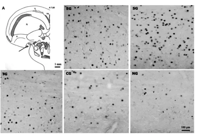
the other groups (P < 0.05) as well as between the groups stimulated with tone alone or tone-shock, TG and EG, and the control groups CG and NG (P< 0.05). A clear difference in the number of Zenk-labeled cells was readily observed in the digital images of frontal sections of the TnA in each group of pigeons (Figure 2).
Discussion
The present study shows that training with tone-shock pairings as well as with tone or shock presented alone triggered an enhanced expression of the Zenk protein in the TnA of pigeons. These data indicate an expression of Zenk that was driven by experience - that is, by the training conditions that were conducted for each group. Moreover, the different groups showed differences in the density of Zenk-positive nuclei, indicating that the type of training af-fected the activation of Zenk in the TnA. Additionally, the freezing behavior data showed that the presentation of an unconditioned aversive stimulus, whether in association with the tone (EG) or not (SG) induced an increase of freezing behavior during the session, whereas the presentation of tone alone resulted in a decrease in freezing during the session, as seen for TG pigeons. On the other hand, CG pigeons showed no freezing during exposure to the experimental chamber.
The fact that the TnA of SG pigeons, which received shock-alone stimulation, exhibited higher expression of Zenk
compared to the other groups suggests that this area may be involved in the processing of aversive stimuli. Furthermore, the TnA may be also involved in the contingent relationship between context and shock, since the presentation of shock in a particular context establishes the condition for aversive contextual conditioning. The increase in freezing observed in SG supports this argument.
In rodents, context conditioning depends on complex hippocampal connections to the amygdala (4,14), suggest-ing that the amygdala has an important role in representsuggest-ing the value of a stimulus. This is in agreement with Campeau et al. (15) who reported elevated c-fos mRNA expression in the amygdala of rats induced by both unconditioned and conditioned fear. There are studies suggesting a reciprocal connection between the TnA and the hippocampal formation in birds, as in mammals, and that the primary target site of the projections from the TnA is the parahippocampal area, and not the hippocampus itself (7).
In addition, discrete tone stimulation can have a novelty value and the exposure to such stimulation induced a high expression of Zenk in this group. Thus, the data suggest that the TnA may also be a critical element for processing of meaningful novel stimuli, a fact that seems to contrast with suggestions that the amygdala may function only in the learning of emotionally salient associations (16). In fact, there are afferent projections reaching the TnA originating from the thalamic auditory nucleus ovoidalis shell and the nucleus subrotundus (17), which may have been related
Zenk expression in the nucleus taeniae following tone and shock 765
to increased Zenk expression in TG animals. However, we do not have a complete knowledge about the intrinsic and extrinsic connections of the arcopallium neurons in birds, or about possible functional subdivisions of the TnA.
Zenk expression in the TnA observed in the present study was greater for SG, EG and TG pigeons than for CG or NG pigeons, indicating that both paired and discrete tone and shock stimulation trigger neuronal responses in this brain area, which mediates changes in the regulation of gene expression. This is in agreement with evidence from research with rodents that has demonstrated that immediate-early genes like zenk (egr-1 or zif268)are in-creased in the amygdala nuclei (lateral, basal and central nuclei) that are known to be involved in fear conditioning, foot shock stress and novelty (18).
References
1. LeDoux JE, Cicchetti P, Xagoraris A, Romanski LM. The lat-eral amygdaloid nucleus: sensory interface of the amygdala in fear conditioning. J Neurosci 1990; 10: 1062-1069. 2. Garcia R, Vouimba RM, Baudry M, Thompson RF. The
amygdala modulates prefrontal cortex activity relative to conditioned fear. Nature 1999; 402: 294-296.
3. Furlong TM, Cole S, Hamlin AS, McNally GP. The role of prefrontal cortex in predictive fear learning. Behav Neurosci,
In fact, to date most fear conditioning studies have
involved rodents and have confirmed the critical role of the
amygdaloid circuitry in processes of conditioning to discrete or paired stimuli (2). Interestingly, the medial nucleus of the rodent amygdala does not appear to be involved in fear conditioning (19). Additional studies will be necessary to better clarify the functional role played by the TnA in fear behavior and, particularly, whether different neuronal populations are involved in unconditioned and conditioned response to aversive/emotional stimuli.
Acknowledgments
Research supported by CNPq, FAPESP, CAPES, and
FAEP/UNICAMP.
2010: 124: 574-586.
4. Maren S. Neurobiology of Pavlovian fear conditioning. Annu
Rev Neurosci 2001; 24: 897-931.
5. Prather MD, Lavenex P, Mauldin-Jourdain ML, Mason WA, Capitanio JP, Mendoza SP, et al. Increased social fear and decreased fear of objects in monkeys with neonatal amygdala lesions. Neuroscience 2001; 106: 653-658. 6. Yamamoto K, Sun Z, Wang HB, Reiner A. Subpallial
amygdala and nucleus taeniae in birds resemble extended amygdala and medial amygdala in mammals in their expres-sion of markers of regional identity. Brain Res Bull 2005; 66: 341-347.
7. Cheng M, Chaiken M, Zuo M, Miller H. Nucleus taenia of the amygdala of birds: anatomical and functional studies in ring doves (Streptopelia risoria) and European starlings (Sturnus
vulgaris). Brain Behav Evol 1999; 53: 243-270.
8. Reiner A, Perkel DJ, Mello CV, Jarvis ED. Songbirds and the revised avian brain nomenclature. Ann N Y Acad Sci 2004; 1016: 77-108.
9. Atoji Y, Saito S, Wild JM. Fiber connections of the compact division of the posterior pallial amygdala and lateral part of the bed nucleus of the stria terminalis in the pigeon
(Columba livia). J Comp Neurol 2006; 499: 161-182.
10. Thompson RR, Goodson JL, Ruscio MG, Adkins-Regan E. Role of the archistriatal nucleus taeniae in the sexual
behav-ior of male Japanese quail (Coturnix japonica): a comparison of function with the medial nucleus of the amygdala in mam-mals. Brain Behav Evol 1998; 51: 215-229.
11. Brito I, Britto LR, Ferrari EA. Classical tone-shock
condition-ing induces Zenk expression in the pigeon (Columba livia) hippocampus. Behav Neurosci 2006; 120: 353-361. 12. Lanahan A, Worley P. Immediate-early genes and synaptic
function. Neurobiol Learn Mem 1998; 70: 37-43.
13. Rasband WS, Bright DS. NIH Image: A public domain im-age processing program for the Macintosh. Microbeam Anal
1995; 4: 137-414.
14. Maren S. Synaptic mechanisms of associative memory in the amygdala. Neuron 2005; 47: 783-786.
15. Campeau S, Hayward MD, Hope BT, Rosen JB, Nestler EJ, Davis M. Induction of the c-fos proto-oncogene in rat amygdala during unconditioned and conditioned fear. Brain Res 1991; 565: 349-352.
16. Cahill L, Weinberger NM, Roozendaal B, McGaugh JL. Is the
amygdala a locus of “conditioned fear”? Some questions and
caveats. Neuron 1999; 23: 227-228.
17. Wild JM, Karten HJ, Frost BJ. Connections of the auditory forebrain in the pigeon (Columba livia). J Comp Neurol 1993; 337: 32-62.
18. Rosen JB, Adamec RE, Thompson BL. Expression of egr-1 (zif268) mRNA in select fear-related brain regions following exposure to a predator. Behav Brain Res 2005; 162: 279-288.
19. Li CI, Maglinao TL, Takahashi LK. Medial amygdala modula-tion of predator odor-induced uncondimodula-tioned fear in the rat.
Behav Neurosci 2004; 118: 324-332.
20. Karten HJ, Hodos W. A stereotaxic atlas of the brain of the