Critical steps in the early evolution
of the isocortex. Insights from
developmental biology
Departamento de Psiquiatría, Facultad de Medicina y Centro de Investigaciones Médicas, Pontificia Universidad Católica de Chile, and Millenium Nucleus for Integrative Neuroscience, Santiago, Chile
F. Aboitiz, J. Montiel and J. López
Abstract
This article proposes a comprehensive view of the origin of the mammalian brain. We discuss i) from which region in the brain of a reptilian-like ancestor did the isocortex originate, and ii) the origin of the multilayered structure of the isocortex from a simple-layered structure like that observed in the cortex of present-day reptiles. Regarding question i there have been two alternative hypotheses, one suggesting that most or all the isocortex originated from the dorsal pallium, and the other suggesting that part of the isocortex originated from a ventral pallial component. The latter implies that a massive tangential migration of cells from the ventral pallium to the dorsal pallium takes place in isocortical development, something that has not been shown. Question ii refers to the origin of the six-layered isocortex from a primitive three-layered cortex. It is argued that the superficial isocortical layers can be considered to be an evolutionary acquisition of the mammalian brain, since no equivalent structures can be found in the reptilian brain. Furthermore, a characteristic of the isocortex is that it develops according to an inside-out neurogenetic gradient, in which late-produced cells migrate past layers of early-produced cells. It is proposed that the inside-out neurogenetic gradient was partly achieved by the activation of a signaling pathway associated with the Cdk5 kinase and its activator p35, while an extracellular protein called reelin (secreted in the marginal zone during development) may have pre-vented migrating cells from penetrating into the developing marginal zone (future layer I).
Correspondence
F. Aboitiz
Departamento de Psiquiatría Facultad de Medicina Universidad Católica de Chile Marcoleta No. 387, 2º piso Casilla 114-D
Santiago 1 Chile
Presented at the IV International UNESCO Course on “What the Developing Cerebral Cortex Tells About the Adult Cortex (and Vice Versa)”, Rio de Janeiro, RJ, Brazil, December 3-7, 2001.
Research partially supported by Fondecyt (No. 1970294), and by the Millenium Nucleus for Integrative Neuroscience. J. Montiel was the recipient of a Conicyt doctoral fellowship.
Received July 12, 2002 Accepted October 7, 2002
Key words
·Cdk5
·Dorsal ventricular ridge ·Reelin
·Tangential migration ·Ventral pallium
Introduction
This paper is concerned with the early stages of mammalian brain evolution. Our strat-egy to approach this issue will be to borrow evidence from developmental biology and from neuroanatomy in order to validate or reject proposals of homology and evolutionary mechanisms. In order to do so, we will first very briefly discuss the phylogenetic relation-ships of terrestrial vertebrates to show the position of mammals in relation to birds and
func-tional sense of the generation of the inverted, inside-out neurogenetic gradient of the mam-malian isocortex.
Phylogenetic relationships and differences in brain structure
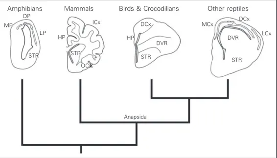
Figure 1 indicates a family tree, or cla-dogram of different vertebrates, together with a diagram of their cerebral hemispheres. As can be seen, amphibians are the sister group of mammals, birds and reptiles all together; mammals are the sister group of birds and reptiles. Note that birds are not the sister group of reptiles. Instead, birds belong to reptiles, and the structure of their brain re-flects this phylogenetic position. This paper will be concerned with the transition be-tween the common ancestor of mammals and reptiles, and modern mammals.
The first reptiles to appear in the fossil record, called anapsids (also termed stem rep-tiles), probably gave rise to all present-day reptiles, including birds (1). Another group of early reptiles, called synapsids, were the an-cestors of the first mammals. The relationship of the synapsids with the stem reptiles is rather obscure. Interestingly, the cranial cavity of both anapsids and synapsids was small and
narrow, suggesting that they had a small brain (2). In both lines, primitive reptiles and mam-mal-like reptiles, the expansion of the brain was a relatively late event. In the mammalian line, the brain became enlarged only when the first true mammals appeared, and was more or less simultaneous with the origin of the middle ear (3,4). Therefore, the expansion of the mam-malian brain took place as a very fast evolu-tionary step, and relatively late in the history of these animals. This is important because it indicates that the enlargement of the brain may have occurred independently in the line lead-ing to reptiles and in the line leadlead-ing to mam-mals. Thus, one important question that we will address in this paper, from the perspective of comparative neuroanatomy, is to what ex-tent it is correct to consider mammals as de-rived from reptiles as we know them today.
Before we deal with the main questions, it will be useful to recall some general aspects of comparative brain anatomy. Figure 1 sche-matically illustrates the structure of the brain in the main classes of terrestrial vertebrates. The brains of amphibians have the simplest organization of all vertebrates and possibly resemble the ancestral amniote brain. Am-phibian brains have no large cell masses, and most cells are located in the periventricular
Figure 1. Phylogenetic relations of terrestrial vertebrates. In each taxon, one cerebral hemisphere of a representative species is de-picted. Medial is to the left of the observer. DCx, dorsal cor-tex; DP, dorsal pallium; DVR, dor-sal ventricular ridge; HP, hippo-campus; LCx, lateral cortex; LP, lateral pallium; MCx, medial cor-tex; MP, medial pallium; STR, corpus striatum; OCx, olfactory cortex; ICx, isocortex.
Amphibians
MP
LCx
Mammals Birds & Crocodilians Other reptiles
LP
STR
HP
ICx
STR
HP
DVR
STR
MCx
DVR
STR
DCx DCx
OCx
zone, there being little cell migration away from the latter. Thalamic sensory projections terminate mostly in the medial pallium and in the corpus striatum (5). The brains of reptiles have a small, three-layered cerebral cortex consisting of a medial part and a dorsomedial part, both of which make up the homologue of the hippocampal formation of mammals; a lateral part which corresponds to the olfactory cortex of mammals, and a very small dorsal cortex, which is possibly homologue to the isocortex. In reptiles, thalamic sensory affer-ents terminate in the dorsal cortex and in a periventricular structure that bulges into the lateral ventriculum and is located immediately above the corpus striatum, called the dorsal ventricular ridge (DVR). The DVR (especially its anterior part, which receives most sensory projections) is the main sensory processing center of the reptilian brain (6). Finally, mam-mals are characterized by the presence of a six-layered neocortex or isocortex, located between the hippocampal (medial) cortex and the olfactory (lateral) cortex. The iso-cortex receives most thalamic sensory pro-jections, and expands significantly in
rela-tion to other brain components.
We will address two main questions in this paper. The first is, which is the structure ances-tral to the isocortex in the reptilian brain?, and the second is, how did the isocortex acquire its six-layered organization? The first question is related to the topographic position of the iso-cortex (and its ancestral structure) in the brain of early reptiles or mammal-like reptiles, while the second question is related to the evolution of the internal structure of the isocortex. In this context, it is important to remember that the isocortex has a conserved laminar organiza-tion among mammals, even though it may expand significantly in some groups like pri-mates. Our second question therefore is re-lated to the steps involved in the acquisition of the six-layered organization that is common to the isocortex of most mammals.
Question 1. Homologues to the isocortex
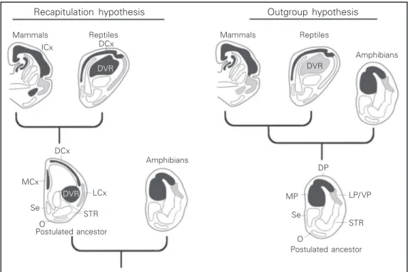
There have been two main hypotheses about the homologies to the isocortex in the non-mammalian brain (Figure 2). The first
Figure 2. Diagrams summariz-ing the essential elements of the recapitulation hypothesis and the outgroup hypothesis. O, olfactory tubercle; Se, sep-tum; VP, ventral pallium. For other abbreviations, see legend to Figure 1.
Amphibians Reptiles
Mammals
Outgroup hypothesis Recapitulation hypothesis
Mammals Reptiles
Amphibians
Postulated ancestor
Postulated ancestor STR LP/ VP MP
Se
O DP
STR LCx MCx
Se
O DCx
ICx DCx
DVR DVR
hypothesis, which was termed the recapitu-lation hypothesis by Northcutt and Kaas (7), asserts that the common ancestor of mammals and reptiles had a reptilian-like brain, with a DVR (or its precursor), and a dorsal cortex. This hypothesis implies that the DVR, together with the dorsal cortex, gave rise to the mammalian isocortex. The second hypothesis, the outgroup hypothesis (7), asserts that the common ancestor of mammals and reptiles had a very simple brain, similar to that of amphibians, with no large cell masses. According to this view, only the dorsal cortex of this common ances-tor became the isocortex. Thus, the main difference between the two hypotheses is whether the DVR of reptiles did or did not give rise to the isocortex.
Recapitulation hypothesis
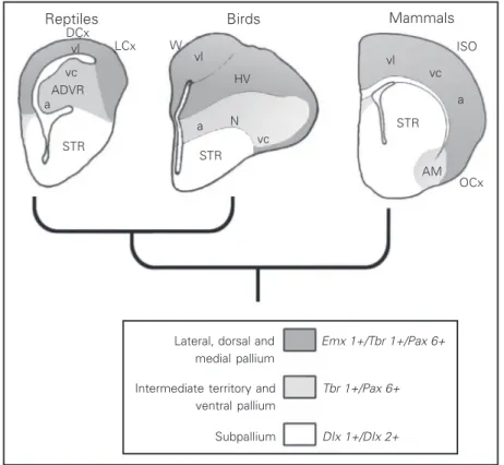
This hypothesis relies mostly on evidence of the termination of thalamic sensory path-ways in the telencephalon. A good example to illustrate this evidence is the visual sys-tem. In most vertebrates, there are two visual pathways. One is the thalamofugal pathway, which in mammals originates from retinal projections to the lateral geniculate nucleus; the pathway then follows to the primary or striate visual cortex (vl in Figure 3). The second pathway, the tectofugal pathway (vc in Figure 3), goes from the retina to the superior colliculus or optic tectum, then projects to the pulvinar nucleus in the thala-mus, and finally projects to the extrastriate visual cortex. The same two pathways can be observed in reptiles and birds, and in most vertebrates. In birds and reptiles, the thala-mofugal pathway (vl in Figure 3) is re-layed in the nucleus equivalent to the lateral geniculate, and terminates in the dorsal cor-tex (the Wulst of birds). On the other hand, the tectofugal pathway (vc in Figure 3) projects to the optic tectum, then projects to a thalamic nucleus called nucleus rotundus, and from there it is directed to the DVR (8,9).
Based on this evidence, it has been pro-posed that the thalamofugal pathway, which ends in the dorsal cortex or Wulst of birds, and in the striate cortex of mammals, indi-cates homology between the striate cortex and the dorsal cortex or Wulst. On the other hand, the tectofugal pathway, terminating in the DVR and in the extrastriate visual cortex, indicates homology between these two struc-tures. It was also claimed that the nucleus rotundus was homologous to the mamma-lian pulvinar nucleus because they are lo-cated at corresponding processing stages in the tectofugal pathway. Furthermore, con-sidering similarities in intrinsic connectiv-ity, it was postulated that the different lami-nae of the extrastriate visual cortex might correspond to different components of the
Reptiles Birds Mammals
DCx
LCx W ISO
OCx vl
vc ADVR a
vl
HV
a
vl
STR a vc
AM STR
STR
Lateral, dorsal and medial pallium
Intermediate territory and ventral pallium
Subpallium
Emx 1+/Tbr 1+/Pax 6+
Tbr 1+/Pax 6+
DIx 1+/DIx 2+
N vc
avian DVR. Thus, there was an equivalent circuitry in the mammalian cortex and in the avian and reptilian DVR, which probably was ancestral to both groups. In addition to this evidence, the finding that the auditory pathway (a in Figure 3) ends in the avian DVR suggested that the mammalian audi-tory cortex derived from a DVR-like struc-ture (8,9).
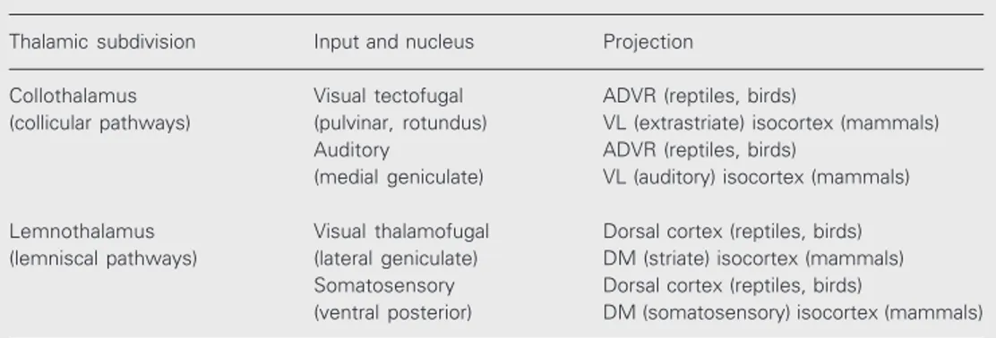
More recently, Butler (10) proposed a subdivision of the thalamic nuclei that some-how agrees with the above view. She classi-fied thalamic sensory nuclei as either col-lothalamic or lemnothalamic (Table 1). Collothalamic nuclei receive sensory pro-jections from the mesencephalon. The pulvi-nar nucleus of mammals and the nucleus rotundus of birds, both of which receive the visual tectofugal pathway, and the medial geniculate and its reptilian equivalent, re-ceiving the auditory pathway, are proposed to be examples of collothalamic nuclei. On the other hand, lemnothalamic nuclei re-ceive projections from lemniscal pathways which bypass the mesencephalon. Examples are the lateral geniculate nucleus, receiving the visual thalamofugal pathway, and the somatosensory nuclei receiving the spinotha-lamic pathway.
Collothalamic nuclei project to the DVR of reptiles and birds, and to what we call the ventrolateral isocortex of mammals. This includes the visual extrastriate isocortex and
the auditory isocortex. At the same time, lemnothalamic nuclei project to the dorsal cortex of reptiles and birds, and to what we call the dorsomedial isocortex of mammals, which includes the visual striate isocortex and the somatosensory isocortex of mam-mals. Overall, considering this connectional evidence, the ventrolateral isocortex, includ-ing the extrastriate and auditory isocortex, would be homologous to the anterior DVR, since both receive collothalamic projections. The dorsomedial isocortex, including the striate isocortex and somatosensory isotex, would be homologous to the dorsal cor-tex of reptiles and birds, since these struc-tures receive lemnothalamic projections. This recapitulationist hypothesis implies that the sensory projection sites and processing cir-cuits have been conserved in reptiles and in mammals, while there have been gross changes in brain topography and cytoarchi-tecture.
Outgroup hypothesis. I. Connectional evidence
Homology of the striate cortex and the somatosensory cortex with the avian or rep-tilian dorsal cortex is well accepted. The Wulst (dorsal cortex) of the pigeon contains regions that can be compared with the mam-malian primary (or striate) visual cortex, with the primary somatosensory cortex and
Table 1. Subdivisions of thalamic nuclei (10).
Thalamic subdivision Input and nucleus Projection
Collothalamus Visual tectofugal ADVR (reptiles, birds)
(collicular pathways) (pulvinar, rotundus) VL (extrastriate) isocortex (mammals) Auditory ADVR (reptiles, birds)
(medial geniculate) VL (auditory) isocortex (mammals)
Lemnothalamus Visual thalamofugal Dorsal cortex (reptiles, birds) (lemniscal pathways) (lateral geniculate) DM (striate) isocortex (mammals)
Somatosensory Dorsal cortex (reptiles, birds)
(ventral posterior) DM (somatosensory) isocortex (mammals)
with a region that might be considered a mixture of somatosensory and motor cortex (11). However, the concept of homology between the DVR and the isocortex has proved to be more controversial. Some early questions regarding this hypothesis (12,13) took issue with the topography of the reptil-ian brain. The position of the DVR in the hemisphere is such that in most reptiles it is separated from the dorsal cortex by the lat-eral cortex (Figure 2). Therefore, the DVR would somehow have to move to a more dorsal position in order to make up part of the isocortex.
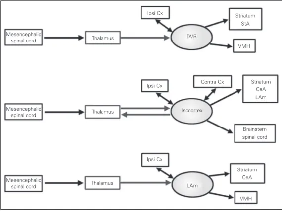
Additional conflicting evidence comes from neuronal connectivity. In amphibians, collothalamic projections end mostly in the corpus striatum (14), which is clearly not a homologue of the DVR or the isocortex of mammals. This indicates that collothalamic pathways may change their preferred termi-nation sites, and therefore they may not be considered a good criterion to establish ho-mology. Furthermore, Bruce and Neary (15) and later Puelles and collaborators (16,17) have indicated that the overall pattern of connections of the DVR resembles more the mammalian laterobasal amygdala than the isocortex. Collothalamic projections also ter-minate in the laterobasal amygdala of mam-mals, but instead of arising from specific nuclei like the pulvinar nucleus, they origi-nate from the intralaminar thalamic nuclei (Figure 4). Thus, the conclusion from these studies has been that the intralaminar tha-lamic nuclei, and not the mammalian pulvi-nar, are comparable to the avian nucleus rotundus.
Finally, another source of evidence has to do with the connections of the extrastriate cortex. This is very old evidence but it has never been considered enough in this con-text. In mammals, the striate visual cortex connects strongly to the extrastriate visual cortex, while in reptiles there is little connec-tivity between the dorsal cortex and the DVR (6). Furthermore, in mammals, the
extrastri-ate and the auditory cortices project, via a sequence of cortico-cortical connections, to the entorhinal cortex and to the hippocampus. On the other hand, in reptiles the anterior DVR (receiving thalamic sensory afferents) projects to the posterior DVR (also termed the reptil-ian amygdala) (6), but is poorly connected to the hippocampus (18-20).
Outgroup hypothesis. II. Developmental evidence
Some relatively recent reports (21-23) concerning the topography of gene expres-sion patterns in the early development of the telencephalon revealed a highly conserved, mosaic-like, organization of the different tel-encephalic components (pallium and sub-pallium). A novel component, located in the equatorial region of the hemispheres, was described in these studies which was termed intermediate territory (21) or, per-haps more appropriately, ventral pallium (22,23). The ventral pallium gives rise to the DVR (especially the anterior part) of reptiles and to the lateral basal amygdala of mam-mals, which implies that these adult struc-tures derive from the same embryonic re-gion. This is a strong argument in favor of homology between the DVR and parts of the mammalian amygdala. Interestingly, based on purely descriptive considerations, Holm-gren (24) had already considered that the amygdalar complex was comparable to the DVR of reptiles, and he termed these two things the hypopallium.
lium and subpallium. This territory has pal-lial markers like Pax 6 and Tbr 1, but does not express the Emx 1 marker. Apparently, the ventral pallium has a different fate in the development of the mouse and the chick (21). In the mouse, the ventral pallium disap-pears relatively soon in development, and gives rise only to early-produced neural popu-lations. However, in the chick the ventral pallium may expand and give rise to a large part of late-produced elements of the DVR. Summarizing these results, the avian and reptilian DVR is comparable to parts of the amygdala of mammals (these two make up the ventral pallium in reptiles and mammals, respectively), and the dorsal cortex of rep-tiles and birds is embryologically compa-rable to the mammalian isocortex (both de-rive from the dorsal pallium). A consequence of this conclusion is that in mammals, col-lothalamic projections reach the dorsal pal-lium, while in reptiles, the same projections terminate in the ventral pallium. Therefore, this hypothesis implies that there has been an important change in brain connectivity in the
origin of mammals.
Tangential migration
The developmental evidence about gene expression patterns strongly confirms the concept based on brain topography, indicat-ing that the lateral cortex is interposed be-tween the dorsal cortex and the DVR. As mentioned above, in order to be correct the recapitulation hypothesis requires that dur-ing development the DVR be displaced dorsalwards, merging with the developing dorsal cortex. May cells from the ventral pallium migrate into the dorsal pallium and contribute to the isocortex? There is now substantial evidence that Dlx-positive GABAergic cells from the embryonic gan-glionic eminences may migrate into the iso-cortex (25,26), and that many pioneer neu-rons of the preplate possibly migrate tangen-tially from the ganglionic eminences (27). However, the ganglionic eminences do not correspond to the ventral pallium, but rather belong to the embryonic subpallium.
Sec-Mesencephalic
spinal cord Thalamus
Ipsi Cx Striatum
StA
Striatum CeA LAm VMH
Contra Cx Ipsi Cx
Thalamus
Brainstem spinal cord
Striatum CeA
VMH LAm
Ipsi Cx
Thalamus
Isocortex DVR
Figure 4. Schematic patterns of connectivity of the reptilian dor-sal ventricular ridge (DVR), the isocortex and the lateral dala (LAm). CeA, central amyg-dala; StA, striatal amygamyg-dala; VMH, ventromedial hypothala-mus; Cx, cortex.
Mesencephalic spinal cord
ond, the kind of cells that migrate, mostly GABAergic interneurons, do not correspond to those specified by the equivalent circuit hypothesis (this implies only excitatory ele-ments) (8). No evidence has been found as yet for a massive migration of excitatory cells from the ventral pallium into the dorsal pallium, as the recapitulation hypothesis would imply.
The Dlx-positive cells that migrate from the ganglionic eminence into the isocortex keep expressing their subpallial marker. Like-wise, if there was such a migration from the ventral pallium into the isocortex, this might make the auditory and extrastriate visual corti-cal areas largely negative to Emx 1, a fact that has not been observed. Recent evidence indi-cates that Emx 1 is expressed in most pyrami-dal cells of the isocortex (28), which is not expected if some cells arise from the Emx 1 -negative ventral pallium. Furthermore, in dis-sociated cortical cultures, this gene is expressed by most glutamate-containing neurons (28), suggesting that the (excitatory) cells that make up the equivalent circuits in mammals and birds (8,9) may express different markers (they are Emx 1-positive in the isocortex and Emx 1 -negative in the DVR). It could still happen that some precursor cells migrate from the ventral pallium very early into the dorsal cortex, and then they acquire Emx 1 expression. To be fair, it would be very hard to prove that this process does not occur. Undoubtedly, the incorpora-tion of GABAergic cells from the subpallium was probably an important element in isocor-tical origins. Nevertheless, this sort of migra-tion of inhibitory interneurons has been ob-served in birds (29), suggesting that this mech-anism predates the origin of mammals.
A new type of brain
The evidence reviewed suggests that in mammals a noticeable expansion of the dorsal pallium has taken place, perhaps to some ex-tent at the expense of the development of the ventral pallium. On the other hand, in reptiles
Another important point raised by the evidence reviewed is that this implies differ-ent patterns of brain connectivity between reptiles and mammals. In reptiles, the col-lothalamic projections and the lemnothalamic projections are largely separate. Collotha-lamic input enters the DVR, which is mainly connected to the hypothalamus, while the lemnothalamic input enters the dorsal cor-tex, which projects to the hippocampus, and establishes association networks with the olfactory cortex (6). On the other hand, in mammals there is a confluence of collotha-lamic and lemnothacollotha-lamic inputs, especially in the visual system (there is a strong projec-tion from the striate cortex to the extrastriate cortex), and both the lemno- and collotha-lamic pathways project to the hippocampus and the amygdala. Summarizing, in reptiles there seems to be a separation of the col-lothalamic and the lemnothalamic process-ing systems, the former related to the amyg-dala and the latter to the hippocampus. In mammals, both collothalamic and lemnotha-lamic projections end in the dorsal pallium, and from there both pathways project to the hippocampus and to the amygdala.
The role of olfaction in the origin of the mammalian brain
What adaptive mechanisms and func-tional constraints may have driven the change in brain organization in the evolution of mammals? One possibility is that olfaction, which was a highly developed sense in the early mammals (2), was a key factor in deter-mining a different evolutionary direction in mammalian brain evolution. In both mam-mals and reptiles, the olfactory system is highly connected to the hippocampus (me-dial cortex); furthermore, the dorsal cortex of reptiles receives olfactory projections and sends projections to the medial cortex (hip-pocampus) (6). Thus, the dorsal cortex ap-pears as an important element in olfactory-hippocampal interactions. In addition, the
dorsal cortex and the hippocampus of rep-tiles participate in spatial learning, and can make use of non-spatial cues (like odors and other stimuli) in orientation (33). Consider-ing the relative importance of olfaction in the behavior of early mammals, we have proposed (12) that the olfactory-hippocam-pal-dorsal cortex circuit may have been put to use by the first mammals to make rela-tively elaborate, largely olfactory-based, rep-resentations of behavioral space, in which specific odors labeled particular places and routes. In mammals, there is strong evidence that odor information participates in spatial and episodic learning (34), and these may have been important functions of olfaction in the first mammals. Nevertheless, the contribution of the visual system became undoubtedly helpful in the elaboration of more precise maps of space, especially when mammals invaded diurnal niches after the decline of dinosaurs. The dorsal cortex, re-ceiving visual information from the thala-mofugal visual pathway, may have become an important sensory processing system in the early mammalian brain (12). This may have triggered expansion of this structure, and the eventual incorporation of sensory information from collothalamic systems.
of the mammalian brain is the confluence of the collothalamic and the lemnothalamic pathways in order to process spatial infor-mation which, among other things, partici-pated in spatial learning and episodic memory. In this process, the hippocampus may have become a fundamental component in which both types of sensory pathways eventually converged. Strictly speaking, this particular proposal is consistent with both the recapitulation and the outgroup hypoth-eses, since the merging of the two pathways is independent of the embryonic origin of the ventrolateral isocortex. However, it may be more parsimonious to consider that for this confluence to occur, only the axonal projec-tions changed their route instead of produc-ing a massive cellular migration that dragged the collothalamic axons to a more dorsal position. Perhaps a middle-way possibility would be that only some cells, recipient of thalamic afferents in the ventral pallium, migrated dorsally to the dorsal pallium, car-rying with them the thalamic afferents. This would not need a massive tangential migra-tion, and many components of the equiva-lent circuits (9) would be derived from the dorsal pallium in mammals. However, as mentioned, there is no evidence for this mi-gratory process.
Question 2. Laminar development of the isocortex
The second part of this paper relates to the internal structure of the isocortex. There are a number of differences between the reptilian cortex and the mammalian isocor-tex. First, the reptilian cortex is much smaller, has three layers, and has a tangential organ-ization of inputs in layer I, above the cortex. The mammalian isocortex is a highly expan-sive structure, has 6 layers, and afferents come radially from the white matter under-neath (6). Second, the reptilian cortex devel-ops according to an outside-in neurogenetic gradient, in which early-produced neurons
are located above late-produced neurons (37); in the isocortex there is an inside-out neuro-genetic gradient in which early-produced cells are located in inferior layers, while subsequently produced cells are located in more superficial layers (38). In the following sections, we will review some morphologi-cal and genetic aspects of cortimorphologi-cal develop-ment in order to propose some plausible evolutionary scenarios for the origin of the developmental and structural characteristics of the isocortex.
Comparative cortical development
As a brief reminder, mammalian isocor-tical development is characterized by the presence of an early preplate, which is a population of cells that is subsequently di-vided into a deep subplate and a superficial marginal zone by the arrival of cells belong-ing to the cortical plate. Most of these preplate cells die in late cortical development. A characteristic cell type of the marginal zone is the Cajal-Retzius cell which secretes reelin, an extracellular protein involved in the con-trol of neuronal migration (39,40).
One unsolved question is whether there is a preplate in the reptile. An early popula-tion of somatostatin-positive and NPY-posi-tive cells has been detected in the developing cortex of reptiles (40,41). However, these cells do not express reelin or calbindin, which are markers of the mammalian preplate. If there is something like a preplate in the reptiles, it has important differences with what we see in the mammal. Furthermore, it is not known whether the preplate is split by the arrival of the cortical plate, leaving a subplate-like structure underneath.
cor-respond to an embryonic adaptation (43). Furthermore, the subplate is important in many aspects of the development of the cor-tical plate (44). Comparative studies (45,46) of the cerebral cortex of reptiles and of mam-mals have concluded that cells in the inferior layers of the isocortex (output layers V and VI) resemble most the cells of the reptilian cortex. However, in the reptile there are no cell types strictly comparable to those in the granular layer (IV, thalamo-recipient) and supragranular layers (II and III, involved in local and cortico-cortical connections) of the isocortex. Thus, granular and supragranu-lar layers may be an evolutionary acquisition of the mammalian brain. Since these layers are also the latest produced cells of the iso-cortex, this may imply an additional devel-opmental step in the development of the mammalian cortex. Related evidence is the fact that deep cortical layers mostly derive from the ventricular zone (the deepest, pro-liferative region of the hemisphere), and they are characterized by the presence of the
marker Otx 1. Cells belonging to supragranu-lar layers derive from the subventricusupragranu-lar zone (which is above the ventricular zone), and are positive to the Svet 1 gene. In the cortex of the small eye mouse (Pax 6 mutant), only deep layers are formed and the Svet 1 -posi-tive superficial layers do not develop (47). Apparently, there is an inability of the pre-cursors of the superficial layers to differenti-ate and migrdifferenti-ate into the subventricular zone. This evidence suggests that the superficial and the deep layers of the isocortex may be specified by different genetic mechanisms.
Mutants of cortical development
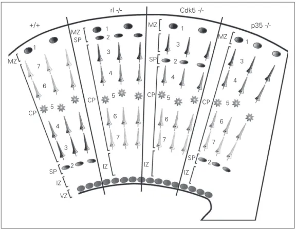
Several genes have been identified which participate in the regulation of cortical de-velopment. Below we will discuss two dis-tinct signaling pathways that participate in the regulation of cell migration, which can relate to the evolutionary origin of the iso-cortex. These are the reelin/mDab pathway, and the Cdk5/p35 pathway (see Figure 5).
+/+
rl -/- Cdk5
p35
-/-MZ
MZ
MZ
MZ SP
SP
SP
SP
IZ
IZ IZ
IZ
VZ CP
CP CP CP
7
7 7
7 6 6
6 6
1
5
4
3
2
1
5 4 3 2
1
5 4
3
2
1
5 4
3
2
The reeler mutant is characterized by an inverse layering of the cortex, in which cells normally belonging to deep layers are lo-cated superficially, and cells normally des-tined to superficial layers are located deeply. An important characteristic of the isocortex of the reeler mouse which is not always emphasized enough is that in the adult, cor-tical plate cells are located immediately be-low the subpial surface, there being no cell-poor layer I in this mutant. This does not happen in other regions of this mutant like the dentate gyrus and the hippocampus, in which a superficial marginal zone can be observed. The reeler phenotype is caused by mutations in a gene coding for an extracellu-lar glycoprotein called reelin, which is se-creted by the Cajal-Retzius cells of layer I (48). Reelin binds to several receptors in the migrating cells (including N-cadherin tors, VLDL receptors and integrin recep-tors), and triggers an intracellular cascade mediated by the protein mDab. Reelin is a serine protease (49), and it has been postu-lated that this proteolytic activity is central to its neuronal function. One hypothesis (50-52) is that normally, by binding to these receptors, reelin detaches the neuron from the radial glia that serves as a substrate for migration, consequently arresting migration of the cell. The separation of the cell from the radial glia permits younger cells to mi-grate past the older neurons, using the same radial glia as a substrate, until they contact reelin molecules as they get to the border of layer I. In this way, younger cells are able to migrate past older ones, generating the in-side-out neurogenetic gradient. If reelin is inactive as in the reeler mouse, neurons do not detach from the radial glia, and keep migrating until they accumulate in the sub-pial layer. Presumably, the inverted outside-in gradient of the reeler is produced because younger cells are not able to migrate past the older neurons that remain attached to the radial glia. In the reeler mutant, neurons are unable to migrate through the subplate and
(54), which opens the possibility that this molecule may have some role in early migra-tional stages.
The next set of mutations relate to the cyclin-dependent kinase Cdk5 and its acti-vator p35. Mutants for Cdk5 have a pheno-type somewhat similar to the reeler one in that cells arrange in an outside-in pattern and most neurons arrange below the subplate, forming an underplate (actually, many cells can be observed in the intermediate zone, forming clusters that apparently displace very slowly along the radial glia; 55). However, there are two important differences with the reeler phenotype: first, layer VI cells (the earliest produced) are able to cross the subplate, correctly splitting the preplate, and second, in the adult there is a normal layer I, indicating that cells do not penetrate into the marginal zone and thus may respond nor-mally to reelin. The p35 mutant has a milder phenotype, as in this animal cortical plate cells migrate better and all are able to move past the subplate, splitting the preplate. How-ever, within the cortical plate, cells arrange in an inverted outside-in pattern. As in the Cdk5 mutant, in the adult p35 mutant there is a normal layer I. Summarizing, early-pro-duced layer VI cells do not need either Cdk5 or p35 to reach their proper position and cross the subplate. Late-produced cells from layers II-V need Cdk5 but not p35 to cross the subplate, and need both Cdk5 and p35 to cross through previous cortical layers within the cortical plate. There is another Cdk5 activator, p39, whose mutation produces a very mild phenotype. The p35/p39 double mutant has been claimed to totally reproduce the phenotype of the Cdk5 mutant (56). This suggests that Cdk5, activated by p39, helps cells from layers II-V to migrate through the intermediate zone and to cross the subplate. In this context, it has been observed that the POU domain transcription factors Brn 1 and Brn 2 critically control the initiation of radial
migration by regulating the Cdk5-regulatory elements p35 and p39 (57).
Recently, two modes of cell migration have been observed in the developing cere-bral cortex (58): one is called translocation, and is observed in early-produced cells of the cerebral cortex, including those of layer VI, and also in the latest stages of migration of late-produced cells within the cortical plate. In this modality, the apical process of the cell contacts the subpial layer, and the cell body is dragged toward the surface while the apical process shortens. The other mode, locomotion, consists of the continuous lengthening and shortening of the apical pro-cess as the cell migrates along the radial glia. It is highly interesting that in the Cdk5 and the p35 mutants, only cells that move by locomotion are affected; cells in layer VI and other early-produced cells that migrate mainly by translocation are unaffected (51, 59). This suggests that Cdk5 and its activa-tors p35 and p39 participate in helping mi-gration by locomotion. It is then possible that Cdk5, activated by p35, allows migrat-ing neurons to move by locomotion within the cortical plate, thus allowing the genera-tion of the inside-out gradient. It has also been found that Cdk5 and p35 inhibit N-cadherin-dependent cell aggregation in mi-grating cortical neurons, which may also be part of a mechanism that permits migrating neurons to move past other cells within the cortical plate (50-52).
Evolutionary origin of the inside-out neurogenetic gradient
permitted long-distance migration by loco-motion of the new cell types. Furthermore, p35 was probably fundamental to permit cells to migrate past older cells in the cortical plate, generating the outside-in gradient. However, these cells activated by Cdk5 may have needed to be prevented from migrating into the marginal zone. The role of reelin may have precisely been to stop these cells before they penetrate into layer I. Although recent findings by Magdaleno et al. (53) and Kim et al. (54) indicate that the developmen-tal role of reelin may be more complex than a simple stop signal, the evidence points to a critical role of reelin in the final stages of neuronal migration, perhaps specifically in the generation of a cell-free marginal zone. It is interesting that reelin is quite poorly ex-pressed in the developing cortex of reptiles (60), and that in the reeler mutant there is a normal layer I in the hippocampus and den-tate gyrus, suggesting that in all these cases there is some other factor beside reelin that prevents cells from entering the marginal zone.
Yet there are some open questions. One is that, like other cortical plate cells, the cells produced early from layer VI (which are Cdk5-independent) seem to depend on reelin to avoid penetrating into the marginal zone. If these cells migrate according to a mechan-ism different from the younger cells of the cortical plate, they might not depend on reelin as a stop signal. Rather, they might be prevented from migrating into the marginal zone by some mechanism similar to that found in reptiles (and perhaps also in the hippocampus and dentate gyrus). This might be because the ancestral stop signal was totally replaced by reelin, and is no longer present in the isocortex. Another point is that layer V cells may actually represent an inter-mediate stage between the new phenotypes of the granular and supragranular layers and those of the earliest-produced cells of layer VI. Layer V cells are produced relatively early in development, derive from the
ven-tricular zone, and have phenotypes similar to those of the reptilian cortex. However, they do depend on Cdk5/p35 to migrate and reach their proper position.
Some why questions
Why did all these changes in isocortical organization come about? Why was there an inverted layering in the isocortex, and why do axons have a radial entrance to it? Up to this point, we have discussed the develop-mental mechanisms involved in isocortical origins, but to make sense of the evolution-ary transition, it is important to discuss the adaptive or functional context in which this process occurred. Below, we will outline some hypotheses attempting to shed some light on this issue.
As discussed before, the dorsal cortex of reptiles has most afferents located in the marginal zone, and the apical dendrites of the pyramidal cells make synapses with these afferents. In most mammals, afferents enter the cortex radially from the underlying white matter. However, in some so-called primi-tive mammals like the hedgehog, afferents enter radially to the cortex, but eventually they penetrate into layer I and run tangen-tially for some distance (6). This pseudo-tangential organization may represent a ves-tigial reminder of the ancestral condition of reptiles.
In a next stage, the early mammalian cortex began to expand in the tangential direction. In such a situation, tangentially running afferents would have to run increas-ingly long distances from the lateral aspect of the cortex (where they enter into the mar-ginal zone) to the site in which they establish their sites of termination. In addition, the marginal zone might have been soon crowded with thalamocortical axons. Perhaps the sim-plest solution, which possibly occurred very early in cortical evolution, was to run axons into the subcortical white matter and let them enter the cortex radially. In this pro-cess, the origin or differentiation of the subplate may have been a pivotal element in attracting axons to the subcortical matter. During development, subplate cells send pio-neer axons which establish contact with tha-lamic axons, guiding them into the subplate and allowing them to establish transient syn-aptic contacts with subplate cells before the cortical plate is ready to receive these axons. Later, thalamic axons are able to penetrate radially into the isocortex, possibly aided by reciprocal connections between the subplate and cortical layer IV (44). According to these hypotheses, the participation of the subplate in axonal guidance and its elaboration into a complex cell layer may have occurred in evolution after the origin of the superficial layers of the isocortex. Nevertheless, during development the subplate appears earlier than the cortical plate. The lack of concordance between the proposed phylogenetic sequence and the ontogenetic processes may be ex-plained by the fact that the subplate is an embryonic adaptation, rather than an adult adaptation like the superficial isocortical lay-ers.
In this way, the inside-out neurogenetic gradient may have been associated with the radial expansion of the isocortex (origin of the superficial layers), while the radial or-ganization of thalamic inputs was possibly more related to the tangential expansion of the early isocortex. In this latter process, the
subplate may have been a fundamental ele-ment in attracting axons through a shortcut via the developing intermediate zone.
Conclusions
This paper has attempted to provide a coherent scenario for the early evolution of the isocortex. The first main point expressed here is that the isocortex possibly arose from expansion of the ancestral dorsal cortex, and may have not arisen from the DVR. The possibility of the ventral pallium (DVR) con-tributing to the isocortex depends on demon-strating a sufficiently massive migratory pro-cess from the former to the latter, something that has not been observed. Perhaps one possibility to reconcile the two hypotheses would be that, although the ventrolateral isocortex may derive mostly from the dorsal pallium, a population of thalamo-receptive cells has migrated dorsally from the ventral pallium, dragging with them the collotha-lamic afferents. In any case, this alternative also relies on the demonstration of a tangen-tial migratory pathway from the ventral pal-lium to the dorsal palpal-lium.
dramatic developmental transformations. The second main point of this paper is that the superficial isocortical laminae are evolutionarily new components of the iso-cortex, and their origin, together with the tangential expansion of the early isocortex, triggered the development of the inverted neurogenetic gradient of mammals and the radial entrance of thalamocortical axons, re-spectively. At least two signaling pathways, one mediated by reelin and the other by Cdk5/p35, may have been fundamental in the origin of the inside-out gradient. Particu-larly, the activator p35 was fundamental in allowing migrating cells to locomote within the cortical plate, thus producing the charac-teristic inside-out gradient of the isocortex, while reelin was recruited to prevent cells from migrating into the marginal zone.
These considerations also raise several intriguing questions. For example, a lot more research is needed into the ventral pallium and its developmental fate. It will also be
References
1. Carroll RL (1988). Vertebrate Paleontol-ogy and Evolution. Freeman Press, New York, NY, USA.
2. Hopson JA (1979). Paleoneurology. In: Gans CC, Northcutt RG & UlinskiPS (Edi-tors), Biology of the Reptilia. Vol. 4. Aca-demic Press, New York, NY, USA.
3. Rowe T (1996). Coevolution of the mam-malian middle ear and neocortex. Science, 273: 651-654.
4. Wang Y, Hu Y, Meng J & Chuankui L (2001). An ossified Meckel’s cartilage in two cretaceous mammals and origin of the mammalian middle ear. Science, 294: 357-361.
5. Neary TJ (1990). The pallium of anuran amphibians. In: Jones EG & Peters A (Edi-tors), Cerebral Cortex. Vol. 8B. Plenum Press, New York, NY, USA.
6. Ten Donkelaar HJ (1998). Reptiles. In: Nieuwenhuys R, Ten Donkelaar HJ & Nicholson C (Editors), The Central Ner-vous System of Vertebrates. Springer-Verlag, Berlin, Germany.
7. Northcutt RG & Kaas JH (1995). The emer-gence and evolution of mammalian
neo-cortex. Trends in Neurosciences, 18: 373-379.
8. Karten HJ (1969). The organization of the avian telencephalon and some specula-tions on the phylogeny of the amniote telencephalon. Annals of the New York Academy of Sciences, 167: 164-179. 9. Karten HJ (1997). Evolutionary
develop-mental biology meets the brain: the ori-gins of mammalian neocortex. Proceed-ings of the National Academy of Sciences, USA, 94: 2800-2804.
10. Butler AB (1994). The evolution of the dorsal pallium in the telencephalon of am-niotes: cladistic analysis and a new hypo-thesis. Brain Research. Brain Research Reviews,19: 66-101.
11. Medina L & Reiner A (2000). Do birds pos-sess homologues of mammalian primary visual, somatosensory and motor cortices?
Trends in Neurosciences,23: 1-12. 12. Aboitiz F (1992). The evolutionary origin
of the mammalian cerebral cortex. Bio-logical Research, 25: 41-49.
13. Aboitiz F (1995). Homology in the evolu-tion of the cerebral hemispheres: the case
of reptilian dorsal ventricular ridge and its possible correspondence with mamma-lian neocortex. Journal of Brain Research, 4: 461-472.
14. Wilczynski W & Northcutt RG (1983). Con-nections of the bullfrog striatum: afferent organization. Journal of Comparative Neurology, 214: 321-332.
15. Bruce LL & Neary TJ (1995). The limbic system of tetrapods: a comparative analy-sis of cortical and amygdalar populations.
Brain, Behavior and Evolution, 46: 224-234.
16. Guirado S, Dávila JC, Real MA & Medina L (2000). Light and electron microscopic evidence for projections from the thalamic nucleus rotundus to targets in the basal ganglia, the dorsal ventricular ridge, and the amygdaloid complex in a lizard. Jour-nal of Comparative Neurology, 424: 216-232.
17. Yoon MS, Puelles L & Redies C (2000). Formation of cadherin-expressing brain nuclei in diencephalic and alar plate divi-sions. Journal of Comparative Neurology, 427: 461-480.
interesting to study the possibility of dorsal-izing, or ventralizing factors, in telencephal-ic development, like BMP 4 and Sontelencephal-ic hedge-hog, which are respectively known as dor-salizing and ventralizing factors in the neu-ral tube and in the telencephalon. Further-more, the point that lemnothalamic and col-lothalamic pathways are probably largely separated in reptiles may imply that these animals process information somewhat dif-ferently than mammals do.
18. Montero V (1993). Retinotopy of cortical connections between the striate cortex and extrastriate visual areas in the rat.
Experimental Brain Research,94: 1-15. 19. Rosa MGP & Krubitzer LA (1999). The
evolution of visual cortex: where is V2?
Trends in Neurosciences, 22: 242-248. 20. Aboitiz F (1999). Evolution of isocortical
organization. A tentative scenario includ-ing roles of reelin, p35/cdk5 and the subplate zone. Cerebral Cortex, 9: 655-661.
21. Smith-Fernández A, Pieau C, Repérant J, Boncinelli E & Wassef M (1998). Expres-sion of the Emx-1 and Dlx-1 homeobox genes define three molecularly distinct domains in the telencephalon of mouse, chick, turtle and frog embryos: implica-tions for the evolution of telencephalic subdivisions in amniotes. Development, 125: 2099-2111.
22. Puelles L, Kuwana E, Puelles E & Ruben-stein JLR (1999). Comparison of the mam-malian and avian telencephalon from the perspective of gene expression data. Eu-ropean Journal of Morphology, 37: 139-150.
23. Puelles L, Kuwana E, Puelles E, Bulfone A, Shimamura K, Keleher J, Smiga S & Rubenstein JLR (2000). Pallial and subpal-lial derivatives in the embryonic chick and mouse telencephalon, traced by the ex-pression of the genes Dlx-2, Emx-1, Nkx-2.1, Pax-6 and Tbr-1. Journal of Compara-tive Neurology,424: 409-438.
24. Holmgren N (1925). Points of view con-cerning forebrain morphology in higher vertebrates. Acta Zoologica, 6: 413-477. 25. Parnavelas JG (2000). The origin and
mi-gration of cortical neurones: new vistas.
Trends in Neurosciences, 23: 126-131. 26. Marin O & Rubenstein JL (2001). A long,
remarkable journey: tangential migration in the telencephalon. Nature Reviews. Neuroscience, 2: 780-790.
27. Deng J & Elberger AJ (2001) The role of pioneer neurons in the development of mouse visual cortex and corpus callosum.
Anatomy and Embryology, 204: 437-453. 28. Chan CH, Godinho LN, Thomaidou D, Tan SS, Gulisano M & Parnavelas JG (2001). Emx1 is a marker for pyramidal neurons of the cerebral cortex. Cerebral Cortex, 11: 1191-1198.
29. Cobos I, Puelles L & Martinez S (2001). The avian telencephalic subpallium origi-nates inhibitory neurons that invade tan-gentially the pallium (dorsal ventricular ridge and cortical areas). Developmental Biology, 239: 30-45.
30. Chapouton P, Gärtner A & Götz M (1999).
The role of Pax6 in restricting cell migra-tion between developing cortex and basal ganglia. Development, 126: 5569-5579. 31. Stoykova A, Treichel D, Hallonet M &
Gruss P (2000). Pax 6 modulates the dor-soventral patterning of the mammalian te-lencephalon. Journal of Neuroscience, 20: 8042-8050.
32. Britto J, Tannahill D & Keynes R (2002). A critical role for sonic hedgehog signaling in the early expansion of the developing brain. Nature Neuroscience, 5: 103-110. 33. Day LB, Crews D & Wilczynski W (2001).
Effects of medial and dorsal cortex le-sions on spatial memory in lizards. Behav-ioural Brain Research, 118: 27-42. 34. Eichenbaum H (1998). Using olfaction to
study memory. Annals of the New York Academy of Sciences, 855: 657-669. 35. Ulinski PS (1990). The cerebral cortex of
reptiles. In: Jones EG & Peters A (Edi-tors), Cerebral Cortex. Vol. 8B. Plenum Press, New York, NY, USA.
36. Ulinski PS (1983). Dorsal Ventricular Ridge: A Treatise on Brain Organization in Reptiles and Birds. John Wiley & Sons, New York, NY, USA.
37. Goffinet AM, Daumerie C, Langerwerf B & Pieau C (1986). Neurogenesis in reptil-ian cortical structures: 3H-thymidine
auto-radiographic analysis. Journal of Compara-tive Neurology, 243: 106-116.
38. Rakic P (1974). Neurons in rhesus mon-key: systematic relation between time of origin and eventual disposition. Science, 183: 425-427.
39. Rakic P (1995). A small step for the cell, a giant leap for mankind: a hypothesis of neocortical expansion during evolution.
Trends in Neurosciences,18: 383-388. 40. Nacher J, Ramírez C, Molowny A &
López-García C (1996). Ontogeny of somatosta-tin immunoreactive neurons in the medial cerebral cortex and other cortical areas of the lizard Podarcis hispanica. Journal of Comparative Neurology, 374: 118-135. 41. Cordery P & Molnár Z (1999). Embryonic
development of connections in turtle pal-lium. Journal of Comparative Neurology, 413: 26-54.
42. Marín-Padilla M (1978). Dual origin of the mammalian neocortex and evolution of the cortical plate. Anatomy and Embryol-ogy, 152: 109-126.
43. Kostovic I & Rakic P (1990). Developmen-tal history of the transitional subplate zone in visual and somatosensory cortex of macaque monkey and human brain. Jour-nal of Comparative Neurology,297: 441-470.
44. Allendoerfer KL & Shatz CJ (1994). The
subplate, a transient neocortical structure: its role in the development of connec-tions between thalamus and cortex. An-nual Review of Neuroscience, 17: 185-218.
45. Ebner FF (1969). A comparison of primi-tive forebrain organization in metatherian and eutherian mammals. Annals of the New York Academy of Sciences, 167: 241-257.
46. Reiner A (1991). A comparison of neuro-transmitter-specific and neuropeptide-specific neuronal cell types present in the dorsal cortex of reptiles with those pres-ent in the isocortex of mammals. Brain, Behavior and Evolution, 38: 53-91. 47. Tarabykin V, Stoykova A, Usman N &
Gruss P (2001). Cortical upper layer neu-rons derive from the subventricular zone as indicated by Svet1 gene expression.
Development, 128: 1983-1993. 48. Curran T & D’Arcangelo G (1998). Role of
reelin in the control of brain development.
Brain Research Reviews, 26: 285-294. 49. Quattrocchi CC, Wannenes F, Persico
AM, Ciafré SA, D’Arcangelo G, Farace MG & Keller F (2002). Reelin is a serine prote-ase of the extracellular matrix. Journal of Biological Chemistry, 277: 303-309. 50. Aboitiz F (2001). The evolution of cortical
development. Trends in Neurosciences, 24: 202-203.
51. Aboitiz F, Morales D & Montiel J (2001). The inverted neurogenetic gradient of the mammalian isocortex: development and evolution. Brain Research Reviews, 38: 129-139.
52. Aboitiz F, Montiel J & López J (2002). A hypothesis on the early evolution of the develoment of the isocortex. Brain Re-search Bulletin, 57: 481-483.
53. Magdaleno S, Keshvara L & Curran T (2002). Rescue of ataxia and preplate split-ting by ectopic expression of Reelin in reeler mice. Neuron, 33: 573-586. 54. Kim HM, Qu T, Kriho V, Lacor P,
Smal-heiser N, Pappas GD, Guidotti A, Costa E & Sugaya K (2002). Reelin function in neu-ral stem cell biology. Proceedings of the National Academy of Sciences, USA, 99: 4020-4025.
55. Pearlman AL, Faust PL, Hatten ME & Brunstrom JE (1998). New directions for neuronal migration. Current Opinion in Neurobiology, 8: 45-54.
57. McEvilly RJ, de Diaz MO, Schonemann MD, Hooshmand F & Rosenfeld MG (2002). Transcriptional regulation of corti-cal neuron migration by POU domain fac-tors. Science, 295: 1528-1532.
58. Nadarajah B, Brunstrom JE, Grutzendler J, Wong RO & Pearlman AL (2001). Two
modes of radial migration in early devel-opment of the cerebral cortex. Nature Neuroscience, 4: 143-150.
59. Gilmore EC & Herrup K (2001). Neocorti-cal cell migration: GABAergic neurons and cells in layers I and VI move in a cyclin-dependent kinase 5-incyclin-dependent manner.
Journal of Neuroscience, 21: 9690-9700. 60. Bar I, Lambert de Rouvroit C & Goffinet A
(2000). Reelin mRNA expression during embryonic brain development in the turtle