Bo1m Inst. oceanogr., S Paulo, 34(único):55-69. 1986
EARLY DEVELOPMENT OF
Thyn~~~op~ lep~dopo~de~(PISCES: GEMPYLIDAE)
Gosuke SAT01
&
Yasunobu MATSUURA21 Empresa Catarinense de Pesquisa Agropecuaria S.A. (EMPASC), Estação Experimental de Itajar,(Caixa Postal 277, 88300 Itajar, Se)
2 Instituto Oceanografico da Universidade de são Paulo, (Caixa Postal 9075, 01051 são Paulo, SP)
Synopsis
Thy~~op~ lep~dopo~d~ larvae were caught with Bongo nets in the upper 200 m of
the ocean from the coast of southern Brazil during 1975-1978. Based on a serie of 271 specimens ranging from 2.5 to 24.0 mm body lenght, morphological and
osteological development of the larva e and juveniles is described. Small larvae (2.5-12.0 mm NL) can be distinguished from all known gempylid larvae by the presence of a distinct melanophore at the base of the dorsal and anal fins. The larvae have well developed dorsal and ventral spines. It is the only gempylid with six preopercular spines and non-serrated dorsal and ventral spines during the
larval stage.
Descriptors: Fish 1arvae, Larva1 deve1opment, Animal morpho1ogy; Osteo1ogy~
Morphogenesis, Thy~~op~ lep~dopo~d~~ Gempy1idae, Southern B r a z i 1 i a n coa s ta
Descritores: Larvas de peixes, Desenvolvimento 1arva1, Morfologia animal. Osteo1ogia, Morfogenia, Thy~~op~ lep~dopo~d~~ Gempy1idae, Costa sul: Brazi 1.
Introduction
The larval morphology of gempylid fishes has been de s c ribed by many authors (Voss, 1954; Jager, 1955; Jones, 1960;
Strasburg, 1964; Yevseyenko &
Serebryakov, 1974; Nakamura & Paxton, 1977; Nishikawa
&
Nakamura, 1978;Gorbunova, 1977, 1982; Nishikawa, 1982,
1984a,b,c.). Collette Ú ai. (1984)
treated the larval morphology and
phyletic relationships of alI gempylids, but still our knowledge of the early life history of gempylids is incomplete.
Nishikawa (1984a) recently described
T. lepidopoides larvae based on only five specimens (one of them partially damaged and deformed), but his
description is precarious. We feel that it is necessary to give a more detailed description of this species.
In this paper we describe morphological and osteological
development of
T.
lep~dopo~d~ based on 271 specimens collected in the south Brazilian waters.Publ .
n. 617do
In~t.oceanogn. da
U~p.Material and methods
Larvae used in this study were collected during seven survey cruises conducted off the southern Brazilian coast from 1975 to 1978. The plankton samples were collected with Bongo nets and preserved in 10% formalin. After sorting, fish eggs and larvae were identified to family group o 'r lower taxon. From the larval specimens identified as
Gempylidae, Thy~~op~ lep~dopo~d~
56 Bolm Inst. oceanogr., S Paulo, 34(Gnico}: 1986
For our osteological study, larvae were c1e ared and stained with alcian blue and alizarin red following the double staining technique of Dingerkus &
Uhler (1977). All specimens studied for fin ray and bone development were
maintained in 100% glycerin and studied under 25X and 50X magnification.
Illustrations were made using a camera lucida. For cleared and stained
specimens, colorless and alcian blue stained structures are shown in white and alizarin red stained structures are stippled. The cartilaginous structures were considered to be ossifying if they were stained with alzarin rede
Osteological terms and abbreviations used in this study follow those used by Potthoff
et
alo
(1980).Results Morphology
Larvae are unique in the family
Gempylidae in having no serrated dorsal and ventral fin spines. In other
respects they show the typical gempylid larval form, such as a well-developed dorsal and ventral fin spines,
conspicuous head spination, and base of spinous dorsal fin longer than soft dorsal fin base.
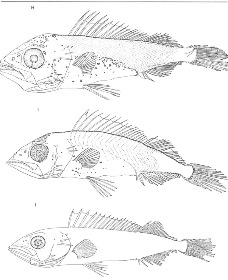
Preflexion stage larvae have aslender and laterally compressed body (Fig. lA, B,C). The notochord flexion of this species starts at 7.0 mm and ends at 12.0 mm, thus the specimens smaller than 7.0 mm are preflexion larvae, those between 7.0 and 12.0 mm are flexion larvae and those larger than 12.0 mm are postflexion larvae.
Flexion larvae have a robust body form due to an increase in body depth and enlargement of the gut (Fig. lD, E, F, G). By the end of the flexion stage, all fin counts are as in adulto During postflexion stage, larvae reaquire a slender, elongated body form as a result of rapid growth in length (Fig.
lH, I, J).
Pigmentation isuniquely characterized by the presence of two melanophores at 2.5 mm NL: one on the base of dorsal fin at about 13-14 th myomere and another on the base of anal fin at about 2l-24th myomere. These melanophores remain at
the same position throughout early
development up to 12.0 mm NL and are not seen in any other known gempylid larva.
Besides the two melanophores
mentioned above, preflexion làrvae have several melanophores ove r the midbrain which posteriorly spreading over the forebrain. Another patch of pigment is present over the dorsal part of gut. Pigment on the dorsal fin membrane firstly appears between the first and second dorsal spines at 6.2 mm NL (Fig. lC) and later between the second and third dorsal spines at 7.5 mm NL (Fig. lD). At this size the gut pigment spreads over the dorso-lateral region. Flexion larvae have similar pigment patterns, but some melanophores appear on the preopercle. Three pigment spots occur on the posterior portion of the upper jaw at 10.7 mm NL (Fig. lG).
In postflexion stage, two
melanophores observed on the bases of dorsal and anal fins at early stages are now obscured medially by a row of
melanophores which develop along the dorsal and anal fin bases. Melanophores over the head and gut regions increase in number with growth.
Osteology
P~eopeneulan ~pination
The first preopercular spine appear at 4.5 mm NL, on the posterior edge. Following this, several spines appear on the preopercle and at the late preflexion stage, these spines become five principal spines and three spinules on the preopercular crest (Fig. 2A, B). During the flexion stage, the
preopercular spines attain it's maximum number (=six) (Fig. 2C, D, E). Counting dorsal to ventral, the fourth spine positioned at the angle of preopercle, is the longest one. No serrations occur on the preopercular spines. The spines number 1 and 2 gradually disappear until 21.9 mm SL (Fig. 2F, G) and alI of them disappear in adults. There are two spines on the postero-dorsal part of the
opercle and two tiny spines on the
temporal region, the latters originating from posttemporal bone and the formers from supracleithrum. The supraorbital crest develops during the flexion stage.
Fotunation
06
dOMal and anal M~ and~uppofl.Li.ng bon~
SATO
&
MATSUURA: Thy~itop~ tepidopoid~: deve10pment57
A
B
D
58
E
~;. Q- '~ . &:: ~.\ ~.!:.1 .
~'!l t> (')
o \;
fi. "0 '"
---t< <iI O" '"
-~D\
'l:'" ~i
'r,o:
"" ~,. i -}-
. ~ . -.-"' \ .~' ..•:-:
:.-
. : ~/ ,~. .. , . . . # 4O.
.
,
G
Bo1m Inst. oceanogr., S Paulo, 34(único) 1986
.,
.
,,-... , ..
... 1 .-~ . ' ~ .. , .. -:""' ..
.:. ~ .
SATO
&
MATSUURA: Thy~~op~ tep~dopo~d~: developmentH
J
Fig. 1. Development of larvae of Th~~top~ tep~dopoid~ collected from southern Brazil. A, 4.6 mmNL; B, 5.7 mmNL; C, 6.2 mmNL; D, 7.5 mmNL;
E, 8.0 mmNL; F, 9.8 mmNL; G, 10.7 mmNL; H, 13.4 mmSL; I, 17.0 mmSL; J, 22.0 mmSL. Scale bar: 1.0mm.
60
Bolm Inst. oceanogr., S Paulo, 34(único): 1986A
Bc
Imm
F
6
Imm
o
\ ~
,
5
G
. . ~
E
6
4
Fig. 2. Development of preopercular spines of Thy~itop~ lep~dopo~d~. A, 5.1 mmNL; B, 6.4 mmNL;
c,
7.1 mmNL; D, 8.3 mmNL; E, 9.4 mmNL; F, 13,8 mmNLj G, 21.9 mmSL.(Table 1 and Fig. 3A). The first carti1aginous dorsal pterygiophore appears at about 7.0 mm NL (Fig. 3C). The 7.9 mm NL larva has the anterior 14 spines and 4 cartilaginous
pterygiophores be10w the anterior dorsal spines (Fig. 3D). The 8.3 mm NL larva has 16 dorsal spines and 17 soft rays
(Fig. 3E). The same figure shows that two anterior pterygiophores are ossified and a third one is ossifying.
The number of dorsal spines increases
with growth and attains the complete number of XVII at about 10 mm NL, but the comp1e tion of dorsal soft rays occurs 1ater at 12.5 mm SL. Figure 3F shows we11 deve10ped anterior dorsal fin
elements and vertebral column of the 13.8 mm SL larva. The carti1aginous predorsa1 bone first1y appears at this size. Norma11y one pterygiophore inserts into each interneura1 space up to the 13th neural spines, then the number of pterygiophores in each
interneura1 space varies from the one to three without any defini te pattern.
SATO
&
MATSUURA: Thy~itop~ fep~dopo~d~: development 61Tabte 1. Fin spine and ray counts of cleared and stained larvae of Thy~itop~ fep~dopo~d~
Dorsal fin Anal fi n Caudal fin Vertebral
Length (mm)
Spines Soft rays Spines 50ft rays Soft rays number
n x S n
x
S nx
S nx
S nx
S nx
S5.01- 6.00 08 5.00. 1.07
-6.01- 7.00 10 8.50 7.27 - 01 16.00
7.01- 8.00 22 14.14 1.83 05 12.20 4.09 03 1. 33 0.58 03 12.00 3.46 16 14.19 2.95 22 7.68 5./16
8.01- 9.00 11 16.00 1. 41 10 111.70 7.45 10 1.10 0.32 10 11.60 3.69 . 10 18.10 0.32 11 21.00 1. 79
9.01-10.00 17 16.82 0.39 17 17.18 1.67 17 1. 94 0.24 17 17.18 1. 55 17 20.24 1.39 17 26.12 1.11
10.01-11.00 10 17.00 0.00 10 18.60 1. 26 10 2.00 0.00 10 19.00 1.05 10 22 .20 0.79 10 29.50 1.96
11.01-12.00 08 17.00 0.00 OA 10.25 1. 28 08 2.00 0.00 08 18.38 1. 92 OA 23.00 1.60 08 28.63 4.57
lJ.Ol-14.00 07 17.00 0.00 02 20.00 0.00 02 2.00 0.00 02 21.00 0.00 01 77 .00 02 33.00 0.00
16.01-17.00 01 17.00 - 01 21.00
-
01 2.00 01 72.00-
01 79.00 OL 33.0025.01-26.00 OI 17 .00
-
01 71.00 01 2.00 01 21.00-
01 33.00 01 33.0028.01-29.00 01 18.00 01 19.00
-
01 2.00 01 20.00-
01 33.00 01 33.00Note: n number of larvae x ; mean
5 ; standard dev;at;on dashed
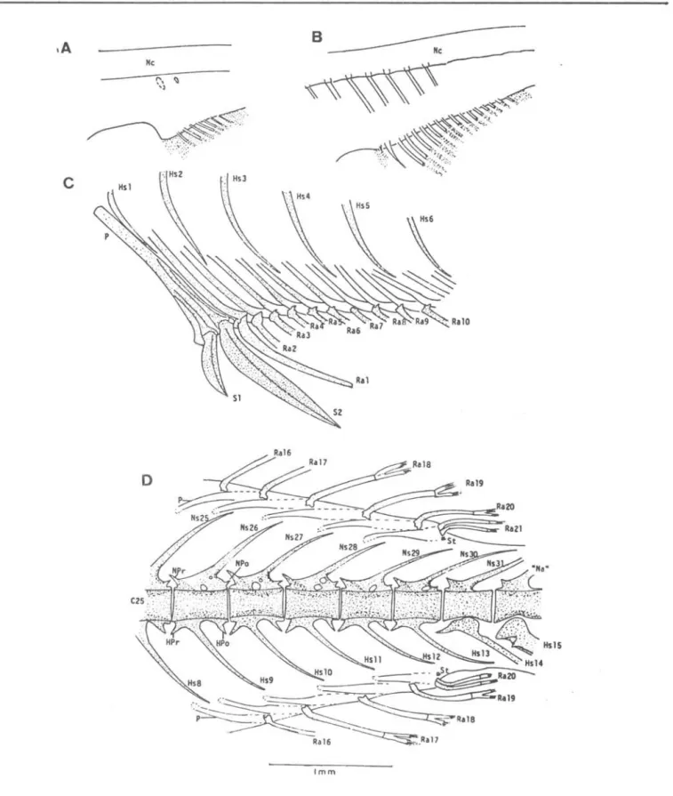
spines are supported by a single strong pterygiophore which inserts in front of the first haemal spine (Fig. 4C). At this size, only the first pterygiophore is fully ossified and the following six pterygiophores are ossifying o The number of pterygiophores inserting into each interhaemal space varies from two to three (Fig o 4C, D).
Figure 4D shows the posterior part of the dorsal and anal fins and vertebral column of 21.9 mm SL specimen. At this size the posterior pterygiophores of both fins are still cartilaginous. The posterior four or five soft rays of both fins have bifurcated tips and they will later transform into finlets in adults o One tiny cartilaginous stay is present at the base of the last double soft ray of dorsal and anal fins.
Development of the vertebcal column starts with the appearance of the bud of the first cartilaginous neural arch at 6.5 mm NL
(Fig. 3B). With growth, more neural arches are added in a posterior
direction. Ossification ofthe vertebral column starts at about 8.0 mm NL
anteriorly at the base of the neural
1; nes ;nd;cate caudal flex;on
arches (Fig. 3D) and proceeds quickly into posterior direction with growth. For example; in the 8.3 mm NL larva, 15 vertebrae are forming, of which five centra are ossified and other five are ossifying.
The first haemal arch appears on the 9th vertebra (Fig. 3E). At 13.8 mm SL alI 33 vertebra are ossified and 13 ribs which articulate on the precaudal
vertebrae are formed. The first rib is articulated at antero-dorsal part of the third centrum and the position of
articulation gradually goes down to the ventral part of centrum up to 6th rib
(Fig. 3F). The 13th - 19th ribs are losely articulated below theparapophysis of the haemal arches o At this size two epineural spines appear on the first and second neural spines and two
62
Bolm Inst. oceanogr., S Paulo, 34(Gnico}: 1986SI
B~
c
SI
fi ~sNS
~~~,=NS~ __________ __
S2
o
--E SI
Imm H.
Imm
Fig. 3. Development of spinous dorsal fins and supporting bones and anterior part of the vertebral column in
T.
{epidopoide~ larvae. Scale bar: 1.0 mm. A, 5.1 mmNL; B, 6.4 mmNL; C, 7.1 mmNL; D, 7.9 mmNL; E, 8.3 mmNL; F, 13.7 mmSL. Ce, centrum; Dr, distaI radial; EpN, epineural; Ha, haemal spine; 1mB, intermuscular bone; NPo, neural postzygaphosis; NPr, neural prezygapohysis; NSI, 1st. neural spine; P, pterygiophore;Pd, predorsal bone; Ribl, 1st. rib; SI' 1st. spiny ray in the dorsai
SATO & M.A:TSUURA:
ThyMilopJ.J lep,i..dopoideó:
developmentMe
c
Imm
Fig.
4.
A, B and C show development of the anterior part of the anal fin and supporting bones and D shows the posterior part of the anal and dorsal fin and a part of the vertebral column. A, 7.9 mmNL; B, 8.3 mmNL; C, 21.9 mmSL; D, 21.9 mmSL. HSl, 1st. haemal spine; "Na", spec i a 1 i zed neu ra 1 a rch; NPo, neu ra 1 pos tzygapophys i S; NP r, neural prezygapophysis; Nc, notochord; Ns2S ' 25th neural spine;P, pterygiophore; Ral, 1st. anal (or dorsal) fin ray; SI' 1st. anal spine; St, stay.
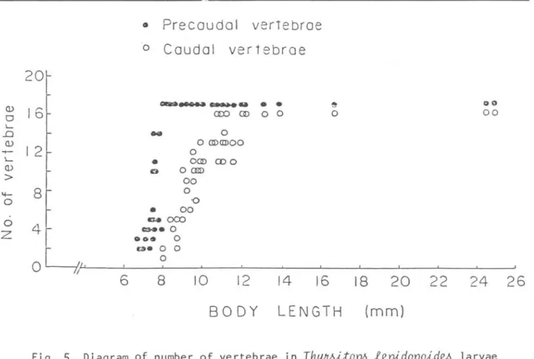
64 Bolm Inst. oceanogr., S Paulo, 34(único): 1986
• Precaudal verTebrae
O
Caudal vertebroe
20,
~~--
..
•
•
~ 0 0Q)
16
em ClD o o o 00o
~
..o
..
oQ) o ClDCIDOO
-+-
12
o~
•
OClD moQ)
o OQlD
> 00
<I-
8
oo
o•
00o
c.-oco
4r
ce.. oZ .o~ o
O~!!
13- o oo
6
8
10
12
1416
18
20
22
24
26
80 DY
LENGTH
(mm)
Fig.
5.
Diagram of number of vertebrae in Thy~itop~ lepidopoid~ larvae between6
and26
mm length.Pec.tofLal. giJtdle
a.n.d
~LL6pe~olÚumThe c1eithrum is one of the first structures to appear as a thin sliver behind the head at 2.9 mm NL. At 5.0 mm NL the posttempora1, suprac1eithrum and origin of the carti1aginous cora co-scapu1ar are visib1e (Fig. 6A). The c1eithrum is on1y one bone, but due to a twist at the midd1e portion, it seems to be comprised of two bones (Fig. 6A, B,
C) •
Uptake of alizarin red of the
pectora1 gird1e starts from the central portion of the c1eithrum at about 6.5 mm NL. At this size, severa1 pectora1 rays are formed and two postc1eithra appear behind the c1eithrum (Fig. 6B). The posttempora1 which is articu1ated on the suprac1eithrum posterior1y, has a tiny spine and the suprac1eithrum a1so has a spine on its posterior median edge. These two spines are exposed out of the skin and are visib1e in the temporal region (Fig. 1C).
At 8.3 mm NL (Fig. 6C), a11
components of the suspensorium (post-temporal, suprac1eithrum and
postc1eithra) are formed and ossified.
Two postc1eithra are connected to each other and on the dora1 side of the dorsal postc1eithrum, a sma11 process appears, which becomes disk-1ike in juveni1e and adu1t specimens (Fig. 6D, E, F). The number of pectora1 fin rays
~n the 8.3 mm NL larva is 13 and origins of 3 radia1s are visib1e.
Formation of pectora1 fin rays starts from the dora1 part and proceeds
ventrad. At 12.0 mm NL a11 15 rays and the coracoscapu1ar carti1age are formed. In the 1argest specimen examined the second tiny spine on the posterior part of the posttempora1 appeared (Fig. 6E), however these spines at 1arva1 and juveni1e stages in the temporal region disappear in the adu1t stage (Fig. 6F).
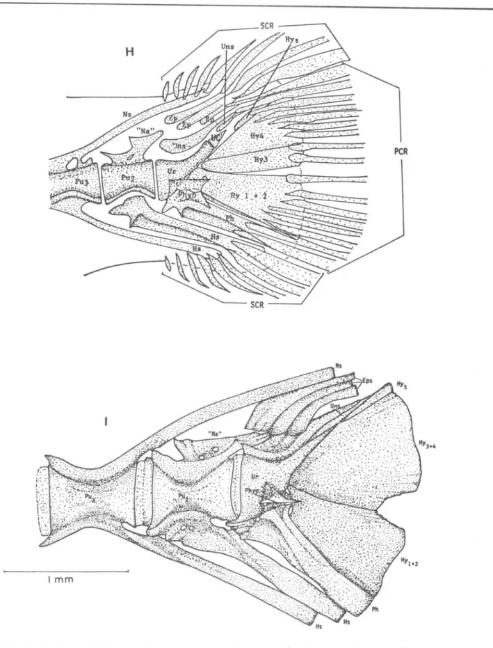
Ca.udal c.omplex
Deve10pment of the caudal fin starts at about 6.5 mm NL. Before notochord
f1exion five caudal rays and
SATO
&
MATSUURA: Thy~~op~ tep~dopo~de4: development 65A B c o
E
Fig.
6.
Left lateral view of the peetoral girdle inT.
tep~dopo~de4showing ontogeny. A, 5.3 mmNL; B, 7.1 mmNL; C, 8.3 mmNL; D, 13.7 mmSL;
E,
27.5 mmSL. F, 242.0 mmSL (adult). Cl, eleithrum;Cor, eoraeoid; PstCl, posteleithrum; RI' radial 1; Se, scapular; ScF, seapular foramem; SC1, supraele i thrum; Pt, posttemporal.
parhypural are developed (Fig.7B). Uptake of alizarin red starts at the bases of caudal fin rays at about 8.0 mm NL. The cartilaginous haemal spine are visible on the preural centra 2 and 3, preceding the parhypural. When the notochord is almost flexed, the principal caudal rays (9+8) are formed and the urostyle starts to ossify. Following this stage, ossification extends to the preural centrum 2, the hypurals and the parhypural.
Cartilaginous neural spines and the specialized neural arch also start to appear (Fig. 7E).
At 13.8 mm SL ossification proceeds to the last haemal spine and preural centra 3 and 4. The larger anterior
uroneural appears above the urostyle at this stage. At 21.9 mm SL , most
components of the caud a l complex are ossified. except the three epurals
(Fig. 7G). Other structures which appear at this stage are: the smaller posterior uroneural, the parhypurapophysis at the base of the parhypural and the
well-developed specialized neural arch. All five hypurals are still separated. At 27.5 mm SL (Fig. 7H), the first and the second hypurals are fused and three epurals are ossified. We could not find any radial cartilage as found in Pagn~
majon
1arvae (Matsuoka, 1982), nor procurrent spur of Johnson (1975) .66 Bolm Inst. oceanogr., S Paulo, 34(único): 1986
c
'o
peR
SATO
&
MATSUURA: Thy~itop~ {ep~dopo~de4; developmentFig.
7.
Development of the caudal complex in Thy~~op~ {epidopoid~ larvae. A, 6.4 mmNL; B, 7.1 mm NL; C, 8.3 mmNL; D, 9.4 mmNL; E, 11.4 mmNL; F, 13.7 mmSL; G, 21.9 mmSL; H, 27.5 mmSL; 1,242.0 mmSL {adult}. Ep, epural; Hy, hypural; Hs, haemal spine; "Na" specialized neural arch; Nc, notochord; Ns, neural spine; Ph, parhypural; peR,principal caudal rays; Phyp, parhypurapophysis; PU2, preural centrum 2; Uns, uroneurals; Ur, urostyle; SCR, secondary caudal rays.
68
Bolm Inst. oceanogr. , S Paulo, 34(Gnico}: 19869 + 8 principal and 16 secondary caudal rays ar e supported by elements of the urostyle and preural centra 2 and 3.
Discussion
The larvae were identified as belonging to the family Gempylidae based on the following characters: well-developed dorsal and ventral spines, the
preopercular spination and general body shape (Voss, 1954). Identification of
T.
lepidopoid~ larvae was made onspecimens larger than 11.0 rnrn, since at this size alI meristic characters are already formed. After identifying the larger specimens, we followed a
sequencial developmental series down to 2.5 rnrn NL. The larvae smaller than 6.0 rnrn NL can be confused with those of scombrid larvae (e.g.
ThunnUó,
Eu:thynnUó, Ka..:t.6uwonUó, Aux,w)
,
but we can distinguish them by myomere count. The S~ombnolabnaxhetenolep,w
larvae have similar myomere count (= 30) and body form, but theT.
lepidopoid~larvae develop dorsal and ventral fin spines since early stage and they have conspicuous pigments on the bases of dorsal and anal fins during preflexion and flexion stages.
After analysing a large series of gempylid larvae, Voss (1954) concluded that the spinous structure of the preopercular margin is one of the most important larval characters for species identification. From known gempylid larvae, six species have serrated preopercular spine or spines
(Lepido~ybium,
Neoepinnula, Epinnula,
Rexea,
NealotUó
and Viplo~pinUó) and five have no serrated spines(ThyMUop~, ThYMU~, Pnomethi~hthy~,
N~ian~hUó and
GempylUó)
(Colletteet
al.,
1984). The number ofpreopercular spines of gempylid larvae ranges from one t o s i x and only
ThyMUOp6
has six preopercular spines during larval stage. However, the upper two short spines disappear during the juvenile stage.Acknowledgements
The authors wish to thank Dr. William J. Richards and Dr. Thomas Potthoff of the National Marine Fisheries Service, for revisions and criticaI reading of the manuscript. The financiaI support of
this work carne from the Financiadora de
Estudos e Projetos (FINEP). The junior author received the research
fellow ship of the Conselho Nacional de Desenvolvimento Científico e Tecnologico
(CNPq). This is a contribution n9 617 of the Instituto Oceanografico da Uni-versidade de são Paulo.
References
COLLETTE, Bo B.; POTTHOFF, T.; RICHARDS, W. J.; UEYANAGI, S.; RUSSO, J. L. &
NISHlKAWA, Y. 1984. Scombroidei: development and relationships.
In:
Moser, H. G., ed.-in-chief-Ontogeny and sistematics of
fishes. Based on an International Syrnposium Dedicated to the
Memory of Elbert Halvor
Ahlstrom. The Symposium was held August 15-18, 1983, La Jolla, Calif. New York, American Society of Ichthyologist and Herpetologists, p. 511-620.
DINGERKUS, G.
&
UHLER, L. D. 1977. Enzyme clearing of alcian blue stained whole small vertebrates for demonstration of cartilage. Stain Technol., 52(4):229-232.FRITZSCHE, R. A.
&
JOHNSON, G. D. 1980. Early osteological development of white perch and striped bass with emphasis on identification of their larvae. Trans. Am. Fish. Soc., 109:397-4060
GORBUNOVA, N. N. 1977. Larvae and juveniles of some species of
trichiuroid fishes (Trichiuroidae: Gempylidae and Trichiuridae). Trudy Inst. Okeanol. Akad. Nauk, 109:133-148. (In Russian).
1982. Larvae of trichiroid fishes from collection of the International Mexican Biologi-cal Center (Gempylidae, Trichiuridae, Pisces). Trudy Inst. Okeanol. Akad. Nauk, 118:85-106. (In Russian).
JAGER, B. V. D. 1955. The South African pilchard (Sandinop~ o~ellata).
The development of the snoek
SATO
&
MATSUURA: Thy~itop~ lep~dopo~deJ: development 69JOHNSON, G.D. 1975. The procurrent spur: an undescribed perciform caudal character and its phy10genetic
imp1ications. Occ. Papo Ca1if. Acad. Sci., (121):1-23.
JONES, S. 1960. On the snake mackere1,
GempylUó
~eJtpeYL6 Cuvier from the Laccadive Sea. J. mar. bio1. Ass. India, 2(1):85-88.MATSUOKA, M. 1982. Deve10pment of vertebral co1umn and caudal ske1eton of the red seabream Pag~ majon~
Jap. J. Ichthyo1., 29(3):285-294. (In Japanese).
MOSER, H. G.
&
AHLSTROM, E. H. 1970. Deve10pment of 1antern fishes of fami1y Myctophidae in the Ca1ifornia Current. BulI. Los Ang. Cty Mus. nato Hist., (7):1-145.NAKAMURA. I.
&
PAXTON, J. R. 1977. A juveni1e gempy1id fish, Nealo~Uó~pe6, from eastern Austra1ia.
Aust. Zool., 19(2):179-184.
NISHlKAWA, Y. 1982. Early development of the fishes of the fami1y Gempylidae. I. Larvae and juveni1es of escolar
Lep~docyb~um
6lavobnunneum
(Smith). BulI. far Sea Fish. Res. Lab., (19): 1-14.NISHlKAWA, Y. 1984a. Post1arvae and juveni1es of Thy~ito~ lep~dopo~d~
Cuvier (Pisces: Gempy1idae). BulI. far Seas Fish. Res. Lab., (21):9-17.
1984b. Post1arva1 development of the gempy1id fish
Pana~plo~pinUó gna~ (Brauer).
BulI. far Seas Fish. Res. Lab.,
(21):1-8.
NlSHlKAWA, Y. 1984c. Additiona1 description of 1arvae of N~oep~nnula
o~e~ (Gi1christ and Von Bonde)
(Pisces: Gempy1idae). BulI. far Seas Fish. Res. Lab., (21):19-24.
&
NAKAMURA, I. 1978.Post1arvae and juveni1es of the gempy1id fish, Neoep~nnuta o~e~
(Gi1christ and Von Bonde), from the north Arabian Sea. BulI. far seas Fishs. Res. Lab., (16):75-91.
POTTHOFF, T.; RICHARDS, W. J.
&
UEYANAGI, S. 1980. Deve10pment of
Scombnolabnax
h~eJtolep~ (Pisces, Scombro1abracidae) and connnents on fami1ia1 re1ationships. BulI. mar. Sci., 30(2):329-357.STRASBURG, D. W. 1964. Post1arva1 scombrid fishes of the genera
Aca~hocyb~u.m,
Nealoru,
andV~plo~p~nUó from central Pacific Oceano Pacif. Sci., 18(2):174-185.
VOSS, N. A. 1954. The post1arva1 deve10pment of the fishes of the fami1y Gempy1idae from the Florida Current. I. N~~chUó Johnson and
GempylUó
Cuv. and VaI. BulI. mar. Sci. Gu1f Caribb.,4(2):120-159.
YEVSEYENKO, S. A.
&
SEREBRYAKOV, V. P. 1974. Larvae of V~plo~p~nUóm~~~ Mau1 (Pisces,
Gempy1idae) from the northwestern At1antic. J. Ichthyo1., 14(1): 92-98.
(Received 28-Sept-19~6;