http://periodicos.uem.br/ojs/acta ISSN on-line: 1807-8672
Doi: 10.4025/actascianimsci.v40i1.35686 AQUICULTURE
Influence of rearing temperature on muscle growth and adipose
tissue in Nile tilapia (
Oreochromis niloticus
) strains
Rondinelle Artur Simões Salomão1*, Vander Bruno dos Santos2 and Edson Assunção Mareco3
¹Centro de Aquicultura, Universidade Estadual Paulista ‘Júlio Mesquita Filho’, Via de Acesso Prof. Paulo Donato Castellane, s/n, 14884-900, Jaboticabal, São Paulo, Brazil. ²Agência Paulista de Tecnologia do Agronegócio, Presidente Prudente, São Paulo, Brazil. ³Universidade do Oeste Paulista, Presidente Prudente, São Paulo, Brazil. *Author for correspondence. E-mail: [email protected]
ABSTRACT. This study aimed to evaluate the effect of temperature on the tissue growth of Red, GIFT and Supreme tilapia strains. Fingerlings of approximately 1.4 g from male populations were reared for a period of 60 days, in three recirculating systems, each system containing 6 water tanks of 0.5 m³, at three temperatures (22, 28 and 30ºC). Ten fish of each recirculation system and each temperature studied were sampled. Fishes have been anesthetized, euthanized and weighed. The samplings have been done from the dorsal musculature, caudal, visceral and ventral subcutaneous adipose tissue. Histological sections were prepared and evaluated to determine the diameter of the cells. The experimental design was completely randomized factorial 3 X 3 (strain X temperature). Analysis of variance was performed and means were compared. Higher weights were found for fish reared at 30°C (56.23 g) compared to those grown at 28 (42.12 g) or 22°C (10.52 g). The cooled water temperature (22°C) may have contributed to the hypertrophy of white muscle fibers and visceral adipocytes from tilapia, compared to 28 or 30ºC. Red Tilapia, GIFT and Supreme strains show differences in muscle growth and adipose tissues in response to temperature of cultivation.
Keywords: adipocytes, white fibers, red fibers, GIFT strain, red strain, supreme strain.
Influência da temperatura de cultivo no crescimento muscular e do tecido adiposo em
linhagens de tilápia do Nilo (
Oreochromis niloticus
)
RESUMO. Objetivou-se avaliar o efeito da temperatura no crescimento dos tecidos de tilápias do Nilo das linhagens Vermelha, GIFT e Supreme. Alevinos de aproximadamente 1,4 g, oriundos de populações de machos revertidos, foram mantidos em três sistemas de recirculação. Cada sistema contendo contém seis caixas d’água de 0,5 m³, em três temperaturas (22, 28 e 30ºC). Foram coletados dez peixes de cada tratamento. Os peixes foram insensibilizados, eutanasiados e pesados. Foram realizadas amostragens das regiões da musculatura dorsal, do pedúnculo caudal, do tecido adiposo subcutâneo ventral e visceral. Lâminas histológicas foram confeccionadas e avaliadas para determinar o diâmetro das células. O delineamento experimental foi inteiramente casualizado em esquema fatorial 3 X 3 (linhagem X temperatura). A análise de variância foi realizada e as médias foram comparadas. Maiores pesos foram encontrados para os peixes cultivados a 30ºC (56,23 g), quando comparados àqueles cultivados a 28 (42,12 g) ou 22ºC (10,52 g). A temperatura resfriada da água (22ºC) pode ter contribuído para a hipertrofia das fibras musculares brancas e dos adipócitos viscerais das tilápias, quando comparada a 28 ou 30ºC. As linhagens de tilápia Vermelha, GIFT e Supreme apresentam diferenças de crescimento do tecido muscular e adiposo em resposta à temperatura de cultivo.
Palavras-chave: adipócitos, fibras brancas, fibras vermelhas, linhagem GIFT, linhagem vermelha, linhagem supreme.
Introduction
Tilapias are among the most important species of freshwater fish in tropical climate used for the production (Charo-Karisa et al., 2006). The intensification of production systems has resulted in a rapid expansion in the cultivation of tilapia and new strains have been introduced in Brazil and worldwide.
GIFT (Genetically Improved Farmed Tilapia) strain arrived in Brazil in 2005, from the World Fish
Ramírez-Paredes, Garduño-Lugo, & Muñoz-Córdova, 2011), however these strains have low production in response to its slower growth, when compared to the common ones (Macaranas et al., 1997; Moreira, Moreira, & Hilsdorf, 2005; Ng & Hanin, 2007; Ramírez-Paredes et al., 2011).
Tilapia strains have different growth rates and this can change the dynamics of muscle growth or fat tissues, affecting carcass quality and meat. In most species of fish, skeletal muscle can represent up to 60% of the animal body weight (Sanger & Stoiber, 2001). This significant percentage of muscle mass is not only a mechanical adaptation, specific to aquatic life, but also serves as an important source of proteins to be used in human food (Weatherley & Gill, 1985).
Many fish show seasonal changes in growth rate, and one of the main factors involved in these changes is the water temperature. Temperature can cause changes in skeletal muscle cellularity (number and size of muscle fibers), by altering the rate of proliferation and differentiation of myogenic cells, although the exact effect of cultivation temperature may vary between species and the seasonal period studied (Vieira & Johnston, 1992; Usher, Stickland, & Thorpe 1994; Johnston, 1999;).
In general, the development and growth rate of fish increases when the water temperature is raised to a temperature near the optimum temperature for growing species. Furthermore, since different species of fish have been introduced in various geographic regions of the world, the adaptation of the animals to the water temperature in their new habitats have received special attention (El-Sayed, 2006).
The development of adipose tissue in fish is considered fundamental the same as occurred in mammals. However the postembryonic development of adipose tissue is influenced by environmental and metabolic factors (Imrie & Sadler, 2010). Nevertheless, fish stock lipid in adipose tissue located around the digestive tract, under the skin, the meat between the myomeres and in the myomeres (Fauconneau, Corraze, Lebail, & Vernier, 1991). The most common measurements of growth of domestic animals are based on measuring the changes in weight. However, during growth, the animals not only increase in weight but also change the proportions in which tissues are deposited. The objective of this study was to evaluate the effect of temperature on the growth of Red, GIFT and Supreme tilapia strains and under morphological aspects of muscle and adipose tissue.
Material and methods
The experiment was conducted in the Sao Paulo Agency for Agribusiness Technology (APTA),
headquarter in Presidente Prudente, São Paulo State, Brazil.
It was used fingerlings of tilapia (Oreochromis niloticus) of Red, GIFT and Supreme strains. The fish, from a population of sexually reversed males, were kept in three recirculation systems, each system containing six water tanks of 0.5 m³. Each tank received 70 fingerlings weighing approximately 1.4 g, in a density of 140 fish m-3. The temperature of the water in recirculation systems was kept at 22, 28 and 30 ºC. The dissolved oxygen and water temperature were monitored daily, the limnological parameters of the water as pH, alkalinity, hardness, ammonia, nitrite and nitrate were monitored weekly. The experiment was conducted for 60 days.
All tilapias were fed the same commercial feed, specific for each growth phase, with the amount being supplied at 10% of the biomass of the tank and also according to the water temperature.
At the end of the experimental period, samples of 10 fish were collected after 24 hours fasting period. They were anesthetized by heat shock (ice and water) and euthanized. The sampled fish were weighed (g), measured for standard length (cm) and tissue samples were removed for morphometric analysis.
Samples of white muscle were removed in the middle portion of the body below the dorsal fin for subsequent analysis of white fibers (section that only contain a portion of white muscle). The red muscle samples were taken near the caudal fin, the tail areas mostly contain red muscles fibers. Samples of adipose tissue were removed in the visceral and ventral region, as illustrated in Figure 1.
Figure 1. Location of tissue samples in Nile tilapia on the left longitudinal view highlighting regions of collecting samples of muscle and adipose tissue in the middle portion of the body below the dorsal fin (MND) and the region immediately posterior to the anal fin (MNA) on the right, cross section of the body, highlighting again the sample of muscle tissue taken at the middle region of the body just below the dorsal fin (MND); visceral adipose tissue samples (AV) and subcutaneous adipose ventral (AS).
Historesin). Transverse sections of 4 μm thick were obtained in microtome and stained with hematoxylin and eosin (HE) to evaluate the tissues morphology. Using an image analysis system (Leica Qwin, Germany), it was calculated an area of 200 muscle fibers per animal, and the diameter of each fiber was determined using the following formula: D = 2A0, 5π -0, 5 (Valente et al., 1999).
Data were analyzed as a completely randomized design with treatments in a 3 x 3 factorial (temperature X strain), with 10 repetitions each, with each fish as the experimental unit. Variance analysis was made by GLM procedure using SAS for Windows version 9.2. The mean values were compared by Tukey test at 5% significance.
Results and discussion
The means of limnological parameters during the experimental period are presented in Table 1. All parameters were considered appropriate for the culture of tilapia (El-Sayed, 2006).
Table 1. Mean of limnological parameters of water recirculation systems of tilapia cultivation at different temperatures.
Parameter Temperature
22ºC 28ºC 30ºC
Oxygen (mg L-1) 5.24 5.15 4.71
pH 7.05 7.16 7.08
Alkalinity (mg L-1) 114.56 100.24 110.98
Hardness (mg L-1) 229.12 273.87 297.14
Ammonia (mg L-1) 0.03 0.03 0.05
Nitrite (mg L-1) 1.3 3.3 2.75
Nitrate (mg L-1
) 50 70 68.5
Variance analysis showed a significant effect of temperature on the length and weight of fish after 60 days of culture (Table 2). Higher weights were found for fish reared at 30ºC compared to those grown at 28 or 22ºC. The difference of 2ºC in temperature cultivation increased the weight of the fish at 33.5%. It can be observed less weight of fish cultured at 22ºC and low survival rate.
Table 2. Initial and final weight (after 60 days of culture), coefficient of variation and survival rate of Nile tilapia strains grown at different temperatures.
Temperature Lineage weight (g) Survival rate (%) Initial CV (%) Final CV (%)
22ºC
Red 1.47 a 36.05 8.38 35.22 90.88 GIFT 1.48 a 32.43 10.63 42.08 92.05 Supreme 1.43 a 21.68 12.57 22.52 96.93
Mean 10.52 c
28ºC
Red 1.57 a 18.47 34.49 17.79 97.69 GIFT 1.88 a 43.34 50.19 34.32 97.69 Supreme 1.67 a 13.17 41.68 13.43 100.00
Mean 42.12 b
30ºC
Red 1.80 a 27.22 45.92 26.25 98.46 GIFT 1.76 a 36.93 66.93 18.65 98.30 Supreme 1.73 a 22.54 55.82 15.53 100.00
Mean 56.23 a
Values followed by the same letter in the column are not significantly different by Tukey test (p < 0.05).
Sifa, Chenhong, Dey, Gagalac and Dunham (2002) observed that two wild strains of tilapia, found in China, had better cold tolerance than the GIFT strain, and the authors attributed this to the following factors: the recent introduction of the GIFT strain in China did not allow the selection of this strain to low temperatures, African strains introduced in China for several years, were exposed to low water temperatures and thus allowed a kind of natural selection. Another possibility for this low cold tolerance may be due to possible contamination with Oreochromis mossambicus (which has low cold tolerance) while selecting the GIFT strain.
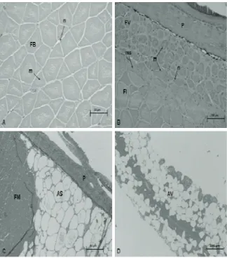
Images of cross sections of the lateral muscles collected below the dorsal fin (MND), in the region of the anal fin (MNA), subcutaneous ventral adipose tissue (AS) and visceral fat (AV) are shown in Figure 2.
Figure 2. Photomicrographs of cross-sectional images of tissue samples from GIFT tilapia strain of 60 days of cultivation A) White muscle in the dorsal region B) Red muscle in the caudal tail C) Subcutaneous ventral adipose tissue D) Visceral adipose tissue. Abbreviations: (AS) subcutaneous adipose (AV) visceral adipose (FB) white fibers (FM) muscle fibers (FI) intermediate fibers (FV) red fibers (m) myofibrils (ms) myosepta (n) nucleus (P) skin. Coloration HE and toluidine blue Increase 40 X and 10x image D.
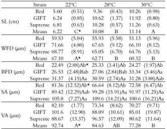
diameter when compared to culture at 28 and 30ºC. The strain x temperature interaction was significant for the diameter of the red fibers. In the GIFT strain, the temperature heated to 30ºC provided greater diameter (33.34 μm) of these fibers when compared to 28 or 22ºC, but this was not observed in the Red and Supreme strains. The Red strain showed smaller diameter in the red fibers at the three growth temperatures, although at 22 and 28ºC it was not different to GIFT strain.
Table 3. Mean and standard deviation of standard length (SL), diameter of white muscle fibers (WFD) and red muscle fibers (RFD), subcutaneous (SA) and visceral adipocytes (VA) in tilapia strains grown at different temperatures for 60 days.
Strain 22ºC 28ºC 30ºC
SL (cm)
Red 5.60 (0.51) 9.36 (0.43) 10.26 (0.98) GIFT 6.24 (0.85) 10.62 (1.37) 11.92 (0.80) Supreme 6.81 (0.63) 10.28 (0.57) 11.26 (0.63)
Means 6.22 C* 10.08 B 11.14 A
WFD (μm)
Red 59.53 (5.04) 55.93 (5.58) 51.13 (5.96) GIFT 71.66 (4.80) 67.65 (9.12) 66.10 (8.12) Supreme 68.77 (8.91) 65.05 (6.70) 64.76 (5.13)
Means 67.10 A* 62.71 B 60.32 B
RFD (μm)
Red 22.49 (2.88)Ab* 25.33 (3.41)Ab 24.27 (1.97)Ab GIFT 26.53 (2.48)Bab 27.06 (2.84)Bab 33.34 (3.46)Aa Supreme 31.37 (4.15)Aa 30.59 (2.74)Aa 31.28 (3.88)Aab
SA (μm)
Red 81.36 (12.52)Ab* 66.64 (8.12)Ab 72.58 (6.47)Ab GIFT 89.42 (12.29)Aab 99.28 (15.91)Aa 91.97 (11.25)Aa Supreme 105.8 (7.27)Aa 109.6 (14.21)Aa 100.6 (16.21)Aa
VA (μm)
Red 82.10 (1.77) 73.34 (8.62) 70.27 (9.71) GIFT 101.6 (12.90) 88.09 (10.61) 85.01 (19.87) Supreme 88.67 (13.37) 96.57 (12.09) 80.62 (11.64)
Means 92.74 A* 84.63 AB 77.28 B
*Means followed by the same capital letters in the row and small letters in the column are not different by Tukey test at 5%.
The analysis of cells from subcutaneous adipose tissue of each fish was performed and showed significant interaction of strain x temperature at 60 days (Table 2). The lowest diameter adipocyte was found in Red strain in the three temperatures, although at 22ºC this strain was similar to GIFT. The temperature influenced the diameter of visceral adipocytes at 60 days of cultivation and this effect was similar between tilapia strains. Fish cultivated at 22ºC showed larger diameter (92.74 μm) when compared to the ones cultured at 30ºC (77.28 μm). Among the strains, the Red one had a smaller diameter (73.71 μm) compared with GIFT (91.99
μm) and Supreme (88.13 μm) ones.
With few exceptions, most species of fish tend to have indeterminate growth. In these animals, muscle growth occurs through two processes: hypertrophy and hyperplasia, which contribute throughout the growth period of post-embryonic skeletal muscle. The growth ‘hyperplastic’ refers to muscle increase in the number of muscle fibers due to the formation of new fibers. The production of new myotubes and muscle fibers continues until the fish reach about 40% of the maximum length of the body (Johnston, 2004).
In hyperplasia, the fusion between satellite cell activated results in the formation of new myotubes on the surface of existing fibers, with subsequent differentiation into muscle fibers (Stellabotte & Devoto, 2007).
During the hypertrophic growth, satellite cells activated fuse with existing muscle fibers by increasing the number of myonuclei and synthesis of myofibrils, leading to an increase in the diameter or area of muscle fibers (Johnston, 1999). Muscle fibers grow by hypertrophy throughout the period of post-larval growth, until they reach a maximum functional diameter which is around 100 micron (white fibers). The rate of hypertrophic growth varies with the rate of somatic growth and in different stages of life of the animal.
The white muscle is approximately 70% of the total volume of muscle tissue (Sanger & Stoiber, 2001). This ratio varies along the length of the fish with a higher proportion in the anterior region of the animal and a large decline towards the caudal region. When compared with the red fibers, these have larger diameters (between 50 and 100 micron) and lower supply of blood capillaries, also have lower concentration of myoglobin and fewer mitochondria (Sanger & Stoiber, 2001). This type of muscle is recruited in sudden movements of swimming, how to capture food and escaping from predators (Altringham & Johnston, 1981).
The red musculature normally corresponds to 5 to 15% relative to the entire myotomal muscle of the fish (Altringham & Johnston, 1981). This muscle is found in the surface region below the dermis, in a higher proportion in the tail region and close to the lateral line. This compartment is composed of red muscle fibers, slow twitch and oxidative metabolism, have small diameter (between 25 and 45 μm), excellent blood supply, high concentration of myoglobin, many mitochondria in the subsarcolemmal region and between the myofibrils, and many droplets lipids (Altringham & Johnston, 1981, Johnston, 1981, Sanger & Stoiber, 2001). The red fibers are recruited when performing slow movements and support, such as migration (Bone, 1978).
adipose tissue harboring perivascular and stromal precursor cells such as fibroblasts, pre-adipocytes with mitotic capacity and committed to differentiation into adipocytes.
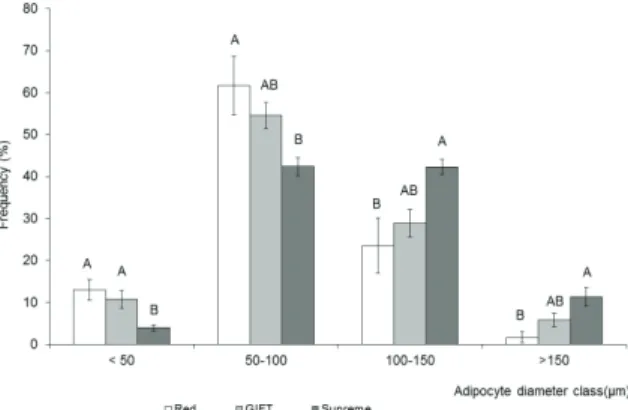
Figure 3 shows the frequency distribution in diameter classes of ventral subcutaneous adipocytes of tilapia cultured at 22ºC. It was observed that the Red strain showed higher frequency adipocytes in the < 50 μm and 50-100 μm classes, whereas the Supreme strain showed a higher frequency of adipocytes in major classes of diameters, 100-150
μm and > 150 μm. The GIFT strain was in middle position. Similar results occurred with fish reared at 28 and 30ºC.
Figure 3. Frequency distribution of subcutaneous ventral adipocytes of tilapia strains cultured at 22ºC.
Fauconneau et al. (1997) analyzed four distinct regions of fat deposition, perivisceral, subcutaneous ventral, dorsal subcutaneous and red muscle of different strains of rainbow trout. At each of these sites, fat cells were found with a variety of size 5-200 microns. The relative proportions of large and medium-sized cells were dependent on the location of the adipose tissue. In the ventral adipose tissue, the population of large cells and their average sizes were larger than in the dorsal adipose tissue. No difference was found in mean size of muscle fibers and fat cells between strains. However, strains with high growth rates were also characterized by a high rate of fat deposition. This may explain the larger diameter and the higher frequency in large adipocytes in GIFT and Supreme strains when compared with Red one.
Conclusion
Red, GIFT and Supreme tilapia strains show differences in growth of muscle and adipose tissues in response to growth temperature. The cold temperature (22ºC) may have caused hypertrophy of white muscle fibers and visceral adipocytes in fingerlings.
These strains present differentiated morphological aspect in red fibers and subcutaneous ventral adipocytes diameters. Furthermore, heating to 30ºC may have contributed to the hypertrophy of red fibers in the GIFT strain.
Acknowledgements
The authors acknowledge the financial support given by Fapesp, São Paulo State, Brazil.
References
Altringham, J. D., & Johnston, I. A. (1981). Quantitative histochemical studies of the peripheral innervation of cod (Gadusmorhua) fast myotomal muscle fibres. Journal of Comparative Physiology A, 143(1), 123-127.
Bentsen, H. B., Eknath, A. E., Vera, M. S. P., Danting, J. C., Bolivar, H. L., Reyes, R. A., … Gjerd, B. (1998). Genetic improvement of farmed tilapias: Growth performances in a complete diallel cross experiment with eight strains of Oreochromis niloticus. Aquaculture, 160(1-2), 145-173.
Bone, Q. (1978). Locomotor muscle. Fish Physiology, 7(1), 361-424.
Charo-Karisa, H., Komen, H., Rezk, M., Ponzoni, R. W., Van Arendonk, J. A. M., & Bovenhuis, H. (2006). Heritability estimates and response to selection for growth of Nile tilapia (Oreochromis niloticus) in low-input earthen ponds. Aquaculture, 261(1), 479-486.
El-sayed, A. F. M. (2006).Tilapia culture. London, UK: Cabi publishing.
Fauconneau, B., André, S., Chmitilly, J., Lebail, P. Y., Krieg, F., & Kaushik, S. J. (1997). Control of skeletal muscle fibers and adipose cells size in the flesh of rainbow trout. Journal of Fish Biology, 50(2), 296-314. Fauconneau, B., Corraze, G., Lebail, P. Y., & Vernier, J.
M. (1991). Lipid storage in fish: cellular, metabolic and hormonal control. Inra, Production Animal, 3(1), 369-381.
Gjoen, H. M. (2004). A new era: The merging of quantitative and molecular genetics - prospects for tilapia breeding programs. In R. B. Bolivar, G. C. Mair, & K. Fitzsimmons (ed.), Proceedings of the sixth international symposium on tilapia in aquaculture (p. 379-399). Manila, PH: Bureau of Fisheries and Aquatic Resources.
Imrie, D., & Sadler, K. C. (2010). White adipose tissue development in zebrafish is regulated by both developmental time and fish size. Developmental Dynamics, 239(11), 3013-3023.
Johnston, I. A. (1981). Quantitative analysis of muscle breakdown during starvation in the marine flatfish
Pleuronectesplatessa. Cell and Tissue Research, 214(2), 369-386.
Johnston, I.A. (1999). Muscle development and growth: potential implications for flesh quality in fish.
Johnston, I. A. (2004). Mechanisms of muscle development and responses to temperature chan in fish larvae. American Fisheries Society Symposium, 40(1), 85-116.
Leaver, M. J., Bautista, J. M., Björnsson, B. T., & Jönsson, E. (2008). Towards fish lipid nutrigenomics: current state and prospects for fin-fish. Aquaculture, 16(sup1), 73-94. Macaranas, J. M., Mather, P. B., Lal, S. N., Vereivalu, T.,
Lagibalavub, M., & Capra, M. F. (1997). Genotype and environment: a comparative evaluation of four tilapia stocks in fiji. Aquaculture, 150(1-2), 11-24. Majumdar, K. C, Nasaruddin, K., & Ravinder, K. (1997).
Pink body colour in tiiapia shows single gene inheritance. Aquaculture research, 28(1), 581-589. Moreira, A. A., Moreira, H. L. M., & Hilsdorf, A. W. S.
(2005). Comparative growth performance of two nile tilapia (chitralada and red-stirling), their crosses and the israeli tetra hybrid nd-56. Aquaculture Research, 36(11), 1049-1055.
Ng, W-K., & Hanim, R. (2007). Performance of genetically improved Nile tilapia compared with red hybrid tilapia fed diets containing two protein levels.
Aquaculture Research, 38(9), 965-972.
Ramírez-Paredes, J. G, Garduño-Lugo, M., & Muñoz-Córdova, G. (2011). Productive performance of a new synthetic red tilapia population ‘pargo-unam’ compared with that of wildtypenile tilapia (Oreochromis niloticus l.). Aquaculture Research, 43(6), 870-87.
Sanger, A., & Stoiber, W. (2001). Muscle fiber diversity and plasticity. Fish Physiology, 18(1), 187-250.
Serlachius, M., & Andersson, L. C. (2004). Up regulated expression of stanniocalcin-1 during adipogenesis.
Experimental Cell Research, 296(1), 256-264.
Sifa, L., Chenhong, L., Dey, M., Gagalac, F., & Dunham, R. (2002). Cold tolerance of three strains of Nile tilapia, Oreochromis niloticus. China Aquaculture, 213(1), 123-129.
Stellabotte, F., & Devoto, S. H. (2007). The teleost dermomyotome developmental dynamics. The American Association of Anatomists, 236(9), 2432-2443. Usher, M. L., Stickland, N. C., & Thorpe, J. E. (1994). Muscle development in atlantic salmon (Salmo
salar) embryos and the effect of temperature on
muscle cellularity. Journal of Fish Biology, 44(6), 953-964.
Valente, L. M., Rocha, E., Gomes, E. F. S., Silva, M. W., Oliveira, M. H., Monteiro, R. A. F., & Fauconneau, B. (1999). Growth dynamics of white and red muscle fibers in fast- and slow- growing strains of rainbow trout. Journal of Fish Biology, 55(4), 675-691.
Vieira, V. L. A., & Johnston, L A. (1992). Influence of temperature on muscle-fiber development in larvae of the herring Clupea harengus. Biology, 112(2), 333-341. Weatherley, A. H., & Gill, H. S. (1985). Dynamics of
increase in muscle fibers in fishes in relation to size and growth. Experientia, 41(3), 353-354.
Received on March 1, 2017. Accepted on June 19, 2017.