Steal coronary-subclavian syndrome: case report and
literature review
Síndrome do roubo coronário-subclávio: relato de caso e revisão da literatura
Jean Carlo Miiller1, Patrick Cardoso Candemil2, João Marcelo Goncalves da Rocha Loures3, Fabrício Martins Zucco3,
Walmor Erwin Belz3, Nilceu Gomes da Rocha Loures3, Marco Rodrigo Ortiz3, Rogério Sanchez Garcia4
Introduction
Advances in Interventional Cardiology have turned angioplasty with stenting into an increasingly attractive option for patients with coronary atherosclerotic disease1.
hus, patients that are referred to myocardial revascula-rization (MR) usually have more severe coronary disease, frequently associated with atherosclerosis in other vascular territories2.
he phenomenon of coronary-subclavian steal is dei-ned as reverse blood low in a coronary artery through an internal mammary artery grat (IMAG), towards the dis-tal subclavian artery, and it happens in patients with severe
stenosis or total occlusion of the proximal portion of the latter1. Despite being rare2, its morbidity is signiicant, with
possible complications3 that can lead to the
coronary-sub-clavian steal syndrome (CSSS). It should be in patients af-ter MR using the inaf-ternal mammary araf-tery grat as bypass, who develop recurrent chest pain, especially when trigge-red by physical efort2.
Case report
A 68-year-old male patient, with hypertension and dys-lipidemia had a history of MR using IMAG 12 years earlier. He presented with pain in the let hemithorax at moderate Abstract
he phenomenon of coronary-subclavian steal is deined as the reversed blood low in a coronary artery, through internal mammary artery graft towards medial-distal subclavian artery, which happens due to severe stenosis or total occlusion of the proximal portion of the latter. It is a rare but signiicant cause of cardiac ischemia after coronary artery bypass surgery and it can cause a syndrome of the same name and with typical manifestations. We have reported the case of a patient with this disease, who underwent percutaneous angioplasty with stent implantation, and we also reviewed the literature on the subject.
Keywords: subclavian artery; subclavian steal syndrome; coronary-subclavian steal syndrome.
Resumo
O fenômeno do roubo coronário-subclávio é deinido como o luxo sanguíneo invertido de uma artéria coronária, por meio de enxerto de artéria mamária interna em direção à subclávia médio-distal, e ocorre devido à estenose signiicativa ou oclusão total da porção proximal desta última. É uma causa rara, mas signiicante, de isquemia cardíaca após cirurgia de revascularização miocárdica e pode originar uma síndrome de mesmo nome e com manifestações típicas. Relatou-se o caso de um paciente com esta enfermidade, que foi submetido à angioplastia percutânea com implante de stent. Também revisou-se a literatura a respeito.
Palavras-chave: artéria subclávia; síndrome do roubo subclávio; síndrome do roubo coronário-subclávio.
Study carried out at the Service of Angiology, Vascular and Endovascular Surgery of Hospital Santa Isabel de Blumenau – Blumenau (SC), Brazil.
1 Angiologist in the Angiology, Vascular and Endovascular surgery team at Hospital Santa Isabel de Blumenau; Professor of Angiology in the Medical Course of Fundação Universitária de Blumenau (FURB) – Blumenau (SC), Brazil.
2 Head of the Angiology, Vascular and Endovascular team at Hospital Santa Isabel de Blumenau – Blumenau (SC), Brazil; Professor of Angiology and Vascular Surgery at the Medical Course of FURB – Blumenau (SC), Brazil.
3 Members of the Angiology, Vascular and Endovascular team at Hospital Santa Isabel de Blumenau – Blumenau (SC), Brazil. 4 Medical student at FURB – Blumenau (SC), Brazil.
Financial support: none.
efort, especially in activities involving the upper limbs,
suggestive of angina pectoris, associated with mild
dysp-nea and sweating. He denied limb claudication, dizziness or syncope. He did not present with murmurs, and had wide and symmetrical pulses in the upper and lower limbs. Blood pressure was 150/75 mmHg in the right upper limb, and 80/50 mmHg in the let upper limb.
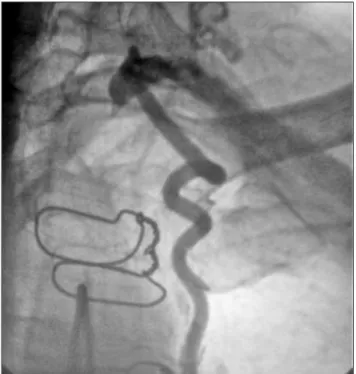
At irst, he underwent myocardial radionuclide ima-ging, which pointed to an ischemic area on the anterior wall. He then underwent percutaneous coronary angio-plasty that showed the reverse blood low in an anterior descending artery (Figure 1), even at rest. he reverse blood low fed the IMAG and reached the distal let sub-clavian artery (LSA), which presented proximal occlusion (Figures 2 and 3). Color Doppler ultrasonography of the cervical arteries showed let subclavian artery steal.
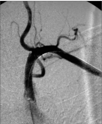
Despite the occlusion, it was decided to perform per-cutaneous angioplasty with stenting of the let subclavian artery, through retrograde puncture of the right common femoral artery and the let brachial artery, by the Seldinger technique. Ater the procedure, patency of the occluded segment was observed (Figure 4). he patient’s symptoms disappeared in the postoperative period. Control radionu-clide myocardial imaging showed improvement of the is-chemic pattern.
Discussion
he internal mammary artery, or internal thoracic ar-tery, was irst used as MR bypass in 19704, and it is currently
the grat of choice, used in about 90% of the patients sub-mitted to this procedure2. It has several advantages
descri-bed in literature, such as: to provide higher patency rates in comparison to saphenous vein grat5 ; to rarely be involved
by atherosclerotic disease5 – even in these cases, high
ra-tes of success with angioplasty are reported6 –; and rarely
present occlusion, that usually happens in the distal anas-tomosis with the coronary artery by a local proliferative reaction7.
he prevalence of let subclavian artery (LSA) stenosis,
mostly found in the proximal segment (85% of the cases)7,
ranges from 0.5 to 6.8%2 in the general population; 3.5 to
5.3% in potentially surgical coronary patients, and from 11.8 to 18.7% in individuals with peripheral arterial disease (PAD). hus, the latter has the highest predictive value for LSA stenosis3,8.
CSSS was irst described in the 1970s, by Tyras and
Barner5, and it is considered an unusual complication
of MR9, with incidence of 0.5 to 2% of the total number of
Figure 1. Coronarography showing retrograde low in the internal
mam-mary artery with illing of the left distal and axillary subclavian arteries.
Figure 2. Angiography shows the obstruction of the left subclavian
operated patients4. However, its incidence is supposedly
in-creasing, because the age of patients submitted to coronary bypass has been increasing, and the risk factors for corona-ry disease are the same for LSA stenosis10.
he etiology of CSSS is almost invariably atherosclero-tic, even though cases of patients with Takayasu’s arteritis10
or IMAG malformations, such as the presence of arteriove-nous istula, have been described11.
he physiopathology is similar to the Subclavian Steal
Syndrome (SSS), described in 1961 by Reivich12, in which
the vertebral artery presents reverse low towards the sub-clavian artery in the presence of stenosis of the proximal LSA. It can be aggravated by the peripheral vasodilation produced by the physical exercise of the afected limb. Symptoms are upper limb claudication and vertebrobasi-lar symptoms (dizziness, vertigo, ataxia and syncope)13. In
CSSS, besides the let vertebral artery, there may be a decre-ase or even low reversal in IMAG12.
Since the IMAG supplies blood low to the coronary artery ater MR, low inversion through the IMAG leads to myocardial ischemia, resulting in ischemic symptoms
and even leading to myocardial infarction10. Patients can
be asymptomatic14, however, the diagnosis should be taken
into consideration in those submitted to MR using IMAG who present with cardiac symptoms such as chest pain and
arrhythmia1; and non-cardiac symptoms, such as dizziness,
vertigo, ataxia and upper limb claudication. he symptoms are usually triggered or aggravated by physical efort9,12. he
onset of the syndrome may occur from 2 to 31 years ater
MR (mean of 14 years)14, which shows that LSA occlusive
lesions developed late ater the operation. he onset of CSSS up to one year ater MRS suggests that LSA stenosis was not observed at the moment of cardiac surgery9.
Physical examination should search for supraclavi-cular murmurs, pulse asymmetry, and especially the ar-terial blood pressure diference between the upper limbs
>20 mmHg, which is the most signiicant inding1. Color
Doppler ultrasonography is a valid method to detect he-modynamically signiicant stenosis in the subclavian area, and the images of computed angiotomography and mag-netic angioresonance can be diagnostic. However, digital subtraction angiography is still the gold standard for this
Figure 3. Angiography in the aortic arch showing obstruction in the
left subclavian artery.
Figure 4. Angiography of post-angioplasty control with stent implant
diagnosis10. Ater contrast injection in the anterior
descen-ding artery, reverse low of IMAG towards the subclavian bed is observed12. Besides, during the procedure, direct
me-asurement of the pressure gradient can be obtained, along with the demonstration of low inversion10.
Diferent types of treatment for CSSS have been des-cribed. he most common procedures in the 1970s and the 1980s were prosthetic or autologous subclavian-subclavian, aorta-subclavian or, most commonly, carotid-subclavian bypass10,12. he latter is contraindicated in cases of critical
stenosis of the carotid segment5.
Alternatively, the proximal third of the IMAG can be
transferred to another donor artery, such as the aorta11.
Dacron prosthesis or polytetraluoroethylene (PTFE) are used as preferential bypasses for open surgery. Autogenous veins, like the saphenous magna, are not a good option due to the high probability of axial torsion and rotation with the movements of the neck, and due to the great diference
between the calibers of both vessels4. he supraclavicular
approach for anastomosis with subclavian artery grat is not free of diiculties and potential complications, due to the proximity to lymphatic channels and local nervous tissues. he infraclavicular approach is simpler, and avoids some of
these potential risks14. he improvement of CSSS symptoms
ater bypass surgery reached 75% in a series of 168 pa-tients5. Possible complications are: stroke, cervical
lympha-tic istula, phrenic nerve paralysis and Horner syndrome5.
he medium and long term patency rate demonstrated in studies is 96% ater four years, and 83% ater eight years of follow-up5,14. he morbidity rate is approximately 25%, and
mortality ranges from 1 to 2%7,13.
Another option for open surgery is the transposition of the subclavian artery to the carotid, which was irst des-cribed in 1964 by Parrot4. It is considered to be an excellent
method to treat stenosis and proximal LSA occlusions due to the lack of synthetic material and the performance of a single anastomosis, with higher long term patency rates than the carotid-subclavian bypass4. However, the transposition
requires the temporary LSA constriction, which ceases the low in the IMAG. his can lead to transitory myocardial ischemia and cause complications13.
Since the 1990s, percutaneous transluminal angioplas-ty is considered to be an efective method to treat for LSA
stenosis9. Followed by the stent placement, the technique
provides more anatomical and physiological results when
compared to open surgery7,12, and it associated with low
morbidity, zero mortality and short hospital stay2,3,7. he
short term technical success is >90%3 and ive-year patency
rates higher than 90%2,3 have been reported.
Some factors may be obstacles for angioplasty, such as cases of densely calciied chronic plaques14, signiicant
ste-nosis or LSA occlusion4,9,12. De Vries et al. reported 100%
success rate for stenosis, and only 65% for occlusions15.
Besides, when the stenosis is too close to the origin of the vertebral artery, the stent may occlude it14.
Postangioplasty thrombosis is rare2; however, long term
in-stent stenosis was described as frequent by Schilliner et al., reaching 40.7% in ive years14-16. Even then, the
efective-ness of angioplasty with stent and open surgery is compara-ble3, with less complications in the angioplasty group,
whi-ch leads to the conclusion that this should be considered as the primary choice of therapy3,9.
In cases of urgent MR with known LSA stenosis, car-diac surgery can be combined with the carotid-subclavian bypass14, or the right internal thoracic artery can be used to
supply the coronary artery2. Angioplasty is the method of
choice2 on elective patients and those with LSA stenosis.
References
1. Kursaklioglu H, Kose S, Iysoy A, et al. Coronary-Subclavian steal syndrome presenting with ventricular tachycardia. Yonsei Med J. 2009;50(6):852-5.
2. Gomes V, Roman M, Barcellos C, et al. Prevalência de estenose da artéria Subclávia em pacientes candidatos à cirurgia de revasculari-zação do miocárdio: Registro multicêntrico. Rev Bras Cardiol Invas. 2008;16(3):307-11.
3. Noord B, Lin A, Cavendish J. Rates of symptom reoccurrence after endovascular therapy in subclavian artery stenosis and prevalence of subclavian artery stenosis prior to coronary artery bypass graf-ting. Vasc H R Management. 2007;3(5):759-62.
4. Guardado J, Goulao J, Pereira H, et al. Síndrome do roubo do Miocárdio, a propósito de um caso clínico. Rev Por Cardiol. 2005;24(2):253-8.
5. Ngueyn NH, Reeves F, herasse E, Latour Y, Genest J. Percutaneous transluminal angioplasty in coronary-internal thoracic-subclavian steal syndrome. Can J Cardiol. 1997;13(3):285-9.
6. Prifti E, Bonachi M, Frati G, Giunti G. Reoperative revascularization of an occluded left subclavian artery and left internal mammary artery ostial stenosis. Eur J Cardio-thoracic Surg. 2002;21:108-10.
7. Dolz LM, Sanchez E, Almenar L, Arnau M, Osa A, Alencia M. Angina refractaria por robô subclávio-coronario tratada mediante angioplastia y stent. Rev Esp Cardiol. 2001;54:920-3.
8. Westerband A, Rodriguez J, Ramaiah V, Dietrich E. Endovascular therapy in prevention and management of coronary-subclavian steal. J Vasc Surg. 2003;38:699-704.
9. Argiriou M, Fillias V, Exarhos D, et al. Surgical treatment of coronary subclavian steal syndrome. Hellenic J Cardiol. 2007;48:236-9.
11. Jofresa Ab, Ortuno FM, Esena F, Nodar J, Navarrete CO, Tello VM. Angioplastia transluminal percutânea em pacientes com esteno-sis de la artéria subclávia e injertos de artéria mamaria. Rev Esp Cardiol. 2002;55(5):537-40.
12. Sadek MM, Ravindran A, Marcuzzi DW, Chisholm RJ. Complete oc-clusion of the proximal subclavian artery post-CABG: Presentation and treatment. Can J Cardiol. 2008;24(7):591-2.
13. Fayad G, Modine T, Beregi JP, Koussa M. A new form of coronary subclavian steal syndrome: ‘the spinning wheels” syndrome. Interact Cardiovasc horac Surg. 2008;7:355-7.
14. Al-Jundi W, Saleh A, Lawrence K, Choksy S. A case report of corona-ry-subclavian steal syndrome treated with Carotid to Axillary artery bypass. Case Rep Med. 2009;2009:1-3.
15. De Vries JP, Jager LC, Van den Berg JC, et al. Durability of percuta-neous transluminal angioplasty for obstructive lesions of proximal subclavian artery: long-term results. J Vasc Surg. 2005;41(1):19-23.
16. Schillinger M, Haumer M, Schillinger S, Ahmadi R, Minar E. Risk strati-ication for subclavian artery angioplasty: is there an increased rate of restenosis after stent implantation? J Endovasc her. 2001;8(6):550-7.
Correspondence
Patrick Cardoso Candemil Rua Marechal Floriano Peixoto, 245 – Sala 75 – Centro CEP 89010-500 – Blumenau (SC), Brasil E-mail: [email protected]
Authors’ contributions