Abstract
Objective: To evaluate selenium dietary intake and nutritional status of patients with phenylketonuria. Methods: The study prospectively evaluated 54 children with phenylketonuria, from 4 to10 years old. The study was performed before and after the use of a selenium-supplemented amino acid mixture. The second phase of the study was performed after, at least, 90 days of use of the supplementation. Selenium nutritional status was assessed through the analysis of biochemical parameters: serum free thyroxin and selenium and glutathione peroxidase in erythrocytes. Selenium dietary intake was evaluated by the administration of the Food Frequency Questionnaire.
Results: Mean age of the children was of 7.0±1.8 years, and 35.2% were female. Mean time of supplementation of selenium, on special formula, was 122.2±25.1 days. The selenium-supplemented amino acid mixture represented 72.9% of the daily supply of the mineral. Upon supplementation, mean concentrations of serum selenium and glutathione peroxidase in erythrocytes increased signiicantly (p < 0.05). The average daily intake of selenium increased signiicantly (p < 0.001), reaching the levels recommended by the Dietary Reference Intakes. The concentration of free thyroxin, in serum, presented signiicant reduction (p < 0.001) in all patients during the second phase of the study, and returned to normal limits in those who had changed levels.
Conclusion: Selenium supplementation through protein replacement is effective to improve and adapt the nutritional status of selenium in patients with phenylketonuria.
J Pediatr (Rio J). 2012;88(5):396-400: Phenylketonuria, selenium, glutathione peroxidase, thyroid hormones.
Copyright © by Sociedade Brasileira de Pediatria
396 Introduction
Phenylketonuria (PKU) is an autosomal recessive disorder characterized by the absence or deiciency of the enzyme
phenylalanine hydroxylase, which prevents the hydroxylation
of phenylalanine (phe) to tyrosine (Tyr). Consequently,
there is an increase of phe in the blood, causing changes
in the central nervous system and irreversible mental retardation, among other manifestations.1
Treatment of PKU is accomplished through the use of phe-restricted diet, with signiicant reduction in the ingestion
of natural proteins. To complete the daily requirement of
Selenium intake and nutritional status of children
with phenylketonuria in Minas Gerais, Brazil
Michelle R. A. Alves,1 Ana L. P. Starling,2 Viviane C. Kanufre,3 Rosângelis D. L. Soares,4Rocksane de C. Norton,5 Marcos J. B. Aguiar,6 José N. Januario7
1. MSc, Saúde da Criança e do Adolescente. Nutritionist, Núcleo de Ações e Pesquisa em Apoio Diagnóstico (NUPAD), Faculdade de Medicina (FM), Universidade Federal de Minas Gerais (UFMG), Belo Horizonte, MG, Brazil. Adjunct professor, Faculdade de Nutrição, Pontifícia Universidade Católica de Minas Gerais (PUCMinas), Belo Horizonte, MG, Brazil.
2. PhD, Saúde da Criança e do Adolescente. Adjunct professor, Departamento de Pediatria, FM, UFMG, Belo Horizonte, MG, Brazil. Associate investigator, Academic director, NUPAD, FM, UFMG, Belo Horizonte, MG, Brazil.
3. MSc, Saúde da Criança e do Adolescente. Clinical nutritionist, Serviço de Nutrição, Hospital das Clínicas, UFMG, Belo Horizonte, MG, Brazil. Nutritionist, NUPAD, FM, UFMG, Belo Horizonte, MG, Brazil.
4. PhD candidate, Saúde da Criança e do Adolescente. Clinical nutritionist, Serviço de Nutrição, Hospital das Clínicas, UFMG, Belo Horizonte, MG, Brazil. Nutritionist, NUPAD, FM, UFMG, Belo Horizonte, MG, Brazil.
5. PhD, Gastroenterologia. Nutrologist physician, Sociedade Brasileira de Pediatria. Associate professor, Departamento de Pediatria, FM, UFMG, Belo Horizonte, MG, Brazil. Associate investigator, NUPAD, FM, UFMG, Belo Horizonte, MG, Brazil.
6. PhD, Pediatria. Associate professor, Departamento de Pediatria, FM, UFMG, Belo Horizonte, MG, Brazil. Geneticist, Associate investigator, Vice Director, NUPAD, FM, UFMG, Belo Horizonte, MG, Brazil.
7. MSc, Saúde da Criança e do Adolescente. Assistant professor, Departamento de Clínica Médica, FM, UFMG, Belo Horizonte, MG, Brazil. Director, Associate investigator, NUPAD, FM, UFMG, Belo Horizonte, MG, Brazil.
No conflicts of interest declared concerning the publication of this article.
Suggested citation: Alves MR, Starling AL, Kanufre VC, Soares RD, Norton RC, Aguiar MJ, et al. Selenium intake and nutritional status of children with phenylketonuria in Minas Gerais, Brazil. J Pediatr (Rio J). 2012;88(5):396-400.
Manuscript submitted Nov 22 2011, accepted for publication May 30 2012.
http://dx.doi.org/10.2223/JPED.2217
protein in the diet, a substitute for protein is provided – protein hydrolysate or amino acid mixture – free of or with low concentrations of phe, added by Tyr, vitamins and minerals2; many of them, however, with no selenium supplementation.
Studies have reported a deiciency of selenium in patients with PKU who use this type of product, concluding that
supplementation was needed, not only due to the small quantity contained in food ingested by these patients, but also for a possible low bioavailability of the nutrient.3 The main sources of selenium are foods rich in protein of high biological value, such as meat, eggs, milk, cereals and
oilseeds, which are prohibited for patients with PKU.4,5 Vegetables, which are the only natural protein sources for
people with PKU, have, in general, concentrations lower
than 5 mcg/100 g.5
Supplementation is recommended either through the
use of a speciic product or through the addition of selenium
in the protein substitute, or even through the use of foods rich in selenium.6
Selenium is a cofactor of glutathione peroxidase
(GPX), fundamental for the antioxidant system, and is
involved in the synthesis of the iodothyronine 5’-deiodinase
(selenoenzyme).6 The low intake of this mineral alters the thyroid function, decreasing serum concentrations of
triiodothyronine (T3) and increasing simultaneously, the concentrations of free thyroxine (FT4).7 Therefore, the decrease in the concentration of selenium is accompanied by an increase in FT4 by reducing the activity of the iodothyronine 5’-deiodinase enzyme.
An inadequate intake and low levels of selenium in
the blood, in the long run, were related to the Keshan
syndrome, characterized by cardiomyopathy and various
degrees of cardiac hypertrophy, and the Kashin-Beck
disease, characterized by endemic osteoarthritis presented by necrotic degeneration of chondrocytes, symmetric stiffness and pain in the interphalangeal joints of the hands,
followed by a generalized osteoarthritis, dwarism, and joint deformations as inal results. However, the dietary or
supplement use of selenium does not reverse the clinical scenarios once installed. These syndromes were described
in some regions of China.8,9 Signs and symptoms related
to biochemical deiciency of this mineral, in PKU patients,
have not been described in other countries.7,10
This study aimed to assess the nutritional status of
selenium in patients with PKU, aged between 4 and 10
years, with early diagnosis and treatment, treated by
the Special Genetics Service at Hospital das Clínicas within Universidade Federal de Minas Gerais (UFMG). The eficiency of using a selenium-supplemented amino acid mixture in the recovery of possible mineral deiciencies was also veriied, guaranteeing, thus, a more appropriate
treatment for the patients.
Methods
We conducted a prospective cohort study with 54 patients
with PKU, from 4-10 years old, early diagnosed and treated. The study was divided in two phases: the irst, or phase 1,
happened before the child ingested selenium-supplemented amino acid mixture; the second, or phase 2, happened at least 90 days after the onset of consumption of the mixture supplemented with the mineral.
The assessment of dietary intake of selenium was performed by Quantitative Food Frequency Questionnaire
(QFFQ) adapted for people with PKU. The preparation of the
list of foods present in the QFFQ was based on 24 or 72-hour dietary recalls, obtained from each child’s nutritional records. The parent or guardian, answered the QFFQ on the day of the consultation, through an interview. The calculation of selenium content in foods was analyzed by
the Diet Pro® 4.0 software.
The selenium content of most foods was obtained from
the tables contained in the Brazilian study of Ferreira et al.5
and the table from the Department of Agriculture in the
United States.11 Dietary intake of selenium was compared
to the Dietary Reference Intake/Recommended Dietary Allowances (DRI/RDA).
The selenium content of the selenium-supplemented amino acid mixture has been removed from the label of the
product used by patients. Children were divided according
to age group, so that the intake of selenium, according to
the recommendation of DRI/RDA, could be assessed.12
In the irst phase of the study, the intake of selenium
indicated only to the content of selenium on the diet of the patient, once the mixture of amino acids was not
supplemented. In the second phase, the amount of selenium
indicated the content of selenium on the diet plus the content of the selenium-supplemented amino acid mixture.
To perform the biochemical tests, we collected from each child: 3.0 mL whole blood in dry tube to obtain serum for measurement of free T4; 4.0 mL of whole blood in heparin
tube for determination of GPX in erythrocytes; 6.0 mL of whole blood in a trace tube (special tube, metal free)
for measurement of serum selenium. The measurement of serum selenium were made by atomic absorption
spectrophotometry (reference value [RV] = 46.0-143.0 mcg/L)13; the measurement of GPX, in erythrocytes by
an enzymatic method (RV = 27.5-73.6 U/G Hb) (Randox Laboratories, Antrim, UK); and the measurements of FT4, by chemiluminescence (RV = 0.54-1.24 nanog/dL) (Unicel DxI 800®, Beckman Coulter, Brea, EUA).
Data were analyzed with the Statistical Package for the Social Sciences 10.0. Categorical variables
Pearson (r) correlation test. The chi-square test was used
to study the association between categorical variables. To compare means, we used Student’s t test. Statistically
signiicance was established at p < 0.05. The McNemar
test was used to assess whether the children were able to reach the reference ranges of biochemical tests,
establishing p ≤ 0.05 as statistically signiicant.
The parents signed an informed consent form, authorizing the child to take part in the study, which
was approved by the Research Ethics Committee of the University, under n. ETIC 468/06.
Results
Among the 54 children, 19 (35.2%) were female, 43 (79.6%) were between 4 and 8 years old, and 11 (20.4%)
were between 9 and 10 years, with mean age of 7.0±1.8 years. The mean time of use of the supplemented amino acid mixture was of 122.2±25.1 days.
A high percentage (67.9%) of GPX below the values of reference was found in the irst phase, while in the second
phase, after the complementation, only 18.5% remained
with values below the recommended (p < 0.001). Regarding serum selenium, it was observed that 98.1%
of patients presented values below the recommendation
in the irst phase, and after the supplementation, 18.5%
reached adequate concentrations. There was, however, a
statistically signiicant increase in the content of serum
selenium, in all patients, when the two phases of the
study were compared (p = 0.0016).
In the irst phase, 16.7% (n = 9) of patients had FT4
above the recommended value. After the supplementation with the mineral, values of FT4, in these cases, reached
normal values. It was also observed a reduction in the
level of the hormone in all patients in the second phase
of the study (p = 0.004).
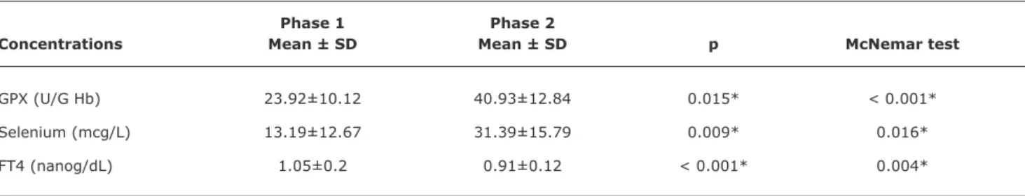
Table 1 - Mean and standard deviation of the concentrations of glutathione peroxidase, serum selenium and free thyroxine of 54 children with phenylketonuria, between 4 and 10 years old, before and after the use of selenium-supplemented amino acid mixture
Phase 1 Phase 2
Concentrations Mean ± SD Mean ± SD p McNemar test
GPX (U/G Hb) 23.92±10.12 40.93±12.84 0.015* < 0.001*
Selenium (mcg/L) 13.19±12.67 31.39±15.79 0.009* 0.016*
FT4 (nanog/dL) 1.05±0.2 0.91±0.12 < 0.001* 0.004*
FT4 = free thyroxine; GPX = glutathione peroxidase; SD = standard deviation. * Statistical significance.
Figure 1 - Contribution of the amino acid mixture in relation to the mean selenium content (mcg/day) in the diet of 54 children with phenylketonuria, in the age groups studied
AA = amino acids.
The mean serum concentrations of GPX, selenium
and FT4, in the two studied phases, are demonstrated
in Table 1. It is observed that there was a statistically signiicant difference, between the two phases in all
parameters assessed.
Only with natural food consumption, there was an average intake of selenium far below the recommended levels. Table 2 presents the mean of selenium content
(mcg/day) supplied by a normal diet (phase 1) and by
adding the mixture of amino acids supplemented with
selenium (phase 2) in both age groups. There was a statistically signiicant increase in the supply of selenium
between the two phases.
Table 2 - Means of selenium micrograms per day (mcg/day) provided by normal diet and by the mixture of amino acids, according to the age groups analyzed
Phase 1 Phase 2 Diet +
Age group Diet Mixture of AA* Mixture of AA* DRI/RDA p
4-8 years (n = 43) 11.5±6.7 28.3±6.2 39.8±8.8 30 < 0.001†
9-10 years (n = 11) 10.7±4.7 38.4±9.2 49.1±11.6 40 0.026†
AA = amino acids; DRI/RDA = Dietary Reference Intakes/Recommended Dietary Allowances. * Mixture of amino acids supplemented with selenium.
† Statistical significance.
acid mixture compared to the average content of selenium
(mcg/day) in the diet of 54 children with PKU, in the age
groups studied, before and after supplementation.
There were no clinical signs and symptoms related to
selenium deiciency.
Discussion
The number of children with PKU participating in this study (n = 54) should be emphasized, because as far as it is known, it is the largest sample of patients with PKU
coming from the same referral center for treatment of the disease, in which the nutritional status of selenium was studied. This number represents 67% of patients, aged
between 4 and 10 years, and 30% of all those with PKU diagnosed and treated early in the state of Minas Gerais, since 1993, when the program was implemented. It is
important to know that the early treatment, in a study conducted in the same reference center, prevented mental retardation in 90.5% of patients.14 It is, therefore, a fairly representative sample of this population. Furthermore, the use of the same patients as controls, in both phases of the study, reduced the bias of the interindividual comparison.
Some studies with phenylketonuric children found
signiicant reduction in the GPX activity in the erythrocyte,
plasma and erythrocyte-selenium concentrations and increased oxidative stress related to the low intake of the micronutrient compared to controls. Supplementation of selenium, in the diet, normalized the concentrations of all biochemical parameters related to the mineral,15,16 as found in the present study.
As the protein substitute is obtained by a bidding, it was not possible to increase the time of use of the selenium-supplemented mixture, due to change for a new product. Studies report that 90 days are enough for selenium serum
concentrations, as well as concentrations of GPX in the
erythrocyte, to increase and even reach normal levels.16
While there has been signiicant increase in the content of selenium and GPX in erythrocytes, as well as signiicant
decrease in FT4, serum concentrations of selenium have not reached the normal range in most patients in this study. Perhaps a longer time of supplementation would be required.
However, we found that this supplementation was suficient to reverse or improve, signiicantly, the nutritional status
of selenium in most children.
This is the only Brazilian study evaluating the thyroid hormone (FT4) from the point of view of selenium deiciency, besides being the only Brazilian study assessing the
supplementation of selenium in children under treatment
for PKU. Concentrations of FT4 above the reference values
were found in some patients, which may be explained by a decrease in the activity of the enzyme 5’ iodothyronine deiodinase, due to reduced concentrations of selenium in the body.17-19 After supplementation with selenium, concentrations of FT4 in these patients, as well as in the
whole group, reduced signiicantly, remaining within an
appropriate range, which may be explained by the greater availability of selenium, resulting from the standardization of
intake, similar to what was veriied in other studies.7,19
The low consumption of selenium, by patients with PKU,
was also reported in reviews by other authors.20,21 In one of
the studies, it was observed selenium deiciency even with
its presence in the composition of the protein substitute.21 The mixture of amino acids was the main source of selenium for the patients studied.
Using the content of selenium of foods from the Brazilian
study5 reduced the bias that could have been produced by the use of foods from composition tables of other countries. Although the food in this study was not primarily grown in the
References
1. Scriver CR, Kaufman S. Hyperphenylalaninemia: phenylalanine hydroxylase deiciency. In: Scriver CR, Beaudet AL, Sly WS, Valle D, Childs B, Vogelstein B, eds. The metabolic and molecular basis of inherited disease. 8th ed. New York: McGraw-Hill; 2001. p. 1667-724.
2. Acosta PB, Yanniccelli S. The Ross metabolic formula system, nutrition support protocols. 4th ed. Columbus: Ross Laboratories, Library of Congress; 2001. 432p.
3. Mira NV, Marquez UM. Importance of the diagnoses and treatment of phenylketonuria. Rev Saude Publica. 2000;34:86-96. 4. Kanufre VC, Santos JS, Soares RD, Starling AL, Aguiar MJ.
Abordagem dietética para fenilcetonúria. Rev Med Minas Gerais. 2001;11:129-34.
5. Ferreira KS, Gomes JC, Bellato CR, Jordão CP. Selenium
concentration in food consumed in Brazil. Rev Panam Salud Publica. 2002;11:172-7.
6. Rayman MP. The importance of selenium to human health. Lancet. 2000;356:233-41.
7. Köhrle J. Selenium and the control of thyroid hormone metabolism. Thyroid. 2005;15:841-53.
8. Food and Agriculture Organization of the United Nations (FAO), World Health Organization (WHO). Vitamin and mineral requirements in human nutrition. 2nd ed. Geneva: World Health Organization. 2004.
9. Brenneisen P, Steinbrenner H, Sies H. Selenium, oxidative stress, and health aspects. Mol Aspects Med. 2005;26:256-67. 10. Jochum F, Terwolbeck K, Meinhold H, Behne D, Menzel H, Lombeck
I. Is there any health risk of low dietary selenium supply in PKU children? Nutr Res. 1999;19:349-60.
11. U.S. Department of Agriculture, Agricultural Research Service. 2011. USDA National Nutrient Database for Standard Reference, Release 24. https://www.ars.usda.gov/SP2UserFiles/
Place/12354500/Data/SR24/nutrlist/sr24w317.pdf. Access: 03/05/2011.
12. Food and Nutrition Board, Institute of Medicine, National Academies. Dietary Reference Intakes (DRIs): Recommended Dietary Allowances and Adequate Intakes, Vitamins. http://iom.edu/
Activities/Nutrition/SummaryDRIs/~/media/Files/Activity%20 Files/Nutrition/DRIs/RDA%20and%20AIs_Vitamin%20and%20 Elements.pdf. Access: 28/04/2011.
13. Iyengar V, Woittiez J. Trace elements in human clinical specimens: evaluation of literature data to identify reference values. Clin Chem. 1988;34:474-81.
14. Castro IP, Borges JM, Chagas HA, Tibúrcio J, Starling AL, de Aguiar MJ. Relationships between phenylalanine levels, intelligence and socioeconomic status of patients with phenylketonuria. J Pediatr (Rio J). 2012 Mar 28. [Epub ahead of print]
15. Sitta A, Barschak AG, Deon M, Terroso T, Pires R, Giugliani R, et al. Investigation of oxidative stress parameters in treated phenylketonuric patients. Metab Brain Dis. 2006;21:287-96. 16. Wilke BC, Vidailhet M, Favier A, Guillemin C, Ducros V, Arnaud
J, et al. Selenium, glutathione peroxidase (GSH-Px) and lipid peroxidation products before and after selenium supplementation. Clin Chim Acta. 1992;207:137-42.
17. Arthur JR, Beckett GJ. Thyroid function. Br Med Bull. 1999;55:658-68.
18. Lombeck I, Jochum F, Terwolbeck K. Selenium status in infants and children with phenylketonuria and in maternal phenylketonuria. Eur J Pediatr. 1996;155:S140-4.
19. Zimmermann MB, Köhrle J. The impact of iron and selenium
deiciencies on iodine and thyroid metabolism: biochemistry and
relevance to public health. Thyroid. 2002;12:867-78.
20. Reilly C, Barrett JE, Patterson CM, Tinggi U, Latham SL, Marrinan A. Trace element nutrition status and dietary intake of children with phenylketonuria. Am J Clin Nutr. 1990;52:159-65. 21. Dobbelaere D, Michaud L, Debrabander A, Vanderbecken S,
Gottrand F, Turck D, et al. Evaluation of nutritional status
and pathophysiology of growth retardation in patients with phenylketonuria. J Inherit Metab Dis. 2003;26:1-11.
22. Artuch R, Colomé C, Sierra C, Brandi N, Lambruschini N, Campistol J, et al. A longitudinal study of antioxidant status in phenylketonuric patients. Clin Biochem. 2004;37:198-203.
23. Barretto JR, Silva LR, Leite ME, Boa-Sorte N, Pimentel H, Puriicação AC, et al. Poor zinc and selenium status in phenylketonuric children
and adolescents in Brazil.Nutr Res. 2008;28:208-11.
24. Gordon SJ, Latham SC, Spink JD, Galbraith AJ. Assessment of cardiac function by M-mode echocardiography in
selenium-deicient phenylketonuric children. J Paediatr Child Health. 1991;27:47-50.
Correspondence:
Michelle Rosa Andrade Alves
Rua Diametral, 116/13, bloco A, Sagrada Família CEP 31030-350 - Belo Horizonte, MG - Brazil Tel.: +55 (31) 3457.1382, +55 (31) 9663.1288 E-mail: [email protected]
One of the positive aspects of this study is the supplementation of selenium by adding it to the amino acid mixture, ensuring access to the mineral for all patients,
from all age groups. It also avoids inadequate handling and
problems in acquisition/purchase of the substance used as a complement, avoiding errors in the calculation of the dose to be used. The increase of the biochemical parameters related to selenium supplementation increases the need
for solid mineral, justiied by the existence of a positive
correlation between serum selenium concentrations and
dosages of GPX in erythrocytes.22
As reported in the literature,16,22,23 no clinical
manifestations of selenium deiciency were found in our patients. Therefore, selenium deiciency in this population is
only biochemical. Since the public system does not provide access to these diagnostic exams, it is not possible to obtain, routinely, an assessment of the nutritional state of selenium in all patients. The literature is unanimous in highlighting the need for selenium supplementation in patients with phenylketonuria, once subclinical manifestations may happen, such as changes in myocardial function observed
in echocardiograms of children with PKU.24
The selenium supplementation in patients with PKU
should be recommended, therefore, preferably through the continuous use of protein substitutes supplemented with selenium in all age groups. A thorough investigation of the intake should be performed, as well as an assessment of the biochemical markers of selenium levels in the body, whenever possible, to avoid further damage to health,