254
O
riginala
rticle Copyright © 2009 by Sociedade Brasileira de Pediatriaabstract
Objective: To characterize the stomatognathic system and stomatognathic functions in patients with mucopolysaccharidosis.
Methods: Cross-sectional and observational study of patients with mucopolysaccharidosis seen at the outpatient clinic at the Medical Genetics Service of Hospital de Clínicas de Porto Alegre. The inclusion criteria were the existence of a biochemical or molecular diagnosis of any type of mucopolysaccharidosis and the agreement to participate in the study by signing an informed consent form. Seventy-eight patients were evaluated through phonoaudiological anamnesis and physical exam.
results: Alterations in at least one item of each structure of the stomatognathic system or stomatognathic function were found in all patients who allowed evaluation of both items on physical examination (n = 76/78). The most frequently compromised structures and functions were respectively the dental arch and the tongue, swallowing and mastication. The only statistically signiicant difference found between types of mucopolysaccharidosis involved the habitual position of the tongue between the teeth (most frequent in mucopolysaccharidosis VI). Among patients with mucopolysaccharidosis I, II or VI who underwent enzyme replacement therapy or not, there was statistically signiicant difference in oral breathing mode (more frequent in the group without enzyme replacement therapy).
conclusions: Alterations in stomatognathic systems and functions are prevalent among individuals with mucopolysaccharidosis, even if enzyme replacement therapy is administered. Such inding suggests that speech therapy follow-up plays a major role in the treatment plan of this group of diseases; this hypothesis should be conirmed by additional studies.
J Pediatr (Rio J). 2009;85(3):254-260: Mucopolysaccharidosis, speech therapy, stomatognathic system.
introduction
Mucopolysaccharidoses (MPS) are genetic diseases
caused by the deicient activity of enzymes that degrade
glycosaminoglycans (GAGs) and by the consequent accumulation of these macromolecules inside lysosomes.1 The
MPS are classiied according to the deicient enzyme as: MPS
I (
α
-L-iduronidase deiciency), MPS II (iduronate-sulfatasedeiciency), MPS III-A (heparan N-sulfatase deiciency),
MPS III-B (
α
-N-acetylglucosaminidase deiciency), MPSIII-C (acetyl-CoA-
α
-glucosaminide acetyltransferasedeiciency), MPS III-D (N-acetylglucosamine 6-sulfatase deiciency), MPS IV-A (N-acetylgalactosamine 6-sulfatase deiciency), MPS IV-B (
β
-galactosidase deiciency), MPS VI (N-acetylgalactosamine-4-sulfatase deiciency), MPS VII(
β
-glucuronidase deiciency), and MPS IX (hyaluronidase deiciency). It is estimated that the combined incidence of this group of diseases is 1:25,000 live newborns.2evaluation of orofacial motricity in patients
with mucopolysaccharidosis: a cross-sectional study
giovana S. turra,1 ida Vanessa D. Schwartz2
1. MSc, Medicine, Medical Sciences, Universidade Federal do Rio Grande do Sul (UFRGS), Porto Alegre, RS, Brazil.
2. PhD, Sciences, Genetics, UFRGS, Porto Alegre, RS, Brazil. Associate professor, Department of Genetics, UFRGS, Porto Alegre, RS, Brazil.
This paper is part of the Graduate Program in Medicine: Medical Sciences, Universidade Federal do Rio Grande do Sul (UFRGS), Medical Genetics Service, Hospital de Clínicas de Porto Alegre, and Department of Genetics, UFRGS, Porto Alegre, RS, Brazil.
Financial support: Rede MPS Brasil, CAPES.
No conflicts of interest declared concerning the publication of this article.
Suggested citation: Turra GS, Schwartz IV. Evaluation of orofacial motricity in patients with mucopolysaccharidosis: a cross-sectional study. J Pediatr (Rio J). 2009;85(3):254-260.
Manuscript received January 12 2009, accepted for publication April 1 2009.
Each type of MPS is associated with great clinical heterogeneity. As to MPS I, for instance, its most severe form (characterized by mental retardation, severe somatic
involvement and death before the age of 10 years) is
usually called Hurler syndrome. Its mild form (characterized by preserved cognition and less somatic involvement) is called Scheie syndrome.3 In all MPS types, the following
indings are frequent: macrocephaly, hepatosplenomegaly,
umbilical and inguinal hernias, bone dysplasia, delayed
motor development, hearing loss, respiratory dificulty, facial
and dental alterations, enlarged tongue, heart disease, and limitation of joint mobility. Corneal clouding is not frequent in MPS II, III and IV; MPS IV and VI are not usually associated with mental retardation.4-6
MPS treatment may be symptomatic or speciic.
Symptomatic treatment addresses the disease complications,
such as the use of medication for heart insuficiency. The speciic treatment seeks to replace the deicient enzyme,
and includes when recommended hematopoietic cells transplantation and enzyme replacement therapy (ERT); the latter is available for MPS I, II and VI.10-12
In the literature there are few studies regarding speech and language evaluation in patients with MPS, and most are focused on aspects related to speech and language development.13-15 The main objective of this study was to describe the alterations found in the stomatognathic system and functions of patients with MPS.
Methods
This is a cross-sectional and observational study, approved by the Research Ethics Committee of Hospital de Clínicas de Porto Alegre (HCPA). The study assessed patients with MPS seen at the MPS outpatient clinics of the Medical Genetics Service at HCPA, which is an international reference center for the diagnosis and treatment of this group of diseases. The patients were invited to participate
in the study at one of their visits to the clinics in 2006/2007
and were included in the study only after signing an informed consent form. Inclusion criterion was existence of biochemical or molecular diagnosis of any type of MPS. The situation of current or past treatment was not part of the inclusion or exclusion criteria.
Assessment comprised phonoaudiological anamnesis,
physical exam, and medical ile review. Anamnesis and
physical exam were based on Bianchini, Junqueira, Morales and Marchesan16-19 and were performed by phonoaudiologist that wrote this paper, not blinded to patients’ treatment status, on a consultation of around one hour. Anamnesis was obtained from the patient or guardian. Physical examination consisted of evaluation of stomatognathic
system structures (lips, tongue, cheeks, mandible, hard
palate, soft palate, palatine tonsils and dental arch) and stomatognathic functions (suction, mastication, swallowing,
breathing and speech). Examination of tongue, cheeks,
palate, palatine tonsils and teeth was performed with
the aid of gloves, spatula and lashlight. Evaluation of
mastication was performed using solid food (bread roll); solid food (bread roll) and liquid (water) in a glass for swallowing; and plastic straw and liquid (water) in a glass for suction.
Data were grouped into a Microsoft Excel table and further analyzed using the Statistical Package for the Social Sciences (SPSS) 14.0 for Windows. Variables with data available to more than 50% of the sample were analyzed. Categorical
variables were summarized by means of frequencies and percentages. Continuous variables with normal distribution were summarized by mean and standard deviation values, while those with asymmetrical distribution were summarized by means of median values and interquartile ranges. MPS
types that individually corresponded to over 10% of the
sample were compared as to the most frequent alterations found in each of the stomatognathic structures and functions assessed. MPS I, II and VI patients on ERT or not on ERT were also compared as to these variables; due to the sample size, the effect of ERT was not analyzed according to the exposed MPS subgroups. Statistical tests used to perform such comparisons were chi-square or Fisher’s exact test applying Bonferroni’s correction.
Patients were not compared in relation to being or not on phonoaudiological treatment because exposure to this type of intervention was not homogenous in the sample, including different professionals and techniques, in addition
to a different number of weekly sessions.
results
Eighty-six patients with MPS were evaluated at the Medical Genetics Service of HCPA during the study period
(MPS I = 15; MPS II = 37; MPS III-B = 2; MPS IV-A = 5; MPS VI = 27). Of these, 78 agreed to participate and their data are summarized in Table 1. As to origin, 74/78 patients were Brazilian: 29 (39.2%) from the South, 26 (35.1%) from the Southeast, 14 (18.9%) from the Northeast, three (4.1%) from the North, and two (2.7%)
from the Midwest. Mental retardation was present in 17/78
(21.8%) patients (MPS I severe form = 4/14 or 28.6%; MPS II severe form = 11/35 or 31.4%; and MPS III-B = 2/2). Major visual deicit was present in 9/78 (11.5%)
patients, all with MPS VI. Two patients had a tracheotomy tube (both had MPS VI). MPS I, II and VI each accounted
for more than 10% of the total sample, and were included in subgroup analysis. During the evaluation, 18 patients were receiving phonoaudiological treatment, and 40 were
given ERT (at the Medical Genetics Service of HCPA).
Anamnesis
Patients gender Mean age at evaluation number of patients
type of MPS (n) (male/female) ± SD (years) in ert*
MPS I 14 5/9 12.6±9.4 11 (10; 6-18)
MPS II 35 35/0 11.9±6.5 19 (25; 18-30)
MPS III-B 2 1/1 11.0±2.8 0
MPS IV-A 4 2/2 11.0±3.4 0
MPS VI 23 13/10 10.4±4.7 10 (19; 14.2-24.5)
Total 78 56/22 11.5±6.4 40 (19.5; 14-26)
table 1 - Characterization of patients with MPS evaluated in this study (n = 78)
ERT = enzyme replacement therapy; MPS = mucopolysaccharidosis; SD = standard deviation. * Median in months of ERT; interquartile range.
patients were breastfed at some time in their lives (median of breastfeeding time = 6 months; interquartile
range = 3-18); of these, four (6.4%) were foreigners.
Median of breastfeeding time considering only Brazilian
patients was 8 months (interquartile range = 3-18). Use of baby bottle occurred in 69/76 (90.8%) patients (median of baby bottle use = 30 months; interquartile range = 15.7-58.5). Use of paciier was reported by 53/77 (68.8%) patients (median of paciier use = 36 months; interquartile range = 24-72).
Information on food consistency consumed until 2 years of age was obtained from 74/78 patients, being adequate in 62/74 (83.8%) The other patients consumed only pasty foods until 2 years of age. At the time of the study, consistency of the food was normal in 66/77 (85.7%)
patients (the others ingested only pasty foods). In terms of deleterious habits, there was presence of onychophagia
in 13/77 (16.9%) patients and bruxism in 7/77 (9.1%). With regard to sleep, 48/73 (65.7%) reported occurrence of drooling, 35/78 (44.9%) reported snoring, and 23/76 (30.3%) reported apnea. As to previous surgeries, 42/76 (55.3%) patients had undergone adenoidectomy, 25/76 (32.9%) myringotomy with placement of ventilating tubes, and 18/76 (23.7%) tonsillectomy.
Physical examination
Cognitive, visual and joint deicits found in many patients made physical examination dificult, especially in items
regarding stomatognathic functions. Physical examination could not be performed in one patient (MPS I severe form); in another patient (also MPS I severe form) any function could be evaluated, only the stomatognathic system was partially evaluated and had normal results. All the other patients had some abnormality on physical exam, which is
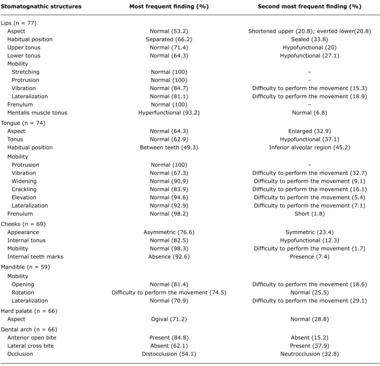
summarized in Tables 2 and 3. The most frequently impaired
structures of the stomatognathic system were the dental
arch and tongue (98.4 and 95.9% of patients, respectively,
had some type of change); the other structures (lips,
cheeks, mandible and hard palate) were each compromised in 70-90% of patients. The most impaired functions were swallowing and mastication (98.5 and 95.3% of patients,
respectively, had some type of change); the other functions
(suction, breathing and speech) were each impaired in 65-80% of patients. Soft palate and palatine tonsils could not be evaluated in more than 50% of the sample. For that reason, such variables were not included in Table 2.
MPS I, II and VI were compared in relation to frequency of the most prevalent changes on physical exam (Table
4). When Bonferroni’s correction was applied, only the habitual position of the tongue between teeth had signiicant
statistical difference (more frequent in MPS VI). In a comparison of groups with and without ERT, after application
of Bonferroni’s correction, there was signiicant statistical
difference only in relation to oral breathing mode (more frequent in the group without ERT).
Discussion
MPS are severe, chronic, multisystemic genetic diseases associated with wide heterogeneity and high morbidity and mortality rate in childhood.1,20 Although clinical suspicion of MPS can be easily performed by a basic healthcare professional when the patient has the complete extent of the
disease (that is, in more advanced stages), its identiication in earlier stages or in mild or atypical cases is more dificult and requires a deeper knowledge of the disease.
The diagnostic path that MPS patients have to follow
is long and expensive: in average diagnosis is performed 4.8 years after symptom onset; during this period, such patients see four to ive specialists in average, require
one to two hospitalizations and are submitted to one or two surgical procedures.21 In addition, conirmation of diagnosis (and type of MPS as each type is caused by
a speciic enzyme deiciency) is dependent on complex
Stomatognathic structures Most frequent inding (%) Second most frequent inding (%)
Lips (n = 77)
Aspect Normal (53.2) Shortened upper (20.8); everted lower(20.8)
Habitual position Separated (66.2) Sealed (33.8)
Upper tonus Normal (71.4) Hypofunctional (20)
Lower tonus Normal (64.3) Hypofunctional (27.1)
Mobility
Stretching Normal (100) –
Protrusion Normal (100) –
Vibration Normal (84.7) Dificulty to perform the movement (15.3)
Lateralization Normal (81.1) Dificulty to perform the movement (18.9)
Frenulum Normal (100) –
Mentalis muscle tonus Hyperfunctional (93.2) Normal (6.8)
Tongue (n = 74)
Aspect Normal (64.3) Enlarged (32.9)
Tonus Normal (62.9) Hypofunctional (37.1)
Habitual position Between teeth (49.3) Inferior alveolar region (45.2) Mobility
Protrusion Normal (100) –
Vibration Normal (67.3) Dificulty to perform the movement (32.7)
Widening Normal (90.9) Dificulty to perform the movement (9.1)
Crackling Normal (83.9) Dificulty to perform the movement (16.1)
Elevation Normal (94.6) Dificulty to perform the movement (5.4)
Lateralization Normal (92.9) Dificulty to perform the movement (7.1)
Frenulum Normal (98.2) Short (1.8)
Cheeks (n = 69)
Appearance Asymmetric (76.6) Symmetric (23.4)
Internal tonus Normal (82.5) Hypofunctional (12.3)
Mobility Normal (98.3) Dificulty to perform the movement (1.7)
Internal teeth marks Absence (92.6) Presence (7.4)
Mandible (n = 59) Mobility
Opening Normal (81.4) Dificulty to perform the movement (18.6)
Rotation Dificulty to perform the movement (74.5) Normal (25.5)
Lateralization Normal (70.9) Dificulty to perform the movement (29.1)
Hard palate (n = 66)
Aspect Ogival (71.2) Normal (28.8)
Dental arch (n = 66)
Anterior open bite Present (84.8) Absent (15.2)
Lateral cross bite Absent (62.1) Present (37.9)
Occlusion Distocclusion (54.1) Neutrocclusion (32.8)
table 2 - Evaluation of stomatognathic structures of patients with MPS*†‡
MPS = mucopolysaccharidosis.
* No variable could be evaluated on physical exam in one patient (see Results). In another patient only the stomatognathic system could be evaluated. Therefore, of the 78 patients included in the study, 77 had their stomatognathic structures evaluated and 76 had their stomatognathic functions evaluated.
† Habitual position represents the position at rest in which lips, tongue and dental arch are found.
‡ Only variables that could be evaluated for more than 50% of the sample are described. As a consequence, mean diagnostic age in Brazilian patients (76 months) is higher than the double of that found in developed countries.21 Since the pediatrician is the health professional most frequently sought by patients/relatives with MPS when clinical manifestations occur,21 it is crucial that such professionals know about the clinical signs associated with this group of diseases, as well as of the currently available possibilities of management and treatment.
Within such context, this study indicates that alterations in lips, tongue, cheeks, mandible, hard palate and dental
arch are frequent in patients with MPS, as well as their functional correspondents (suction, mastication, swallowing,
breathing and speech). Such inding is in accordance with
what is expected from the information available in the literature on orofacial motricity and MPS, which particularly
comes from chapters of specialized books about inborn
MPS i (n = 13/14) MPS ii (n = 35) MPS Vi (n = 23)
Change n (%)* n (%) n (%) p
Hyperfunction of mentalis muscle at rest 12/13 (92.3) 29/32 (90.6) 22/22 (100) 0.398†
Habitual tongue position between teeth 3/12 (25) 13/33 (39.4) 18/23 (78.3) 0.003‡§
Cheek asymmetry 6/10 (60) 23/28 (82.1) 18/22 (81.8) 0.309†
Dificulty in mandible rotation 6/9 (66.7) 17/21 (81) 15/18 (83.3) 0.652†
Ogival palate 9/13 (69.2) 19/28 (67.9) 17/21 (81) 0.569‡
Anterior open bite 10/11 (90.9) 20/27 (74.1) 23/23 (100) 0.017†§
Suction with pressed lips 4/6 (66.7) 21/26 (80.8) 5/11 (45.5) 0.083†
Vertical mastication mode 6/7 (85.7) 24/24 (100) 20/21 (95.2) 0.127†
Hyperfunction of mentalis muscle in swallowing 7/8 (87.5) 27/28 (96.4) 17/17 (100) 0.380†
Oral breathing mode 9/12 (75) 19/33 (57.6) 21/23 (91.3) 0.021‡§
Tongue protrusion in speech 7/10 (70) 21/28 (75) 17/21 (81) 0.780†
Stomatognathic functions Most frequent inding (%) Second most frequent inding (%)
Suction (n = 50)
Eficiency Normal (92) Ineficient (8)
Tongue posture Normal (88.9) Protrusion (11.1)
Lip posture Pressed (71.1) Protrusion (28.9)
Mastication (n = 64)
Symmetry Symmetrical (73.7) Asymmetrical to the left (15.8)
Lip position Separated (65.6) United (34.4)
Speed Slow (89.8) Fast (10.2)
Mandible mobility Vertical (92.6) Normal (7.4)
Bite Anterior (55) Lateral (45)
Swallowing (n = 67)
Mentalis muscle tonus Hyperfunction (96.4) Normal (3.6)
Perioral muscles Normal (53.7) Altered (46.3)
Lingual projection Absent (78.5) Present (21.5)
Compensatory movement Absent (90.9) Present (9.1)
Breathing (n = 74)
Mode Oral (71.6) Nasal (28.4)
Type Costodiaphragmatic (47.7) Upper (32.3)
Nasal cavities Normal (70.1) Obstructed (28.4)
Speech (n = 62)
Tongue protrusion Presence (74.2) Absence (25.8)
Phoneme changes Absence (69.4) Presence (30.6)
Phoneme omission Absence (85.5) Presence (14.5)
table 3 - Evaluation of stomatognathic functions in patients with MPS*†
MPS = mucopolysaccharidosis.
* No variable could be evaluated on physical examination in one patient (see Results). In another patient only the stomatognathic system could be evaluated. Therefore, of the 78 patients included in the study, 77 had their stomatognathic structures evaluated and 76 had their stomatognathic functions evaluated.
‡ Only variables that could be evaluated for more than 50% of the sample are described.
table 4 - Distribution, according to type of MPS, of the most frequent changes found in evaluation of the stomatognathic system and functions
MPS = mucopolysaccharidosis.
* No physical examination item could be evaluated in one patient with MPS I.
† Fisher’s exact test. ‡ Chi-square test.
such information systematically, using a relatively adequate sample size (considering that MPS are rare diseases) and
including determination of frequency of indings between
MPS types and evaluation of ERT.
Although many of the alterations found can be explained by the accumulation of GAG in every tissue and by the
fact that patients with MPS present generalized skeletal
dysplasia often associated with neurological involvement, we put forward the hypothesis, as explained below, that the development of such alterations is also dependent on environmental components (such as maternal breastfeeding
time). This would reinforce the potential beneits of pediatric,
otorhinolaryngologic, speech therapy, and dental care interventions.
Anamnesis data showed maternal breastfeeding time in Brazilian patients shorter than that recommended by
the World Health Organization (24 months), but relatively similar to that of the Brazilian population (10 months).22 This shorter breastfeeding time may be considered as a negative point, since breastfeeding, by exercising orofacial musculature and stimulating respiratory, swallowing, mastication and phonation functions, fosters a proper craniofacial development.
Breastfeeding movements also favor lip seal at rest, the correction of physiological mandibular retrognatism and tongue position in the palatine region of central incisive teeth. This muscular balance enables nasal breathing, which puts pressure onto the palate forcing it to lower and expand and generating space for adequate tooth eruption. Therefore, the high prevalence of habitual position of separate lips and tongue between teeth, hyperfunctional mentalis muscle, distocclusion, ogival palate and anterior open bite found in the sample could be partially explained by the relatively short time of breastfeeding. The relatively long use of the baby bottle could also contribute to these
indings. A study states that the use of the baby bottle has
a negative impact on orofacial development.23 The sample
used paciiers for a long time, and it is known that there is an association between the use of paciier and a shorter
maternal breastfeeding time.24 As previously mentioned, patients with MPS are diagnosed in Brazil with median age of 76 months,21 that is, after the breastfeeding period. However, clinical manifestations usually start on average at the age of 18 months,21 in other words, also after the breastfeeding period. Thus, one would not expect the breastfeeding period of patients with MPS to be different from that of the Brazilian population in general. And this is
accordance with the indings of the present study.
Another interesting inding concerns ingestion of pasty food only until the age of 2 years, which was relatively frequent. We know that this contributes to the occurrence
of malocclusion (open and crossed bite), low-quality mastication (separate lips, very slow mastication) as well as alterations in the development of the stomatognathic
system,25 these alterations were frequent among the patients assessed in the present study .
The association between MPS and occurrence of adenotonsillar hypertrophy, enlarged tongue and limitation
of mouth opening/mandible anomalies is well known.5,26-30 Although it has not been possible to evaluate prevalence
of adenotonsillar hypertrophy, the other indings were
frequent in the sample. They bring patients an increased
risk of oral breathing and obstructive apnea and other sleep
disorders, in addition to a greater need of adenoidectomy and tonsillectomy,26-30 also present in this sample. However, it is important to stress that most patients had normal mandible opening. In the authors’ opinion, prevalence of alterations
in this item, as well as in that relative to suction eficiency, in which most patients were also classiied as normal, is underestimated – evaluations of mandible mobility and suction presented some dificulties on physical examination,
which can be inferred from the lower sample size of these variables in comparison with the others. Therefore, the majority of patients were those who collaborated with the examination, that is, those who potentially had a milder
somatic involvement and absence of cognitive deicit.
The analysis performed to identify the differences between clinical manifestations and the type of MPS showed the presence a higher number of patients with MPS VI with a habitual position of the tongue between the teeth when compared to MPS I and MPS II patients. This
inding may probably be explained by the fact that in the
present sample MPS VI patients were the most severely compromised from the somatic perspective, being even the
only patients with tracheostomy and severe visual deicit. If
Bonferroni’s correction had not been applied, open bite and oral breathing would also be considered more frequent in this type of MPS. This suggests that these three manifestations (habitual tongue position between the teeth, open bite and oral breathing) are related.
Considering patients with MPS I, II and VI submitted or
not to ERT, there was statistically signiicant difference in
relation to oral breathing, which was more frequent in the
group not exposed to ERT. One limitation of such inding is
that data collection and analysis were not blinded. However, it is in accordance with data from the literature, which suggest a positive clinical effect of ERT on upper airways.10-12
A small number of patients were undergoing speech therapy during the study period. Among the possible
reasons for that are dificulty to access this form of treatment; lack of knowledge of the high prevalence of
speech abnormalities in patients with MPS by assistant
physicians; and absence of evidence on the beneits
Correspondence:
Ida Vanessa Doederlein Schwartz Serviço de Genética Médica Hospital de Clínicas de Porto Alegre Rua Ramiro Barcelos, 2350
CEP 90035-003 - Porto Alegre, RS - Brazil Tel.: +55 (51) 2101.8309
E-mail: [email protected] references
1. Beck M. Mucopolysaccharidoses and oligosaccharidoses. In: Fernandes J, Saudubray M, Van der Berghe G, editors. Inborn metabolic diseases, diagnosis and treatment. 3rd ed. New York: Springer; 2000. p. 415-21.
2. Nelson J. Incidence of the mucopolysaccharidoses in Northern Ireland. Hum Genet. 1997;101:355-8.
3. Pastores GM, Arn P, Beck M, Clarke JT, Guffon N, Kaplan P, et al. The MPS I registry: design, methodology, and early indings of a global disease registry for monitoring patients with Mucopolysaccharidosis Type I. Mol Genet Metab. 2007;91:37-47.
4. Ashworth JL, Biswas S, Wraith E, Lloyd IC. Mucopolysaccharidoses and the eye. Surv Ophthalmol. 2006;51:1-17.
5. Onçağ G, Ertan Erdinç AM, Cal E Multidisciplinary treatment approach of Morquio syndrome (Mucopolysaccharidosis Type IVA). Angle Orthod. 2006;76:335-40.
6. Cardoso-Santos A, Azevedo AC, Fagondes S, Burin MG, Giugliani R, Schwartz IV. Mucopolysaccharidosis type VI (Maroteaux-Lamy syndrome): assessment of joint mobility and grip and pinch strength. J Pediatr (Rio J). 2008;84:130-5.
7. Herskhovitz E, Young E, Rainer J, Hall CM, Lidchi V, Chong K, et al. Bone marrow transplantation for Maroteaux-Lamy syndrome (MPS VI): long-term follow-up. J Inherit Metab Dis. 1999;22:50-62. 8. Vellodi A, Young EP, Cooper A, Wraith JE, Winchester B, Meaney
C, et al. Bone marrow transplantation for mucopolysaccharidosis type I: experience of two British centres. Arch Dis Child. 1997;76:92-9.
9. Peters C, Shapiro EG, Anderson J, Henslee-Downey PJ, Klemperer MR, Cowan MJ, et al. Hurler syndrome: II. Outcome of HLA-genotypically identical sibling and HLA-haploidentical related donor bone marrow transplantation in ifty-four children. The Storage Disease Collaborative Study Group. Blood. 1998; 91:2601-8. 10. Wraith JE, Clarke LA, Beck M, Kolodny EH, Pastores GM, Muenzer
J, et al. Enzyme replacement therapy for mucopolysaccharidosis I: a randomized, double-blinded, placebo-controlled, multinational study of recombinant human alpha-L-iduronidase (laronidase). J Pediatr. 2004;144:581-8.
11. Muenzer J, Wraith JE, Beck M, Giugliani R, Harmatz P, Eng CM, et al. A phase II/III clinical study of enzyme replacement therapy with idursulfase in mucopolysaccharidosis II (Hunter syndrome). Genet Med. 2006;8:465-73.
12. Harmatz P, Giugliani R, Schwartz I, Guffon N, Teles EL, Miranda MC, et al. Enzyme replacement therapy for mucopolysaccharidosis VI: a phase 3, randomized, double-blind, placebo-controlled, multinational study of recombinant human N-acetylgalactosamine 4-sulfatase (recombinant human arylsulfatase B or rhASB) and follow-on, open-label extension study. J Pediatr. 2006;148:533-9.
13. Cho YS, Kim JH, Kim TW, Chung SC, Chang SA, Jin DK. Otologic manifestations of Hunter syndrome and their relationship with speech development. Audiol Neurootol. 2008;13:206-12. 14. Wallace SP, Prutting CA, Gerber SE. Degeneration of speech,
language, and hearing in a patient with mucopolysaccharidosis VII. Int J Pediatr Otorhinolaryngol. 1990;19:97-107.
15. Zimmer D, Kraychete B, Acosta A. Phonoaudiological Findings in patients with mucopolysaccharidoses at the professor Edgard Santos Hospital: establishing a follow up protocol. II Congresso Latino Americano de Enfermidades Lisossômicas (COLATEL); 2008 Dez 14-16; Salvador, Brasil. p. 18-9.
16. Bianchini EM. Avaliação fonoaudiológica da motricidade oral: distúrbios miofuncionais orofaciais ou situações adaptativas. Rev Dental Press Ortodon Ortop Facial. 2001;6:73-82.
17. Junqueira P. Avaliação miofuncional. In: Marchesan IQ. Fundamentos em fonoaudiologia: aspectos clínicos da motricidade oral. Rio de Janeiro: Guanabara Koogan; 1998. p.12-21. 18. Morales RC. Terapia de regulação orofacial. São Paulo: Memnon;
1999.
19. Marchesan IQ. Motricidade Oral: visão clinica do trabalho fonoaudiológico integrado com outras especialidades. São Paulo: Pancast; 1993.
20. Neufeld EF, Muenzer J. The mucopolisaccharidosis. In: Scriver CR, Beandet AL, Sly S, Valle D, Childs B, Kinzler KW, et al, eds. The metabolic and molecular basis of inherited disease. 8th ed. New York: McGraw-Hill; 2001. p.3421-52.
21. Vieira T, Schwartz I, Muñoz V, Pinto L, Steiner C, Ribeiro M, et al. Mucopolysaccharidoses in Brazil: what happens from birth to biochemical diagnosis? Am J Med Genet A. 2008;146A:1741-7. 22. Venancio SI. Diiculdades para o estabelecimento da amamentação:
o papel das práticas assistenciais das maternidades. J Pediatr (Rio J). 2003;79:1-2.
23. Carrascoza KC, Possobon RdeF, Tomita LM, Moraes AB. Consequences of bottle-feeding to the oral facial development of initially breastfed children. J Pediatr (Rio J). 2006;82:395-7. 24. Soares ME, Giugliani ER, Braun ML, Salgado AC, Oliveira AP, de
Aguiar PR. Uso de chupeta e sua relação com o desmame precoce em população de crianças nascidas em Hospital Amigo da Criança. J Pediatr (Rio J). 2003;79:309-16.
25. Pena CR, Pereira MM, Bianchini EM. Características do tipo de alimentação e da fala de crianças com e sem apinhamento dentário. Rev CEFAC. 2008;10:58-67.
26. Simmons MA, Bruce IA, Penney S, Wraith E, Rothera M P. O t o r h i n o l a r y n g o l o g i c a l m a n i f e s t a t i o n s o f t h e mucopolysaccharidoses. Int J Pediatr Otorhinolaryngol. 2005;69:589-95.
27. Yeung AH, Cowan MJ, Horn B, Rosbe KW. Airway management in children with mucopolysaccharidoses. Arch Otolaryngol Head Neck Surg. 2009;135:73-9.
28. Salles C, Fernandes L, Acosta A. Prevalência da Hipertroia Adenotonsilar em Portadores de Mucopolissacaridose. II Congresso Latino Americano de Enfermidades Lisossômicas (COLATEL); 2008 Dez 14-16; Salvador, Brasil. p.16-7.
29. Ribeiro EM, de Bruin V, Silva ACB. Characteristics of sleep disorders in patients with mucopolysaccharidoses. II Congresso Latino Americano de Enfermidades Lisossômicas (COLATEL); 2008 Dez 14-16; Salvador, Brasil. p. 50-1.
30. Greck APBS, Torres MFM, da Costa SV, Sá NBC, Acosta AX. Implantação da Assistência Odontológica aos Pacientes com Mucopolissacaridose na Unidade de Referência do Estado da Bahia. II Congresso Latino Americano de Enfermidades Lisossômicas (COLATEL); 2008 Dez 14-16; Salvador, Brasil. p. 14-5. the diagnosis of these patients is established more the
phonoaudiologist will be able to help prevent certain clinical manifestations of this disease. In this manner, guidance to parents, especially in issues such as breastfeeding and mastication of adequate food consistency could perhaps
reduce the frequency of some of the indings of the present study. These hypotheses should be conirmed
by future studies.
In general, the data obtained in this study reinforce the circular relationship “cause-effect-cause-effect” present
in MPS: the high frequency of alterations in structures
of the stomatognathic system is associated with a high frequency of alterations in stomatognathic functions, which in turn can worsen structural alterations. In the authors’ opinion, only a combined and coordinated action by all health professionals involved in providing care to patients
with MPS can break this circular relationship and provide
better quality of life to patients.
acknowledgments
The authors would like to thank CAPES, MPS-Brazil Network, statistical support at GPPG-HCPA, patients
included in the study and professionals at the Medical
Genetics Service of HCPA, especially Dr. Roberto Giugliani