ORIGINAL
RES
EAR
ABSTRACT | Lymphedema is still considered as one of the main sequela resulting from surgical treatment of breast cancer. The aim of this study was to evalua-te the efficacy of a protocol that included use of a hi-gh-voltage electrical stimulation (HVES) associated with therapeutic exercises, self-massage, and self-care for the treatment of lymphedema of the upper limbs in women who underwent surgery for breast cancer treatment. This study included 17 volunteers (60.9_+11.72 years of age) submitted to unilateral mastectomy, with lym-phedema of the upper limb, ipsilateral to surgery. The treatment consisted of application of 14 HVES, to the patients, twice a week, supplemented by guidance on self-care, self-massage, and physical exercises. The evo-lution of this treatment was assessed by perimetry, cal-culation of the volume difference (VD) between the lim-bs, and calculation of the volume increase percentage (VIP) of the affected limb compared to the contralateral limb. Data were analyzed using the statistical method for paired T and dependent variables, which showed a significant reduction of 14.13% (p=0.0067) in VIP and 13.8% (p=0.0089) in VD, as well as perimetry at the fol-lowing three points: 7 cm above the elbow (p=0.0138), 7 cm below the elbow (p=0.0282), and at the wrist (p=0.0476). It was concluded that the use of HVES asso-ciated with the exercises and guidance on self-care and self-massage was effective to reduce the lymphedema in the evaluated group.
Keywords | breast neoplasms; lymphedema; rehabilitation
Linfedema pós-mastectomia: um protocolo de tratamento
Linfedema post-mastectomía: un protocolo de tratamiento
Vanessa Mundim e Barros1, Marislei Sanches Panobianco2, Ana Maria de Almeida3, Elaine Caldeira de Oliveira Guirro4
Study conducted at the Núcleo de Ensino, Pesquisa e Assistência na Reabilitação de Mastectomizadas (REMA); Escola de Enfermagem de Ribeirão Preto, Universidade de São Paulo (EERP/USP) – Ribeirão Preto (SP), Brazil.
1Physical Therapist; Master´s Degree at Public Health Nursing by the Department of Maternal and Child Nursing and Public Health at
EERP/USP – Ribeirão Preto (SP), Brazil.
2Nurse; PhD at Public Health Nursing; Professor at the Department of Maternal and Child Nursing and Public Health at EERP/USP –
Ri-beirão Preto (SP), Brazil.
3Nurse; PhD at Public Health Nursing; Associated Professor at the Department of Maternal and Child Nursing and Public Health at EERP/
USP – Ribeirão Preto (SP), Brazil.
4Physical Therapist; PhD in Biological Sciences and Professor at the Department of Biomechanics, Medicine and Rehabilitation of the
Locomotor System at the Ribeirão Preto Medicine School from USP – Ribeirão Preto (SP), Brazil.
Correspondence to: Vanessa Mundim e Barros – Rua Nonato Matias, 661 – CEP: 38740-000 – Patrocínio (MG), Brazil – E-mail: [email protected]
Presentation: Dec. 2012 – Accepted for publication: Apr. 2013 – Financing source: Conselho Nacional de Desenvolvimento Científico e Tecnológico (CNPq) – Conflict of interest: nothing to declare – Approval at the Ethics Committee n. 0790/2007.
RESUMO | O linfedema ainda é uma das principais se-quelas decorrentes do tratamento cirúrgico do cânc-er de mama. O objetivo do estudo foi avaliar a eficácia de um protocolo que inclui a utilização da estimulação elétrica de alta voltagem (EEAV) associada a exercícios terapêuticos, automassagem e autocuidados no tratamento do linfedema de membros superiores em mulheres submetidas a cirurgia para tratamento do câncer de mama. Participaram do estudo 17 voluntárias (60,9_+11,72 anos) submetidas à mastectomia unilateral, portadoras de linfedema de membro superior, homolate-ral à cirurgia. O tratamento constituiu-se de 14 aplicações da EEAV, duas vezes por semana, complementadas por orientações quanto ao autocuidado, automassagem e exercícios físicos. A evolução do tratamento foi avalia-da por perimetria, cálculo avalia-da diferença de volume (DV) entre os membros, e percentual de aumento do volume (PAV) do membro afetado em relação ao contralateral. Os dados foram analisados por meio do método estatís-tico T pareado para variáveis dependentes e revelaram redução significativa de 14,13% (p=0,0067) do PAV e de 13,8% (p=0,0089) da DV, bem como da perimetria em três pontos: sete centímetros acima do cotovelo (p=0,0138), sete centímetros abaixo do cotovelo (p=0,0282) e no punho (p=0,0476). Pôde-se concluir que a utilização da estimulação elétrica de alta voltagem associada a exer-cícios e orientações foi eficaz na redução do linfedema do grupo avaliado.
INTRODUCTION
It has been estimated that in Brazil during 2012, more than 50,000 new breast cancers1, including different
therapeutic conducts, were reported. However, depend-ing on the severity of the case or on the tumor stagdepend-ing, surgery is still the irst choice for treatment of breast can-cer. In Brazil, one of the factors that raise difficulties for the treatment of breast cancer is assessing the advanced stage of the cancer during diagnosis, decreasing survival chances, and compromising the therapeutic outcomes2.
he development of the sentinel lymph node biopsy technique for treating breast cancers reduced the need of axillary lymphadenectomy, which makes the surgery less agressive3. Although the involvement of the
lym-phatic system in lymphedema development is estab-lished4, a recent study highlighted the involvement of
blood circulation5.
Clinical factors have also been indicated as risks for lymphedema development, such as high body mass in-dex (BMI), blood hypertension, history of infection or inlammation, excessive use of the limb, exposition to high temperatures, local traumas, seroma, appearance of early post-surgery edema, and arterial and venous blood circulatory changes6.
A literature review study showed prevalence of 6% (England, 2003) to 49% (USA, 2001), and incidence of 0% (USA, 2002) to 22% (USA, 2004) of post- mastectomy lymphedema, depending on the criteria adopted for measuring and deining the lymphedema, during the time from surgery to evaluation, and on the characteristics of the studied population7.
he lymphedema may present the following symp-toms: volume increase in the limb, change of skin me-chanical proprieties, sensitive changes, predisposal to systemic and local infections, development of secondary
malign diseases, stifness and decrease in the move-ment amplitude (MA) and, consequently, function-al decrease of the involved upper limb. Besides these physical symptoms, the patient may also present lower-ing of self-esteem, and problems with body image and social acceptability8,9.
For the International Society of Lymphology4, the
main physical therapeutic treatment for the lymphede-ma is the complex physical therapy (CPT), which is a technique that combines manual lymphatic drainage (MLD), functional compression wrapping, therapeutic exercises, skin care, lymphatic self-massage, and use of elastic wrap. However, total reduction of the lymphede-ma and lymphede-maintenance of the result obtained from this treatment, are still a great challenge10.
he high-voltage electrical stimulation (HVES) has been investigated, and it seems to be a new alter-native for the lymphedema treatment11,12. herefore,
the hypothesis of this study is based on the possibility that HVES, when associated with therapeutic exercises, self-massage, and self-care, can be a supplement to treat lymphedema due to mastectomy.
he objective of this study is to assess the efficacy of a therapeutic protocol including use of HVES, associated with therapeutic exercises, self-massage, and self-care to treat lymphedema in women who underwent the unilateral surgery for breast cancer.
METHODOLOGY
Women who participated in the activities at Núcleo de Ensino, Pesquisa e Assistência na Reabilitação de Mastectomizadas (REMA) of Escola de Enfermagem de Ribeirão Preto at Universidade de São Paulo RESUMEN | El linfedema todavía es una de las principales
secue-las derivadas del tratamiento quirúrgico del cáncer de mama. El objetivo del estudio fue evaluar la eficacia de un protocolo que in-cluye la utilización de estimulación eléctrica de alto voltaje (EEAV)
asociada a ejercicios terapéuticos, automasajes y autocuidados en el tratamiento del linfedema de miembros superiores en mu-jeres sometidas a cirugía para el tratamiento de cáncer de mama.
Participaron del estudio 17 voluntarias (60,9±11,72 años) sometidas a mastectomía unilateral, portadoras de linfedema de miembro superior, ipsilateral a la cirugía. El tratamiento consiste en 14 apli-caciones de EEAV, dos veces por semana, complementadas por
orientaciones en el autocuidado, automasaje y ejercicios físicos. La evolución del tratamiento fue evaluada por perímetros, cálculo
de la diferencia de volumen (DV) entre los miembros y porcen-taje de aumento del volumen (PAV) del miembro afectado en relación al contralateral. Los datos fueron analizados por medio del método estadístico T pareado para variables dependientes y revelaron reducción significativa de 14,13% (p=0,0067) del PAV y de 13,8% (p=0,0089) del DV, también en los perímetros en tres puntos: siete centímetros encima del codo (p=0,0138), siete cen-tímetros abajo del codo (p=0,0282) y en la muñeca (p=0,0476). Se puede concluir que la utilización de la estimulación eléctrica de alto-voltaje asociada a ejercicios y orientaciones fue eficaz en la reducción del linfedema del grupo evaluado.
Palabras clave | neoplasias de la mama; linfedema;
Sociodemographic and personal data about breast can-cer treatment and the weekly frequency of self-massage and home exercises were collected from and registered by the patient in a table with daily notes, which were checked by the evaluator on a weekly basis.
Limb perimetry, was always performed by the same technical expert evaluator, in seven points from the elbow line, in every 7 cm, to obtain three points in the arm; the third one was marked from the second point, with the necessary distance to reach the armpit line. Likewise, in the forearm, measures were taken every 7 cm from the elbow line; the third one at a distance, required to reach the wrist line. hese values deined cut-of cones formed in the points of circumferences measure from the seven points included in the arm and forearm. he volume (V) measure of the upper limb was performed indirectly by summing up the approximate volume of the six cut-of cones, which were formed in the points of circumferences measure from the seven points15.
A descriptive analysis was carried out as data frequen-cy distribution for the sociodemographic characterization of the sample and also with reference to the kinds of sur-geries and other treatments for breast cancer.
he contralateral limb was used as a normality parame-ter for the treated limb. To check the eicacy of this study, the irst and last evaluation of the limb were compared, and was selected the statistical method for paired T and depen-dent variables. In all tests, the level of signiicance α=0.05
was applied, and the sample met the normality standards.
RESULTS
Women had a mean age of 60.9±11.72 years, varying from 42 years to 85 years old; 70.6% women were white, 47% women were married, and only one woman had a professional activity besides housework.
Mean time, since surgery, was found to be 2.22±1.70 years, and the most frequent surgical techniques were modiied radical mastectomy (35.3%) and quadran-tectomy (35.3%); 64.7% of the volunteers had under-gone chemotherapy and/or radiotherapy treatment and 52.9% volunteers used or were still using hormone therapy at data collection period.
Time from the appearance of the lymphedema un-til the evaluation date to begin the study was around 1.56±2.95 years, varying from 3 months to 6.5 years.
To analyze the efficacy of this study in reducing the lymphedema, the measures applied before and after the treatment protocol, were paired. Perimetry of each point of the ipsilateral limb at surgery, subtracted from that of the corresponding point of the contralat-(EERP/USP) were invited to join the study. hese
women had also undergone mastectomy and unilateral axillary lymphadenectomy, with mild and moderate lymphedema — mild if the diference is lower than 3 cm, moderate from 3 to 5 cm; and severe when it is higher than 5 cm13. Also, these women should not
present skin injuries in the afected limb, should not be using an elastic wrap, and should not be carrying out radiotherapy and chemotherapy.
he study was approved by the Research Ethics Com-mittee of EERP/USP (protocol number 0790/2007).
Amongst the 193 women, selected through the monthly control form of upper limbs measures and REMA records, 22 women met all the criteria and ac-cepted to participate in the study. From these partici-pants, ive were excluded from the group because they gave up or were unable to come for the follow-up twice a week, per the requirement of the protocol of the study.
he sample was calculated based on the pilot study, with an 80% statistical power and level of signiicance was found to be as α≤0.05 error. he program used was
Statemate 2 (GraphPad Software, version 2.0), with a minimum number of 20 volunteers in the sample.
he protocol of this study included 14 sessions, two of them happened twice a week, with transcutaneous nervous electrical stimulation, high-voltage current, exercises, self-massage, and self-care guidance.
he equipment, Neurodyn High Volt® (IBRAMED),
was used for the electrical stimulation, according to the criteria established in the study performed by Garcia and Guirro11, unipolar technique (negative), 50 Hz, 3:9 s
on/of, 2:1 s rise/decay, synchronically, motor threshold in the highest intensity tolerated by the volunteer for 20 minutes.
he silicone electrodes (5×3 cm) were positioned on the upper limb (anterior face of the forearm and arm) and the dispersive electrode (10×18 cm) was placed at the ipsilateral scapula level.
he physical exercises, per the protocol, were carried out twice a week in groups and were followed in the following three phases: gradual warming of the mus-cle chains; exercises to increase articular amplitude; and muscle stretching and relaxation14.
he volunteers were oriented to perform self- massage once a day, which consisted of making 20 slowly and smooth circular moves in the contralateral axillary region and in the ipsilateral inguinal region, both at surgery. Se-quentially, semi-circled moves were performed, starting in the massaged areas up to the ipsilateral axillary region at surgery, repeating them three times14. he volunteers
eral limb, provided the perimetry diference. herefore, when the diference was above 2 cm, it was considered lymphedema15. he values thus obtained, demonstrated
a signiicant reduction in three points (Table 1).
A mean 41.16 cm3 reduction, i.e., 13.8% (p=0.0089)
of the volume diference (VD) between the limbs ( Figure 1), and the volume increase percentage (VIP) of the limb with lymphedema compared to the contralateral
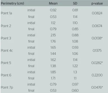
Table 1. Means of perimetry diferences obtained from 17 women between the limb with lymphedema and the contralateral limb, achieved before (initial) and after (final) applying the protocol
Perimetry (cm) Mean SD p-value
Point 1a initial 0.92 0.81 0.0824 final 0.53 1.14
Point 2 initial 1.12 1.10 0.0674 final 0.79 0.85
Point 3 initial 2.15 0.88 0.0138* final 1.76 1.08
Point 4c initial 1.65 0.93 0.1375 final 1.44 1.06
Point 5 initial 1.62 1.14 0.0282* final 1.38 1.22
Point 6 initial 1.85 1.3 0.2200 final 1.71 1.1
Point 7p initial 0.79 0.97 0.0476* final 0.53 0.60
SD: standard deviation; P: significance for the Student’s t-test to paired samples; p*<0.05; 1a: axilla; 4c: elbow; 7p: wrist
Figure 1. Volume diference (cm3) between the limbs before (initial) and
after (final) the protocol use
(VIP), also presented a signiicant reduction (p=0.0067) of around 14.1% (Figure 2).
Home exercises and self-massages were monitored on a weekly basis, and a mean frequency of 4.7±6.28 for self-massages and 9.0±8.4 for weekly exercises was also observed.
DISCUSSION
he main objective of the lymphedema treatment is to reduce volume, restore function, and improve physical appearance of the affected limb16. Although the limb
function was not evaluated, this study showed that in the studied sample there was a reduction of the lymph-edema, after using the proposed protocol.
he most important objective techniques to evaluate lymphedema include perimetry of the af-fected limb in different points and volumetric measures. Although volumetry has the aim of mea-suring irregular edemas like lymphedema17,
pe-rimetry, when performed carefully, supplements the evaluation as it shows the most affected sites, because the lymphedema is not homogeneous18.
In this study, data from the perimetry enabled the mathematical calculation of the limb volume.
Figure 2. Volume increase percentage between limbs before (initial) and after (final) protocol use
380
360
340
320
300
280
260
240
220
200
180
160
V
olume (cm
3)
299,29
258,13
Initial Final
Mean Mean ±SE Mean±1,96 *SE
Volume Difference (VD)
15
14
13
12
11
10
9
8
7
11,82
10,15
Initial Final
Because some statements about the use of HVES were reported in the already published scientiic literature that declared the application of HVES in pro-moting an effect to reduce microcirculation permeabili-ty, decrease the size of capillary pores, and restrain pro-tein movement to the interstitial space11 associated with
the act of bombing the skeletal and lymphatic smooth muscles (also caused by HVES)21, the use of HVES was
made applicable in the protocol of this study. he effect of the use of HVES may have been the cause of lymph-edema decrease.
he HVES parameters used in this study are in con-formance with other studies that were used for reducing lymphedema. he effect of local cathodic stimulation to reduce vase permeability for big molecules was reported in some studies21,22. Griffin et al.23 have also used
ca-thodic HVES, because it seemed to produce a motor reaction with little discomfort.
HVES applied with 50 Hz frequency creates an increase in blood low, which allows the quick removal of toxins and provides better contribution of oxygen24.
Based on these effects, Garcia, Guirro, and Montebello20
have also used 50 Hz frequency in their study.
In this study, placement of electrodes in the forearm anterior region, recruiting lexor muscles, was carried out according to the anatomy of vases and drainage tracking, which were performed mainly by the antero-internal face of the forearm and arm8,20. he principle,
to avoid muscle fatigue when the voltage is high, the current must be modulated in such a way that the pulses should be released in an interrupted manner, in contrac-tion (on) and rest (off ) periods25, was also applied.
he muscle bombing mechanism occurs not only with contractions induced by HVES, but also with the muscle voluntary contractions. herefore, exercises also stimulate bombing, thus increasing the venous and lymphatic f luids. More speciically, they can also recruit sympathetic units, stimulating the contraction of the lymphatic vases19.
Moseley, Carati, and Piller26 consider exercises and
self-massage as a maintenance therapy and emphasize that, despite producing a small volume reduction, they have proved to be more beneicial compared with not performing any other accessible or economically viable treatment for the patient.
Performing home exercises and self-massages were monitored on a weekly basis, and a mean frequency of 4.7±6.28 for self-massages and 9.0±8.4 for exercises was also observed. his demonstrates an association of women to this kind of therapy. Similar observation was found by Mckenzie and Kalda19, because many
wom-en, besides reporting improvement of the lymphedema limb, still continue the exercises even when the research work is completed.
A common alternative to estimate the limb volume consists of calculating circumference measures and volume through geometrical formulae. he cone for-mula represents the limb form in a better way. Sand-er et al.15, after measuring 50 women with upper limb
lymphedema, observed good intra- and inter-evaluator reliabilities between water and geometrical volumes, with intra-class correlation coefficients equal to 0.91 and 0.99, respectively.
In the present study, perimetry and indirect volume were used (cut-off cone formula) for measuring the limbs. Furthermore, in both these measures, changes in the upper limb form were controlled when the con-tralateral limb was applied as a control of the treat-ed limb. Changes inductreat-ed from exercises or from the BMI can happen similarly in both the limbs19. Despite
the fact that this method does not allow to know in which tissue the changes occur, regular exercises are associated with muscular hypertrophy and loss of the adipose tissue.
In the evaluation of each segment in the perim-etry, it was possible to observe a signiicant reduction in the following three points: 7 cm above the elbow (p=0.0138), 7 cm below the elbow (p=0.0282), and at the wrist (p=0.0476). Even though it was non-signii-cant, a decrease was also observed in the other points.
Another study stated a signiicant decrease only 6 cm below the ulna. he justiication for such answer would be the irregular accumulation of the lymph. herefore, the lymphedema justiication could be provided both by a simple change in the circumference, or in the limb total volume20.
he proposed protocol with HVES, exercises, and home guidance also provided a signiicant decrease of 13.8% (p=0.0089) of the VD (initial=299.20±135.32 cm3
and final=258.13±161.78 cm3) and of 14.1%
(p=0.0067) from the VIP (initial=11.82±4.77% and i-nal=10.15±5.60%).
Garcia and Guirro11 observed 15 women with
uni-lateral lymphedema (mild to severe grades) in a study and used the same application parameters of HVES from this study. A signiicant reduction equivalent to 8.53% of the volume, between the 1st and 14th sessions
(2.18 L±0.96 and 1.99 L±0.88) was found by Garcia and Guirro. he severity of the lymphedema also de-creased 4.35% (p<0.05), according to data from the 1st (28.63%±20.50) and 14th sessions (24.28%±19.57).
10. Leal NFBS, Carrara HHA, Vieira KF, Ferreira CHJ. Tratamentos fisiote-rapêuticos para o linfedema pós-câncer de mama: uma revisão de literatura. Rev Lat Am Enfermagem. 2009;17(5):730–6.
11. Garcia LB, Guirro ECO. Efeitos da estimulação de alta voltagem no linfedema pós-mastectomia. Rev Bras Fisioter. 2005;9(2):243–8. 12. Garcia LB, Guirro ECO, Montebello MIL. Efeitos da estimulação elétrica
de alta voltagem no linfedema pós-mastectomia bilateral: estudo de caso. Fisioter Pesqui. 2007;14(1):67–71.
13. Deo SVS, Ray S, Rath GK, Shukla NK, Kar M, Asthana S, et al. Prevalence and risk factors for development of lymphedema following breast cancer treatment. Indian J Cancer. 2004;41(1):8–12.
14. Meirelles MCCC, Mamede MV, Souza L, Panobianco MS. Ava-liação de técnicas fisioterapêuticas no tratamento do linfe-dema pós-cirurgia de mama em mulheres. Rev Bras Fisioter. 2006;10(4):393–9.
15. Sander AP, Hajer NM, Hemenway K, Miller AC. Upper-extremity volume measurements in women with lymphedema: a comparison of measurements obtained via water displacement with geometrical-ly determined volume. Phys Ther. 2002;82(12):1201–12.
16. Koul R, Dufan T, Russell C, Guenther W, Nugent Z, Sun X, et al. Efficacy of complete decongestive therapy and manual lymphatic drainage on treatment-related lymphedema in breast cancer. Int J Radiat Oncol Biol Phys. 2007;67(3):841–6.
17. Bland KL, Perczyk R, Du W, Rymal C, Koppolu P, McCrary R, et al. Can a practicing surgeon detect early lymphedema reliably? Am J Surg. 2003;186(5):509–513.
18. Bergmann A, Mattos IE, Koifman RJ. Diagnóstico do linfedema: análise dos métodos empregados na avaliação do membro superior após linfadenectomia axilar para tratamento do câncer de mama. Rev Bras Cancerol. 2004;50(4):311–20.
19. McKenzie DC, Kalda AL. Efect of upper extremity exercise on secondary lymphedema in breast cancer patients: a pilot study. J Clin Oncol. 2003;21(3):463–6.
20. Garcia LB, Guirro ECO, Montebello MIL. Avaliação de diferentes recur-sos fisioterapêuticos no controle do linfedema pós-mastectomia. Rev Bras Mastol. 2005;15(2):64–70.
21. Hooker DN. Correntes de estimulação elétrica. In: Prentice WE. Moda-lidades terapêuticas para fisioterapeutas. 2a ed. Porto Alegre: Artmed;
2004. p. 76–127.
22. Cook HA, Morales M, La Rosa EM, Dean J, Donnelly MK, McHugh P, et al. Efects of electrical stimulation on lymphatic flow and limb volu-me in the rat. Phys Ther. 1994;74(11):1040–6.
23. Grifin JW, Newsome LS, Stralka SW, Wright PE. Reduction of chronic posttraumatic hand edema: a comparison of high voltage pulsed current, intermittent pneumatic compression, and placebo treatments. Phys Ther. 1990;70(5):279–86.
24. Yang D, Vandongen YK, Stacey MC. Efect of exercise on calf muscle pump function in patients with chronic venous disease. Br J Surg. 1999;86(3):338–41.
25. Howe T, Trevor M. Correntes de baixa frequência–introdução. In: Kitchen S, Bazin S, editores. Eletroterapia: prática baseada em evidências. 11a ed. São Paulo: Manole; 2003. p. 233–40.
26. Moseley AL, Carati CJ, Piller NB. A systematic review of common conservative therapies for arm lymphoedema secondary to breast cancer treatment. Ann Oncol. 2007;18(4):639–46.
Following were the limitations while performing this study: absence of a Control Group, and the use of a measure that is considered not much reliable as the perimetry, even if it was performed carefully, i.e., by only one evaluator and with the same instrument.
CONCLUSION
he results of this study allow us to conclude that the applied protocol, which includes HVES, exercises, self-massage, and guidance about limb care, was effective for reducing lymphedema in the studied popu-lation. New investigations are necessary to deepen such subject for future research.
REFERENCES
1. Instituto Nacional de Câncer José Alencar Gomes da Silva (BR). Coordenação Geral de Ações Estratégicas. Coordenação de Pre-venção e Vigilância. Estimativa 2012: incidência de câncer no Brasil. [In-ternet] Rio de Janeiro: INCA, 2011 [citado em 16 de novembro de 2012]. Disponível em: http://www.inca.gov.br/estimativa/2012/estimativa 20122111.pdf
2. Instituto Nacional de Câncer (INCA). Normas e recomendações do Ministério da Saúde: controle do câncer de mama – documento de consenso. Rev Bras Cancerol. 2004;50(2):77–90.
3. Ridner SH. Pretreatment lymphedema education and identified edu-cational resources in breast cancer patients. Patient Educ Couns. 2006;61(1):72–9.
4. International Society of Lymphology. The diagnosis and treatment of peripheral lymphedema. Consensus document of the International Society of Lymphology. Lymphology. 2003;36(2):84–91.
5. Matheus CN, Guirro ECO. Change in blood flow velocity demonstra-ted by Doppler ultrasound in upper limb after axillary dissection surgery for the treatment of breast cancer. Breast Cancer Res Treat. 2011;127(3):697–704.
6. Rezende LF, Rocha AVR, Gomes CS. Avaliação dos fatores de risco no linfedema pós-tratamento de câncer de mama. J Vasc Bras. 2010;9(4):233–8.
7. Bergmann A, Mattos IE, Koifman RJ. Incidência e prevalência de lin-fedema após tratamento cirúrgico do câncer de mama: revisão de literatura. Rev Bras Cancerol. 2007;53(4):461–70.
8. Camargo MC, Marx AG. Reabilitação Física no Câncer de Mama. 1a ed.
São Paulo: Roca; 2000.