Fisioter. Mov., Curitiba, v. 30, n. 4, p. 745-752, Oct./Dec. 2017 Licenciado sob uma Licença Creative Commons DOI: http://dx.doi.org/10.1590/1980-5918.030.004.AO09
The spasticity in the motor and functional disability
in adults with post-stroke hemiparetic
A espasticidade no comprometimento motor e funcional
de hemiparéticos pós acidente vascular cerebral
Roberta de Oliveira Cacho[a], Enio Walker Azevedo Cacho[a], Anderson Barbosa Loureiro[b],
Gabriele Natane de Medeiros Cirne[a], Silvana Alves Pereira[a], Rodrigo Pegado de Abreu Freitas[a], Núbia Maria Freire Vieira Lima[a], Guilherme Borges[b]*
[a] Universidade Federal do Rio Grande do Norte (UFRN), Santa Cruz, RN, Brazil [b] Universidade Estadual de Campinas (Unicamp), Campinas, SP, Brazil
Abstract
Introduction: Spasticity acts as a limiting factor in motor and functional recovery after Stroke, impairing the performance of daily living activities. Objective: To analyze the influence of spasticity on main muscle groups and to associate it with motor impairment and functional level of chronic hemiparetic patients after stroke. Methods: Twenty-seven chronic hemiparetic patients of both sexes were selected at the Physical Therapy and Occupational Therapy Service of the Unicamp Clinics Hospital. Assessments were carried out in two sessions, in the first one the motor impairment (Fugl-Meyer Assessment - FM) and functional impair -ment (Barthel Index - BI) were evaluated, and in the second, the degree of spasticity of the main muscle groups (Modified Ashworth Scale - MAS). Results: A negative correlation was detected between upper limb spasticity and motor and functional impairment. No muscle group evaluated in the lower limbs showed cor-relation between muscle tone and the level of impairment of the lower extremity on FM and the functional
* ROC: PhD, e-mail: [email protected] EWAC: PhD, email:[email protected] ABL: MS, email: [email protected]
GNMC: Master student, e-mail: [email protected] SAP: PhD, email: [email protected]
746
level measured by BI. Conclusion: Spasticity has been shown to be a negative influence factor in the level of motor and functional impairment of the upper limbs of chronic hemiparetic patients after stroke.
Keywords: Stroke. Hemiparesis. Spasticity.
Resumo
Introdução: A espasticidade atua como um fator limitante na recuperação motora e funcional após o Acidente Vascular Cerebral (AVC), prejudicando a realização das atividades de vida diária. Objetivo: Analisar a influên -cia da espasticidade nos principais grupos musculares e associá-la ao comprometimento motor e ao nível fun-cional de pacientes hemiparéticos crônicos pós-AVC. Métodos: Vinte e sete pacientes hemiparéticos crônicos, de ambos os sexos, foram selecionados no Serviço de Fisioterapia e Terapia Ocupacional do Hospital de Clínicas da Unicamp. As avaliações foram realizadas em duas sessões: na primeira foi avaliado o comprometimento mo-tor (Protocolo de Desempenho Físico de Fugl-Meyer - FM) e funcional (Índice de Barthel - IB), e na segunda, o grau de espasticidade dos principais grupos musculares (Escala Modificada de Ashworth – EMA). Resultados:
Foi detectada uma correlação negativa entre a espasticidade dos membros superiores com o comprometimen-to mocomprometimen-tor e funcional. Nenhum grupo muscular avaliado nos membros inferiores apresencomprometimen-tou correlação entre o tônus muscular e o nível de comprometimento da subseção da extremidade inferior FM e o nível funcional mensurado pelo IB. Conclusão: A espasticidade mostrou ser um fator de influência negativa no nível de com -prometimento motor e funcional dos membros superiores de pacientes hemiparéticos crônicos pós-AVC.
Palavras-chave: Acidente Vascular Cerebral. Hemiparesia. Espasticidade.
Introduction
Stroke hemiparetic patients often develop motor disorders associated with imbalance of neural ac-tivity, such as spasticity, recognized as a component of upper motor neuron syndrome (1). Spasticity is
defined as increased resistance to passive muscle
stretching being dependent on the velocity (2). Spasticity prevalence after stroke varies from 18% (3) to 60% (4), and it is more frequent in the upper limbs than in lower limbs (3).
Clinically, spasticity is related to increased muscle
tone, exaggerated reflexes, pain, and possible joint
contractures. Along with these, impairment in motor
control, muscle strength deficit and balance deficit lead to several disabilities, influencing the rehabilitation
process and performance of functional tasks, as well as the quality of life of individuals with spasticity (5, 6).
The first clinical instrument developed to measure muscle tone was the Ashworth Scale and the Modified
Ashworth Scale (7, 8). They are the most commonly used instruments for this purpose. Despite the short-comings in manual methods (speed, angle, and ac-celeration are not standardized), it is the simplest
and the most widespread, besides being a validated quantitative instrument that is easy to apply (9).
Some authors (10, 11) have reported that
ab-normal reflexes associated with spasticity are the
main determinants of motor impairment. It is cur-rently believed that spasticity itself is one of the contributing factors for motor and functional loss after stroke (12), but it is not an isolated agent, since other primary conditions may be associated, such as muscle weakness (13). Conditions secondary to
upper motor neuron injury, such as pain and muscle
contracture, also impair the appearance and mainte-nance of voluntary movements (6) and they are, to a certain degree, associated with the acute and/or chronic rehabilitation process that the individuals underwent. In the United States and Europe, these patients receive more support during recovery,
es-pecially in the first six or twelve months following
stroke (14), which is not the case in Brazil.
747 Methods
This study was cross-sectional with twenty-sev-en post-stroke hemiparetic patitwenty-sev-ents recruited at the Physical Therapy and Occupational Therapy Service of the Unicamp Clinics Hospital. They were informed and consented to participate in the research, which was approved by the research ethics committee of Unicamp (#110/2004).
The patients selected had a single unilateral stroke sequela, from non-traumatic origin, for a period of more than six months, of both sexes and aged be-tween 35 and 70 years. Participants had to be able to understand simple instructions and not to have associated orthopedic or neurological conditions.
The exclusion criterion was the loss of joint range of
motion due to pain or muscle shortening.
Motor impairment was assessed using the Fugl-Meyer Assessmentscale (FM) (15). This scale has a total score of 66 points for the upper limb and 34 for the lower limb. The items are scored on ordinal scale: 0 = no achievement, 1 = partial achievement and 2 = complete achievement. FM is a standard instru-ment, validated in Brazil and widely used in several research centers (16, 17).
The degree of spasticity was assessed using the
Modified Ashworth Scale (MAS) (7) which included
the following scores: 0 (zero), no increase in muscle tone; 1 (one) slight increase in tone, manifested by minimal resistance at the end of the range of motion
when the affected part is moved in extension or flex -ion; +1 (+ one), slight increase in muscle tone mani-fested by blockade, followed by minimal resistance throughout the remainder of the range of motion (less than half); 2 (two), marked increase in muscle tone,
despite the absence of joint range of motion impair -ment, but the affected parts move slowly; 3 (three), considerable increase of muscle tone hindering pas-sive movement; 4 (four), stiffness in the affected parts
for flexion or extension. The muscle groups evaluated in the upper limb were: flexors, extensors, adductors,
abductors, internal and external rotators of the
shoul-der, elbow flexors and extensors, forearm pronators and supinators, and flexors and extensors of wrist and fingers. In the lower limb: flexors, extensors, ad -ductors, ab-ductors, internal and external rotators of
the hip, knee flexors and extensors, plantar flexors
and dorsiflexors. The presence of spasticity was de
-fined as a score ≥ 1 in a muscle group.
Barthel Index (BI) evaluates activities of daily liv-ing (ADLs), reachliv-ing a total of 100 points for individu-als who are independent to perform the tasks (18). The BI is a frequently used measure with well-es-tablished validity, reliability and acceptability (19).
Evaluations were carried out in two sessions (on
different days). In the first one, FM and BIwere ap -plied, and on the following day the muscular tone evaluation was performed in dorsal decubitus, by passive movement of the upper limb (shoulder, elbow,
forearm, wrist and fingers) and lower limb (hip, knee
and ankle), according to MAS criteria. The evalua-tions were performed by two experienced physical therapists familiar with the scales.
Descriptive statistics of continuous variables were established by mean values and standard deviation. Correlation between scores was then performed
us-ing Spearman Correlation Coefficient. This coefficient
was used due to the absence of normal distribution
and reduced sample size. The significance level ad -opted for the statistical tests was 5% (p < 0.05).
Results
Among the selected patients, 40.7% (N = 11) were female and 59.3% (N = 16) males, mean age of 49.48 (± 11.49) years, and mean time of 3.43 (± 3.18) years post-stroke. Left ischemic stroke had higher preva-lence (40.7%/N = 11). FM mean score in the upper limb was 33.81 (± 19.68), in the lower limb was 18.55 (± 8.34), presenting a total score of 52.37 (± 24.72). In BI, the mean was 91.11 (± 6.41).
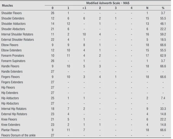
Spasticity in the upper limb was detected in 66.6% (N = 18) of the 27 patients evaluated. Muscle groups of
forearm pronators and flexors of the elbow, wrist and fingers were spastic in more than 60% of the patients
(N = 17) (Table 1). At the lower extremity, spasticity
in plantar flexors occurred in 66.6% of the patients. Six subjects did not present spasticity in any
muscle groups of the evaluated upper limb (FM - up-per limb subsection 61.5 ± 2.96 / BI 98.33 ± 2.58).
Seven subjects scored 0 at Ashworth for all lower limb
748
Table 1 - Frequency and percentage of spasticity in upper and lower limb muscle groups
Muscles Modified Ashworth Scale - MAS
0 1 +1 2 3 4 N %
Shoulder Flexors 26 1 - - - - 1 3.7
Shouder Extenders 12 6 6 2 1 - 15 55.5
Shoulder Adductors 14 12 - 1 - - 13 48.1
Shouder Abductors 21 6 - - - - 6 22.2
Internal Shoulder Rotators 11 2 10 4 - - 16 59.2
External Shoulder Rotators 22 4 1 - - - 5 18.5
Elbow Flexors 9 9 8 1 - - 18 66.6
Elbow Extenders 12 10 4 1 - - 15 55.5
Forearm Pronators 10 11 4 2 - - 17 62.9
Forearm Supinators 26 - 1 - - - 1 3.7
Handle Flexors 9 10 5 3 - - 18 66.6
Handle Extenders 27 - - -
-Fingers Flexors 9 10 3 4 1 - 18 66.6
Fingers Extenders 27 - - -
-Hip Flexors 27 - - -
-Hip Extenders 27 - - -
-Hip Adductors 25 1 1 - - - 2 7.4
Hip Abductors 27 - - -
-Internal Hip Rotators 18 7 2 - - - 9 33.3
External Hip Rotators 23 4 - - - - 4 14.8
Knee Flexors 21 5 1 - - - 6 22.2
Knee Extenders 23 2 1 1 - - 4 14.8
Plantar Flexors 9 11 7 - - - 18 66.6
Flexors Dorsum of the ankle 27 - - -
-Note: n, Number of patients; %, Percentage of patients.
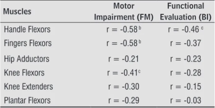
Negative correlation was found between muscle tone of the extensors, adductors and internal
rota-tors of the shoulder, flexors and extensors of the elbow, and flexors of the wrist and fingers, and the
level of motor impairment in the upper limb on FM. Regarding functional level, negative correlation was also observed between the muscle tone of extensors, adductors and internal rotators of the shoulder and
flexors and extensors of the elbow (Table 2).
No muscle group assessed in the lower limbs showed correlation between muscle tone and level of impairment of lower extremity on FM and the func-tional level measured by BI.
Significant positive correlation was found between
total FM and BI (r = 0.6169; p = 0.0001), and between the upper limb FM subsection and BI (r = 0.6747; p = 0.0001).
Table 2 - Statistical significance between the Modified Ash-worth Scale – MAS scores with the Fugl-Meyer As-sessment – FM and the Barthel Index - BI
Muscles Motor
Impairment (FM)
Functional Evaluation (BI)
Shoulder Flexors r = -0.24 r = -0.22
Shouder Extenders r = -0.73 a r = -0.57 b
Shoulder Adductors r = -0.71 a r = -0.60 a
Shouder Abductors r = -0.21 r = -0.37
Internal Shoulder Rotators r = -0.70 a r = -0.69 a
External Shoulder Rotators r = -0.20 r = -0.24
Elbow Flexors r = -0.63 a r = -0.54 b
Elbow Extenders r = -0.71 a r = -0.55 b
Forearm Pronators r = -0.49 b r = -0.48 c
Forearm Supinators r = -0.30 r = 0.15
749
Table 2 - Statistical significance between the Modified Ash-worth Scale – MAS scores with the Fugl-Meyer As-sessment – FM and the Barthel Index - BI
Muscles Motor
Impairment (FM)
Functional Evaluation (BI)
Handle Flexors r = -0.58 b r = -0.46 c
Fingers Flexors r = -0.58 b r = -0.37
Hip Adductors r = -0.21 r = -0.23
Knee Flexors r = -0.41c r = -0.28
Knee Extenders r = -0.30 r = -0.15
Plantar Flexors r = -0.29 r = -0.03
r = Spearman correlation coefficient; a p < 0.001; b p < 0.01;
c p < 0.05
Discussion
Spasticity is one of the main factors contributing to the loss of selective motor control, especially in individuals who manifest severe motor impairment after stroke (12). According to FM scale,upper limbs total motor score of patients in this study shows a
level of impairment classified as severe to moderate
(score between 5 and 46) (20). The lower limb pre-sented FM mean score corresponding to 54.56% of motor recovery. Movement disorders following stroke may also be due to loss of strength and motor ability, because of disconnected interarticular coordination or pathological synergies (21).
In a study on the prevalence of spasticity, 20% of 66 patients evaluated had some level of spastic-ity. This number increased to 34% when
consider-ing hemiparetic subjects (22). Lundströnet al. (3)
observed that 17% of the patients had upper limb spasticity after one year of stroke. In this study, 66.6% of the patients presented upper and/or lower limb spasticity. These values are above the average of other studies and may be explained by the high level of motor impairment (measured by total FM = 52.37 ± 24.52) of the selected patients and by the effects of lesion chronicity. According to Thilmannet al. (23), after three months of stroke, spasticity seems to be also due to intrinsic factors, such as decrease in the number of sarcomeres in
series that result in slow movement and difficulty
in selectivity (co-contraction) (24). In this study, all patients were chronic, that is, with more than
six months of injury. Therefore, we can infer that the difficulty in performing voluntary movements
(Conclusion) (which caused a reduced score in FM scale) may be due to these neuromuscular changes occurring in the chronic phase after stroke, and also to the
con-trol deficit in superior motoneurons firing.
Differently from other studies, the sample stud-ied here showed similar spasticity in both extremi-ties, but it was present in more muscles of the up-per extremity than in the lower one. It is not yet
defined why spasticity affects upper limb motor
performance more than lower limbs (25). It is be-lieved that it may be due to the fact that locomotion is also present in spinal levels and that spasticity may help orthostatism, contributing to maintain body weight (9, 25).
When we selected only patients who did not pres-ent spasticity in any of the muscle groups of the
evalu-ated upper limb (six subjects), we noticed that FM
scores in the subsection of upper limb and BI (FM 61.5 ± 2.96 / BI 98.33 ± 2.58) were higher than the rest of the group (FM 25.90 ± 14.34 / BI 89.4 ± 5.61).
Lundströnet al. (3) and Watkins et al. (26) identi
-fied that the rate of patients dependent on activities
of daily living according to BI was higher in those patients with spasticity when compared to those without spasticity. In contrast, Sommerfeld et al. (12) demonstrated that severe motor and functional problems are observed with the same frequency in
spastic and non-spastic patients. The exact influence
of spasticity on motor impairment and limitations
after stroke is difficult to measure because the level
of spasticity may change according to the positioning and the task demands (12).
The medial descending pathways (medial cortico-spinal, medial and lateral vestibulocortico-spinal,tectospinal and medialreticulospinal) and lateral descending pathways (lateral corticospinal, rubrospinal and lat-eral reticulospinal) commands the axial and distal muscles. After stroke, some of these pathways may be damaged, leading to loss of dexterity, strength and voluntary movement. Several plastic processes
of the central nervous system act after injury in or -der to restore motor and functional control, such as reorganization of intracortical connections,
appear-ance of collateral cortico-spinal projections from
other regions of the primary motor cortex or other areas of the brain,increased cortical activation of descending pathways from the midbrain and
in-creased activity of ipsilateral cortico-spinal projec -tions from the unaffected cortex (27, 28). However,
750
to the cortico-spinal tract is not repaired and/or replaced (29). It explains why spasticity, which is one of the main effects of upper motor neuron
in-jury, was found mainly in distal muscles of the upper
and lower limbs in this study (Table 1). Also on the descending pathways, midbrain motor areas lose
in-fluence of cortical projections after stroke, leading to
abnormal control over the muscles and causing the classic and antigravity pattern of extensor muscles of
the lower limbs and flexors of the upper limbs (28).
This antigravity pattern is present in patients from this study, and may be observed by the higher spas-ticity incidence in muscles that compose this ab-normal synergy.
The level of upper limb motor impairment and the functional capacity of hemiparetic patients in this study were affected by the presence of spastic-ity. Those with spasticity showed decreased FM and BI scores. Lin et al. (25), using different measurement instruments from the ones in this study, reported
significant inverse correlation between the degree of spasticity of the wrist joint and voluntary motor per -formance, including functional tasks such as Box and Blocks test. Watkins et al. (26) also reported that pa-tients with spasticity (measured by MAS) were more functionally impaired than those without spasticity. Welmer et al. (22) correlated upper limb spasticity (measured by MAS) with measures of voluntary mo-tor mobility (Birgitta Lindmark Momo-tor Assessment Scale - LMAS, Rivermead Mobility Index - RMI) and BI, and found moderate to high correlation.
Regarding lower limbs, there was no relationship between spasticity and the level of motor and func-tional impairment of hemiparetic individuals. This
finding is similar to those of Welmer et al. (22) that
reported low correlation between lower limbs spas-ticity with RMI and BI. Katz et al. (29) also observed no correlation between the degree of spasticity of the lower extremity muscles and the scores on FM scale.
Correlations between spasticity and motor and functional impairment in the upper and lower limbs are not similar due to some factors. First, spasticity may manifest differently in different muscle groups, since skill, motor dexterity, and functional require-ments are different for each structure. Second, mea-surement tools fail because they are not able to dif-ferentiate between a functional motor response and a compensatory strategy (30). In addition, it is im-portant to highlight that several factors may
contrib-ute to this event, such as injury time, level of motor
impairment, intrinsic (non-neural) muscular factors, among others (13).
Similar to that found by Oliveira et al. (30), it was observed that a better FM score is also accompanied by an increase in BI, indicating that motor impair-ment is inversely proportional to satisfactory func-tional performance.
Conclusion
Thus, spasticity is characterized as a factor of
neg-ative influence on motor and functional impairment
of the upper limbs of chronic hemiparetic patients after stroke, that is, spasticity acts as a limiting factor for motor and functional recovery.
Acknowledgments
We thank São Paulo Research Foundation (FAPESP) (#06/61199-5) and the National Council
for Scientific and Technological Development (CNPq) (#302189/2004-1) for financial support.
References
1. Ward AB. A Literature review of the pathophysiol-ogy and onset of post-stroke spasticity. Eur J Neurol. 2012;19(1):21-7.
2. Urban PP, Wolf T, Uebele M, Marx JJ, Vogt T, Stoeter P, et al. Occurrence and Clinical Predictors of Spasticity
After Ischemic Stroke. Stroke. 2010;41(9):2016-20.
3. Lundström E, Tere A, Borg J. Prevalence of disabling spasticity one year after first-ever stroke. Eur J Neurol. 2008;15(6):533-9.
4. Wallesch CW, Maes E, Leconte P, Bartels C. Feasibility
study on pharmacoeconomics of botulinum toxin A (Botox) in spasticity following stroke. 3rd European
Botulinum Toxin Symposium Abstracts. 1997;1:4.
5. Williams SA, Reid S, Elliott C, Shipman P, Valentine J. Muscle volume alterations in spastic muscles imme-diately following botulinum toxin type-A treatment in
751 6. Gracies JM, Brashear A, Jech R, McAllister P, Banach
M, Valkovic P, et al. Safety and efficacy of abobotu -linumtoxinA for hemiparesis in adults with upper limb spasticity after stroke or traumatic brain injury: a double-blind randomised controlled trial. Lancet Neurol. 2015;14(10):992-1001.
7. Bohannon RW, Smith MB. Interrater reliability of a Modified Ashworth Scale of muscle spasticity.Phys Ther. 1987;67(2):206-7.
8. Pandyan AD, Price CIM, Barnes MP, Rodgers H. A re-view of the properties and limitations of the Ashworth and modified Ashworth scales as measures of spastic -ity. Clin Rehabil. 1999;13(5):373-83.
9. Wood DE, Burridge JH, van Wijck FM, McFadden C, Hitchcock RA, Pandyan AD, et al. Biomechanical approaches applied to the lower and upper limb for the measurement of spasticity: a systematic review of the literature. Disabil Rehabil. 2005;27(1-2):19-32.
10. Foley N, Pereira S, Salter K, Fernandez MM, Speech-ley M, Sequeira K, et al. Treatment With Botulinum Toxin Improves Upper-Extremity Function Post Stroke: A Systematic Review and Meta-Analysis. Arch Phys Med Rehabil. 2013;94(5):977-89.
11. Brunnström S. Movement Therapy in Hemiple -gia: A Neuropsychological Approach. New York, NY: Harper and Row; 1970.
12. Sommerfeld DK, Eek EUB, Svensson AK, Holmqvist LW, von Arbin MH. Spasticity after stroke: its occurrence and association with motor impairments and activity limitations. Stroke. 2004;35(1):134-9.
13. Ada L, O’Dwyer N, O’Neill E. Relation between spas-ticity, weakness and contracture of the elbow flexors and upper limb activity after stroke: An observational study. Disabil Rehabil. 2006;28(13-14):891-7.
14. Cramer SC, Koroshetz WJ, Finklestein SP. The case for modality-specific outcome measures in clinical trials of stroke recovery-promoting agents. Stroke. 2007;38(4):1393-5.
15. Fugl-Meyer AR, Jaasko L, Leyman L, Olsson S, Steglind, S. The post-stroke hemiplegic patient. 1.a method for evaluation of physical performance. Scand J Rehabil Med. 1975;7(1):13-31.
16. Maki T, Quagliato EMAB, Cacho EWA, Paz PSP, Nilce HN, Inoue MME, et al. Estudo de confiabilidade da aplicação da escala de Fugl-Meyer no Brasil. Rev Bras
Fisioter. 2006;10(2):179-85.
17. Cacho EWA, Melo FRLV, Oliveira R. Avaliação da
recu-peração motora de pacientes hemiplégicos através do Protocolo de Desempenho Físico de Fugl-Meyer. Rev
Neurociênc. 2004;12(2):94-102.
18. Mahoney FI, Barthel D. Functional evaluation: the
Barthel Index. Md State Med J. 1965;14:56-61.
19. Duffy L, Gajree S, Langhorne P, Stott DJ, Quinn TJ.
Reliability (Inter-rater Agreement) of the Barthel
Index for Assessment of Stroke Survivors. Stroke. 2013;44:462-8.
20. Cirstea MC, Ptito A, Levin MF. Arm reaching
improve-ments with short-term practice depend on the se-verity of the motor deficit in stroke. Exp Brain Res. 2003;152(4):476-88.
21. Jarrassé N, Proietti T, Crocher V, Robertson J, Sahbani A, Morel G, et al. Robotic exoskeletons: a perspective
for the rehabilitation of arm coordination in stroke
patients. Front Hum Neurosci. 2014;8:947.
22. Welmer Ak, von Arbin M, Holmquist LW, Sommerfeld
DK. Spasticity and it association with functioning and
heath-related quality of life 18 months after stroke. Cerebrovasc Dis. 2006;21(4):247-53.
23. Thilmann AF, Fellows SJ, Garms E. The mechanism of spastic muscle hypertonus: variation in reflex gain over the time course of spasticity. Brain. 1991;114(Pt
1A):233-44.
24. Lieber RL, Frieden J. Spasticity causes a fundamental rearrangement of muscle-joint interaction. Muscle Nerve. 2002;25(2):265-70.
25. Lin FM, Sabbahi M. Correlation of spasticity with hyperactive stretch reflexes and motor dys -function in hemiplegia. Arch Phys Med Rehabil.
1999;80(5):526-30.
26. Watkins CL, Leathley MJ, Gregson JM, Smith TL, Sharma AK. Prevalence of spasticity post stroke. Clin
752
27. Bradnam LV, Stinear CM, Byblow WD. Ipsilateral motor pathways after stroke: implications for non-invasive brain stimulation. Front Hum Neurosci. 2013;7:184.
28. Turton A. Mechanisms for recovery of hand and arm function after stroke: a review of evidence from stud-ies using non-invasive investigative techniques. Br J Occup Ther. 1998;61(8):359-64.
29. Katz RT, Rovai GP, Brait C, Rymer WZ. Objective quantifi -cation of spasticity hypertonia: correlation with clinical findings. Arch Phys Med Rehabil. 1992;73(4):339-47.
30. Oliveira R, Cacho EWA, Borges G. Post-stroke mo-tor and functional evaluations: a clinical correla-tion using Fugl-Meyer Assessment Scale, Berg Bal-ance Scale and Barthel Index. Arq Neuropsiquiatr. 2006;64(3B):731-5.
Received in 12/16/2015
Recebido em 16/12/2015
Approved in 03/16/2017