INTRODUCTION
The understanding of the physiopathol-ogy of Stress Urinary Incontinence (SUI) has constantly improved over the two last decades, since the presentation of the Integral Theory (1) and TVT introduction (2). In spite of the good
cure rate reported with TVT, severe complications such as visceral and vessels injuries have been described (3). As an alternative to TVT, Delorme introduced the transobturator tape (TOT) (4,5) with successful results. In spite of the low intra-operative complication rate of TOT, most com-plications were related to the blind passage of Introduction and Objectives: The mini sling concept for stress urinary
inconti-nence is an anatomical approach that involves placing a midurethral low-ten-sion tape anchored to the obturator internus muscles bilaterally. They overcome the blind passage of long needles and all the related complications. There are many different devices available and because these are outpatient procedures, primary fi xation plays an important role in the outcome. The objective is to evaluate the primary fi xation of the various devices of attachment of the com-mercially available mini-slings through biomechanical tests.
Materials and Methods: A total of 45 Wistar rats were divided in 3 groups of 15 rats each. They underwent 5 subcutaneous implantation of different mini slings and one polipropilene mesh (control), as follows: TVT-Secur® (Gynecare, USA), Type 1 polypropylene mesh (control); Ophira Mini Sling System® (Promedon, Argentina), Tissue Fixation System® (TFS PTY, Australia), Zipper Sling® and “T device” (Prosurg, USA). The abdominal wall was removed on bloc at different times after implant for biomechanical evaluation, which consisted in applica-tion of unidirecapplica-tional force to the extremity of the fi xaapplica-tion system or mesh, until it was completely removed from the tissue using a tension meter (Nexygen 3.0 Universal Testing Machine - LLOYD Instruments). The force was measured in Newtons (N).
Results: There was signifi cant difference in the resistance to extraction among the different fi xation systems. At 7 days the Ophira Mini Sling System® pre-sented the best fi xation and “T dispositive” the worst.
Conclusion: Ophira mini sling System® presented the best primary fi xation at 7°, 14° and 30° days. The impact of this feature in the clinical setting needs to be verifi ed.
Primary fi
xation of mini slings: a comparative
biomechanical study in vivo
_______________________________________________
Paulo Palma, Rodrigo Teixeira Siniscalchi, Luiz Carlos Maciel, Miguel Angel Bigozzi, Inacio Dal
Fabbro, Cassio Riccetto
Division of Female Urology (PP, MAB, CR), Biomaterial Research Unit (RTS, LCM, CR) and Mechanical Properties of Biological Materials Laboratory (IDF) - University of Campinas - Sao Paulo, Brazil
ABSTRACT ARTICLE INFO
_______________________________________________________________ _____________________
Key words:
urinary incontinence; suburethral sling; outcomes
Int Braz J Urol. 2012; 38: 258-66 ________________
Submitted for publication: April 11, 2011
________________
needles through the obturator foramen (3), which leaded to the creation of small minimally inva-sive devices which avoid this passage and can be implanted under local anesthesia, on an outpa-tient basis and with minimal dissection. Due to a shorter insertion path, it is expected that some complications such as vesical perforation, vas-cular injuries, perineal fasciitis and reduction of postoperative pain in the area of adductor mus-cles can be reduced (6,7). It is possible to suppose that the greater adhesion of the mesh to host tis-sues and the lower the amount of implanted ma-terial lower the risk of extrusion and the rate of sexual discomfort, respectively.
Initial results with some mini sling sys-tems were disappointed although they were ap-pealing. The first results reported with TVT-Secur showed success rates 10% lower than could have been expected with other types of slings (8-10). Therefore, the proposal of biomechanical stud-ies is justified in order to understand the physio-pathological process associated to sling efficacy itself. Up to this moment, no studies have been published in literature comparing the tissue fixa-tion capacity of the different types of mini slings. In this original experimental study, the primary fixation of the different anchoring de-vices of some commercially available mini slings and experimental devices is evaluated “in vivo” through biomechanical tests.
MATERIALS AND METHODS
This study was approved by the Eth-ics Committee for Animal Research of the Uni-versity of Campinas and there is no conflict of interest. Forty-five Wistar rats (weight between 150g and 200g), aged 8 weeks were divided into 3 groups of 15 rats each. Animals were intra-venously anesthetized with sodium pentobarbital at 6% and were positioned in horizontal dorsal decubitus after abdominal trichotomy and asep-sis with Povidone-iodine. A 2 cm transverse inci-sion was then made in the lower abdomen. After the dissection, five different types of mini sling anchoring devices and one polypropylene mesh (control) were implanted between the subcutane-ous cellular tissue and the abdominal muscle
fas-cia, namely: TVT-Secur® (Gynecare, USA), poly-propylene mesh (PP-control), Ophira Mini Sling System® (Promedon, Argentina), Tissue Fixation System® (TFS PTY, Australia), Zipper Sling® and “T device” (Prosurg, USA) (Figure-1).
The five samples of each device were ran-domly implanted in each group (two per animal - one in each side of abdominal wall) (Figure-2). After implantation of the anchoring devices of the mini slings, the skin was sutured, taking care to avoid that the mesh was in direct contact with the skin suture.
The evaluation of the tensile resistance was made in a fresh fragment of the abdomi-nal wall of the rat. After an observation period (7, 14, 30 days) the animals were divided in 3 groups and euthanized, as follows: Group 1 (15 rats euthanized at day 7); Group 2 (15 rats eu-thanized at day 14); and Group 3 (15 rats eutha-nized at day 30).
The abdominal wall was removed and symmetrically divided into 2 blocks containing the implanted anchoring device. Subsequently, approximately 2 mm of the extremity of the de-vices were dissected to be pulled so that they
Figure 1 - Devices.
1°) Ophira mini sling system® (Promedon, Argentina) 2°) Polypropylene Mesh (Promedon, Argentina) 3°) TVT-Secur® (Gynecare, USA)
4°) Tissue Fixation System® (TFS PTY, Australia) 5°) Zipper Sling® (Prosurg, USA)
could be adapted to the fastener of the tension meter (“Nexygen 3.0 Universal Testing Machine” - LLOYD Instruments) which is specially intended for load tests in soft tissues (11). The opposite por-tion of the fragment, containing the abdominal wall without the device, was fixed to the lower fastener of the tension meter and a biomechanical study was performed, in order to measure their tissue adherence at different times.
Next, an increasing load was applied to the extremity of the anchoring device or mesh until it was completely removed from the tissue (Figure-3). The load was measured in Newtons (N), so higher load values show a greater fixation of the device to the tissues. We applied an increasing force (N) and constant speed (2 mm/sec). The strength and the time varied for each test. As the time was not relevant to our study, only the force was measured.
In order to compare the maximum load in relation to the groups over the time (7, 14 and 30
days), the Analysis of Variance (ANOVA) was used and the level of significance adopted was 5%.
RESULTS
There was a significant difference of the maximum load needed for detachment of the mini slings anchoring devices from the tissues.
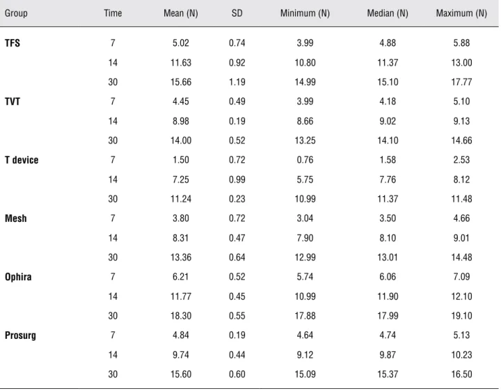
After 7 days of the placement of the im-plant, it was observed that Ophira Mini Sling System® showed the highest maximum load fixa-tion (6.21 ± 0.52 N) and the “T device”, the worst fixation (1.50 ± 0.72 N). There was no significant differences among the other devices.
On 14th day after the implant, it was
load (18.30 ± 0.55 N) and the “T device” showed the worst fixation (11.24 ± 0.23 N) (Tables 1 to 3 and Figure-4).
DISCUSSION
Since the introduction of the mini slings on the market, the failure rate that has been pub-lished by different authors differs markedly. Neu-man published with TVT Secur® a prospective study showing a cure rate of 93.5% at 12-month follow-up [n: 100] (12). In other study Solà Dalenz et al. presented 100% of cure at 2-month follow-up with TVT Secur® [n: 16] (13). Gorlero et al. stud-ied 15 patients, and reported a negative stress test in 86.7% with TVT Secur® at 6-month follow-up (14). In contrast, Meschia and Debodinance pub-lished an objective cure rate of 70.4% and 81% at 6-month up [n: 110] and 15-month follow-up [n: 95] respectively (9,15).
Palma et al. have published preliminary results with arcus to arcus microsling. After 12 months, 88% of patients were dry, 5.5% improved and 5.5% incontinent [n: 20] (16).
The cure rates related to Mini Arc (Ameri-can Medical Systems, USA) are different. Moore et al. reported a negative stress test in 90% of patients
treated at 12-week follow-up [n = 59] (17). Jiménez et al. published satisfactory results with a negative stress test in 90% of patients, with a mean follow-up of 101 days after treatment with MiniArc [n = 41] (18) and Debodinance and Delporte have found an objective cure rate of 75.7% at 2-month follow-up [n = 72] (19). Mini Arc was not included in this experiment because when the study was performed it was not available in the market yet.
One can assume that the rationale for the use of the mini slings is based on its capacity of fixation to the host tissue immediately after the implant, which is probably the main factor for achieving the continence and lowering the risk of vaginal exposure or extrusion.
To date, few cases of vaginal exposure of the mini slings have been described. One case of exposition was reported by Martan et al. (20) and Hazewinkel et al. (21), respectively with TVT Secur® and only two cases were reported by Debodinance et al. with the same mini sling.
tropism, infections and the surgical technique are directly related to the rates of extrusion (26,27).
Since most of the modern slings have been built with materials with well-known biocompat-ibility, the surgeon’s interest has been focused on
the design of the fixation devices and implant instruments like needles and trocars. Although a variety of experimental models can be proposed for the study of such dispositives, one which al-lows for uniaxial stress test seems to be suitable for biomechanical evaluation, since in the vaginal environment the slings are not prone to rotational or centripetal forces.
The trend to perform slings in an outpa-tient basis had been first proposed in the nine-ties, along with the retropubic minimally invasive midurethral slings, but till now it has never been fully adopted because of safety reasons. The mini slings can turn this concept to a real possibility, since most of the clinical data shows a high level of safety. Therefore, as the patient can come back Table 1 - Loads for group and time (days).
Group Time Mean (N) SD Minimum (N) Median (N) Maximum (N)
TFS 7 5.02 0.74 3.99 4.88 5.88
14 11.63 0.92 10.80 11.37 13.00
30 15.66 1.19 14.99 15.10 17.77
TVT 7 4.45 0.49 3.99 4.18 5.10
14 8.98 0.19 8.66 9.02 9.13
30 14.00 0.52 13.25 14.10 14.66
T device 7 1.50 0.72 0.76 1.58 2.53
14 7.25 0.99 5.75 7.76 8.12
30 11.24 0.23 10.99 11.37 11.48
Mesh 7 3.80 0.72 3.04 3.50 4.66
14 8.31 0.47 7.90 8.10 9.01
30 13.36 0.64 12.99 13.01 14.48
Ophira 7 6.21 0.52 5.74 6.06 7.09
14 11.77 0.45 10.99 11.90 12.10
30 18.30 0.55 17.88 17.99 19.10
Prosurg 7 4.84 0.19 4.64 4.74 5.13
14 9.74 0.44 9.12 9.87 10.23
30 15.60 0.60 15.09 15.37 16.50
Table 2 - Results of the ANOVA with repeated measures (in ranks) to Maximum Load.
Source variation p-value
Group 0.0001
Time 0.0001
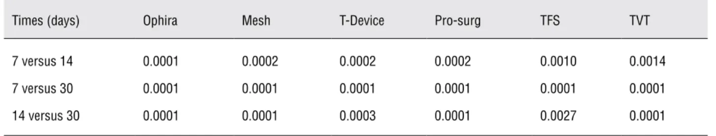
Table 3 - P-values for comparisons along time in each group (Contrast Test).
Times (days) Ophira Mesh T-Device Pro-surg TFS TVT
7 versus 14 0.0001 0.0002 0.0002 0.0002 0.0010 0.0014
7 versus 30 0.0001 0.0001 0.0001 0.0001 0.0001 0.0001
14 versus 30 0.0001 0.0001 0.0003 0.0001 0.0027 0.0001
Figure 4 - Maximum Load - Comparison of different fixation devices.
home in the same day of the procedure, the pri-mary fixation of the sling becomes relevant.
The highest maximum load showed by Ophira Mini Sling System® probably is related to its multipoint fishbone-like polypropylene fixa-tion device. But it is not possible to determine the minimal load which is sufficient to prevent sling misplacing in the clinical setting. TVT-Secur® and Zipper® fixation device slings have not been effec-tive due to the fact that their stabilization on site depends mainly of the in growth of host fibrotic tissue. Previous experimental data showed that in the integration of monofilament polypropylene tapes, the formation of a mature conjuntive tissue around the mesh takes more than ninety days after the implant (28).
Polypropylene is a synthetic, inert, hy-drophobic and non-absorbable material. Implants made of this have shown experimentally better tis-sue integration when compared to other synthetic materials. Other materials and coatings were studied for clinical use but have not demonstrated superior-ity to the polypropylene type I (28).
can infer that for any implant, the duration of each step may vary depending on various factors such as conditions of the host immune system, degree of bacterial contamination site and biocompatibility of implanted material. The complete resolution of the inflammatory response, represented by the recon-stitution of the native tissue, may eventually not be possible due to the maintenance of aggression fac-tors related to the physical and chemical properties of the implant. Thus, what is called integration is the end result of the interaction between the im-plant and the host, which is usually represented by varying degrees of fibrosis (32).
As the interface between the mesh and the host tissues is less extensive than in a conventional transobturator sling, a powerful fixation is necessary to stabilize the sling in the proper position in the early phase of the healing process. So, researchers should find ways to design fine but reliable bioma-terials in order to improve results with minimal risk of recurrence of the incontinence or adverse events.
CONCLUSIONS
Ophira Mini Sling System® showed the best primary fixation at 7, 14 and 30 days. At day 14, Ophira® and TFS® were equally satisfactory. In addition, TVT-Secur® and Zipper® slings have not been effective due to the fact that they depend on tissue integration. These findings could influence clinical practice for outpatient procedures in which an effective primary fixation is needed.
CONFLICT OF INTEREST
None declared.
REFERENCES
1. Petros PE, Ulmsten UI: An integral theory and its method for the diagnosis and management of female urinary inconti-nence. Scand J Urol Nephrol Suppl. 1993; 153: 1-93. 2. Ulmsten U, Henriksson L, Johnson P, Varhos G: An
ambula-tory surgical procedure under local anesthesia for treatment of female urinary incontinence. Int Urogynecol J Pelvic Floor Dysfunct. 1996; 7: 81-5; discussion 85-6.
3. Petros PE, Richardson PA: Midurethral Tissue Fixation System sling -- a ‘micromethod’ for cure of stress incontinence -- pre-liminary report. Aust N Z J Obstet Gynaecol. 2005; 45: 372-5. 4. Palma P, Riccetto C, Dambros M, Fraga R: Pre pubic sparc:
A promising alternative for selected cases of USI. Revista Urología Panamericana. 2003; 15: 19-21.
5. Delorme E: La bandelette trans-obturatrice: un procédé mini-invasif pour traiter l’incontinence urinaire d ‘effort de la femme. Prog Urol. 2001; 11: 1306-13.
6. Urwin GH, Heaton SR: The MiniArc (TM) single-incision sling system for female stress urinary incontinence: early results. Bju Int. 2008; 101: 26.
7. Kennelly M, Moore R, Keil K, Nguyen J, Lukban J: Shortterm assessment of MiniArc (TM) sling for the treatment of female stress urinary incontinence. Int Urogynecol J. 2008; 19: S24. 8. Jiménez Calvo J, Hualde Alfaro A, Raigoso Ortega O, Cebrian
Lo-stal JL, Alvarez Bandres S, Jiménez Parra J, et al.: Our experience with mini tapes (TVT Secur and MiniArc) in the surgery for stress urinary incontinence. Actas Urol Esp. 2008; 32: 1013-8. 9. Meschia M, Barbacini P, Ambrogi V, Pifarotti P, Ricci L,
Spreaf-ico L: TVT-secur: a minimally invasive procedure for the treat-ment of primary stress urinary incontinence. One year data from a multi-centre prospective trial. Int Urogynecol J Pelvic Floor Dysfunct. 2009; 20: 313-7.
10. Roovers J, van Dessel N, Vervest H, den Boon J, Milani F, Hinoul P: TVT-secur: prospective data of outcome, complication risk and patients satisfaction. Int Urogynecol J. 2008; 19: S7-S8. 11. Martins PALS, Jorge RMN, Ferreira AJM: A Comparative
Study of Several Material Models for Prediction of Hyperelas-tic Properties: Application to Silicone-Rubber and Soft Tis-sues. Strain. 2006; 42: 135-47.
12. Neuman M: Perioperative complications and early follow-up with 100 TVT-SECUR procedures. J Minim Invasive Gynecol. 2008; 15: 480-4.
13. Solà Dalenz V, Ricci Arriola P, Pardo Schanz J: Stress urinary incontinence surgical correction with third generation sub-mid-urethra sling: TVT-secur. Actas Urol Esp. 2008; 32: 522-9. 14. Gorlero F, Lijoi D, Glorio M, Mistrangelo E, Nicoletti A, Ferrero
S, et al.: A new technique for surgical treatment of stress uri-nary incontinence: the TVT-secur. Minerva Ginecol. 2008; 60: 459-68.
15. Debodinance P, Lagrange E, Amblard J, Lenoble C, Lucot JP, Villet R, et al.: TVT Secur: more and more minimally invasive. Preliminary prospective study of 110 cases. J Gynecol Obstet Biol Reprod (Paris). 2008; 37: 229-36.
16. Palma P, Riccetto C, Reges R, Fraga R, Miyaoka R, Hermann V, et al.: Arcus to arcus microsling: technique and preliminary re-sults. Int Urogynecol J Pelvic Floor Dysfunct. 2008; 19: 1133-6. 17. Moore RD, Miklos J, Knoll LD, Dupont M, Karram M, Kohli N,
18. Jiménez Calvo J, Hualde Alfaro A, Raigoso Ortega O, Cebrian Lostal JL, Alvarez Bandres S, Jiménez Parra J, et al. Our ex-perience with mini tapes (TVT Secur and MiniArc) in the sur-gery for stress urinary incontinence. Actas Urol Esp. 2008; 32: 1013-8.
19. Debodinance P, Delporte P: MiniArc: preliminary prospec-tive study on 72 cases. J Gynecol Obstet Biol Reprod (Paris). 2009; 38: 144-8.
20. Martan A, Masata J, Svabík K: TVT SECUR System--tension-free support of the urethra in women suffering from stress urinary incontinence--technique and initial experience. Ceska Gynekol. 2007; 72: 42-9.
21. Hazewinkel MH, Schilthuis MS, Roovers JP: Stress urinary incontinence in patients treated for cervical cancer: is TVT-Secur a valuable treatment option? Int Urogynecol J Pelvic Floor Dysfunct. 2009; 20: 357-9.
22. Ghoniem GM, Kapoor DS: Nonautologous sling materials. Curr Urol Rep. 2001; 2: 357-63.
23. Amid PK, Lichtenstein IL, Shulman AG, Hakakha M: Bioma-terials for “tension-free” hernioplasties and principles of their applications. Minerva Chir. 1995; 50: 821-6.
24. Yildirim A, Basok EK, Gulpinar T, Gurbuz C, Zemheri E, Tokuc R: Tissue reactions of 5 sling materials and tissue material detachment strength of 4 synthetic mesh materials in a rabbit model. J Urol. 2005; 174: 2037-40.
25. Bazi TM, Hamade RF, Abdallah Hajj Hussein I, Abi Nader K, Jurjus A: Polypropylene midurethral tapes do not have similar biologic and biomechanical performance in the rat. Eur Urol. 2007; 51: 1364-73; discussion 1373-5.
26. Cosson M: Risk of infection and prostheses: time out or a red flag?. J Gynecol Obstet Biol Reprod (Paris). 2004; 33: 559-60. 27. Versi E, Harvey MA, Cardozo L, Brincat M, Studd JW: Urogeni-tal prolapse and atrophy at menopause: a prevalence study. Int Urogynecol J Pelvic Floor Dysfunct. 2001; 12: 107-10. 28. Riccetto C, Miyaoka R, de Fraga R, Barbosa R, Dambros M,
Teixeira A, et al.: Impact of the structure of polypropylene meshes in local tissue reaction: in vivo stereological study. Int Urogynecol J Pelvic Floor Dysfunct. 2008; 19: 1117-23. 29. Ratner BD, Bryant SJ: Biomaterials: where we have been and
where we are going. Annu Rev Biomed Eng. 2004; 6: 41-75. 30. Xia Z, Triffitt JT: A review on macrophage responses to
bioma-terials. Biomed Mater. 2006; 1: R1-9.
31. Hanson D, Langemo D, Thompson P, Anderson J, Hunter S: Understanding wound fluid and the phases of healing. Adv Skin Wound Care. 2005; 18: 360-2.
32. Morgan JE: A sling operation, using Marlex polypropylene mesh, for treatment of recurrent stress incontinence. Am J Obstet Gynecol. 1970; 106: 369-77.
______________________ Correspondence address:
Dr. Cássio Riccetto Rua Herman Muller, 429 Americana, Sao Paulo, 13465-630, Brazil E-mail: [email protected]
EDITORIAL COMMENT
In this elegant study by Palma et al. authors tested the resistance of different anchoring systems in several commercially available models of mini slings by implanting each anchoring device under the subcutaneous tissue of rat abdominal wall and evaluating them under a tension meter. Ophira® mini sling seems to deliver the most adequate design for the purpose of maintaining the suburethral polypropylene mesh on site with a delicate, fine, multi-spiky fishbone-like format suggesting that the more contact the implant has with host tissue the better is its fixation. This contact must be, however, delivered in an intelligent manner as to provide immediate
adherent capacity to the anchoring system turning it less dependent on tissue interaction which tends to improve throughout time. Under this point of view, it seems logical that the “T” design proposed by Prosurg® would deliver less satisfying results.
Obviously, these results need to be con-firmed in clinical setting, which implies a differ-ent scenario where dynamic and uneven strengths within the pelvic rim pull the sling device and might dislodge it.
Dr. Ricardo Miyaoka
EDITORIAL COMMENT
Following the worldwide trend to adopt minimally invasive procedures, the single incision slings or the mini-slings have been developed. There is no doubt that the main issue of the third generation of the midurethral slings is how to ensure the primary fixation of the tape in order to maintain the sling in proper position while the healing process is completed. Precisely at this point devices differ. This initial biomechanical study has shown that the multipoint fish bone-like polypropylene fixation device is related to a greater initial tensile resistance compared to the others in an in vivo model, but in clinical practice the minimum tensile resistance to stabilize the tape has not been defined yet. On the other hand, it was also observed a significant increase in tensile resistance for all devices during
the first postoperative month. This is a very important information that should be considered when counseling the patients; although these procedures can be performed in an outpatient basis, the post-operative care should be the same as those for the others midurethral slings. Recent literature showed that single incision slings, addressed to obturator internous muscle, are associated with inferior patient-reported and objective cure rates on the short-term follow-up, when compared with others midurethral slings (1). This result suggests that the best local and / or form of anchoring the mesh have not been reached yet. In conclusion, the main issue of the single incision slings persists and more biomechanical and clinical studies are needed to clarify this point.
REFERENCES
1. Abdel-Fattah M, Ford JA, Lim CP, Madhuvrata P: Single-incision mini-slings versus standard midurethral slings in surgical management of female stress urinary incontinence: a meta-analysis of effectiveness and complications. Eur Urol. 2011; 60: 468-80.
Dr. José Tadeu Nunes Tamanini