INTRODUCTION
Lymph is the result of blood plasma ex-travasation from the capillary vessels into the in-terstitial space. Its role is related to the mainte-nance of adequate human body organs and tissues perfusion and depends on the balance between
hydrostatic and coloidosmotic pressures both in-side and outin-side the capillary vessels (1).
Lymphocele is a pseudocystic entity with lymph content covered with a hard fi brous cap-sule. It can be a complication of any surgery in-volving the lymphatic system. The term ‘‘lympho-cele’’ (lymphatic collection without an epithelial Objective: Lymphocele formation following renal transplantation is a frequent
complication and may affect as many as 49% of patients. Operative treatment of symptomatic post transplant lymphocele (PTL) consists of wide drainage of the fl uid collection into the abdominal cavity by excising its wall, connecting the lymphocele cavity to the intraperitoneal space. Laparoscopic fenestration seems to be the best treatment as it combines satisfying success rates with a minimally invasive approach. The aim of the study was to review a single center experience on the laparoscopic treatment of symptomatic PTL and detail relevant aspects of the surgical technique.
Materials and Methods: The data of 25 patients who underwent laparoscopic surgical treatment for a symptomatic lymphocele following kidney transplanta-tion were retrospectively reviewed. Demographic data and surgical results were assessed. Detailed surgical technique is provided.
Results: Between 1996 and 2008, 991 patients received a kidney transplant at our institution. Twenty-fi ve patients (2.52%) developed a symptomatic lymphocele and laparoscopic drainage was performed. The indications for surgical drainage were graft dysfunction (84%), local symptoms (16%) or both (32%). The mean time until surgical therapy was 14.2 ± 6 weeks. Mean hospital stay was 1.5 ± 0.2 days. Postoperative complications occurred in only 2 patients (8%) (one ureteral injury and one incisional hernia) and required reoperation. After a mean follow-up of 36.2 ± 4 months, only 1 patient had a symptomatic recurrence.
Conclusions: Laparoscopic fenestration is an effective surgical technique to treat symptomatic lymphocele following kidney transplantation with low recurrence rate and long standing results.
Laparoscopic treatment of lymphoceles after renal
transplantation
_______________________________________________
Marcelo Lopes de Lima, Cristiano Augusto Calderaro Cotrim, Juliano Cesar Moro, Ricardo Miyaoka,
Carlos Arturo Levi D´Ancona
Division of Urology, Department of Surgery, State University of Campinas (UNICAMP), Campinas, Sao Paulo, Brazil
ABSTRACT ARTICLE INFO
_______________________________________________________________ _____________________
Key words:
Kidney transplantation; lymphocele; laparoscopy; Surgical Procedures; Minimally Invasive
Int Braz J Urol. 2012; 38: 215-21 ________________
Submitted for publication: July 20, 2011
________________
lining) originated from the Japanese literature. Medical and surgical factors are involved in the etiology of the lymphocele. The surgical factor appears to be the most important, special-ly the lesion of the allograft´s special-lymphatic vessels, and also of the perivascular ones near the anas-tomosis (2,3).
Liquid collections around the renal al-lograft are very frequent and may occur in up to 49% of patients. The significant majority of post transplant lymphoceles occurs within the first 3 months after surgery (4). Although most collec-tions are asymptomatic and resolve spontane-ously, some may lead to more serious complica-tions. Some of these patients may have general, vascular or obstructive uropathy repercussions that are secondary to the collection, and require early treatment to preserve allograft function (5). These repercussions cause symptoms that result from compression of the pressure created by the lymphocele on the ureter, bladder, or vascular structures, or from infection of these collections. Intervention is indicated when the compressive effect causes graft dysfunction or other symp-toms including deep vein thrombosis, abdominal pain, or edema of the lower extremity and geni-talia (6).
In this scenario, laparoscopy allows a minimally invasive approach combining image magnification, precise lymphocele drainage and minimal morbidity when proper surgical tech-nique is implemented.
We reviewed our single center 12 - year experience with symptomatic post transplant lymphocele to evaluate the efficacy and safety of laparoscopic procedure for this problem. A com-parison with other centers results is provided.
MATERIALS AND METHODS
After approval by the Institutional Re-view Board, 991 adult patients who underwent kidney transplantation at our institution between March 1996 and July 2008 were evaluated. The information regarding patients and associated surgical procedures were recorded.
In this study, only patients who presented either allograft dysfunction or local symptoms
with perirenal and perivesical fluid collection sur-rounding the kidneys or ureter were considered. They were observed through ultrasonographic evaluation in order to confirm the presence of lymph and the absence of urine. A sample of the fluid collection was acquired through ultrasound guided percutaneous puncture and sent for bio-chemical analysis. Creatinine level similar to se-rum confirmed its lymphatic nature. Computer tomography (CT scan) provided better evaluation of lymphocele collection and its anatomical rela-tions to the surrounding structures.
All drainage procedures were performed under general anesthesia and a single dose of antibiotics (first generation cephalosporin) was given for prophylaxis.
A transperitoneal three-port technique was used. Pneumoperitoneum was obtained with CO2 insuflation up to a pressure of 12mmHg fol-lowing the insertion of a Verress needle through the supraumbilical crease. One 10-mm camera port was placed in the supraumbilical location followed by a second 5-mm working port along the mid clavicular line near the costal margin on the transplant ipsilateral side and a third 5-mm port in the opposite flank side.
The essential steps of the procedure in-clude: identification of the limits of the lym-phocele, laparoscopic needle aspiration for con-firmation; and finally precise incision of the lymphocele cavity. The common wall between the lymphocele and the peritoneal cavity was ex-cised with electrocautery after determining the location of the kidney and other vital structures. The fenestration created between the extraperi-toneal space containing the transplanted kidney associated lymphocele and the peritoneal cav-ity was made as large as possible but with great care to avoid the inferior and lateral peritoneal surfaces where the ureter and renal hylum could be accidentally injured. In this scenario, critical care was taken when performing the lymphocele wall incision (Figure-1). The cutting line was al-ways longitudinal in a cranial to caudal fashion adding extra care to prevent ureteral and vascu-lar injuries.
Figure 1 - Transperitoneal three-port position and lymphocele wall incision.
RESULTS
During the study period, 991 adult renal transplants (523 cadaveric, 468 living) were per-formed (Table-1). The incidence of symptomatic lymphoceles was 2.52%. There were 25 lympho-celes treated (14 women and 11 men). Preopera-tive confirmation of the nature of the fluid col-lection was obtained in all cases by percutaneous aspiration and fluid analysis including creatinine and cytology with differential cells and bacte-rial culture. There was no evidence of infection in any case of our cohort. The indications for surgical drainage were graft dysfunction (21 pts. - 84%), local symptoms (4 pts. -16%) or both (8
pts. - 32%) (Table-1). The mean time from trans-plantation to surgical therapy for lymphocele was 14.2 ± 6weeks.
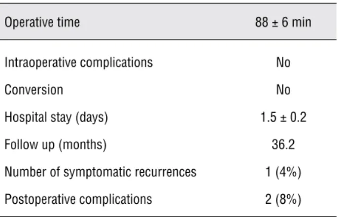
The operative time (including anesthesia time) was 88 ± 6 min. There were no intraopera-tive complications and all cases could be com-pleted with pure laparoscopic technique. Mean hospital stay was 1.5 ± 0.8 days (Table-2).
Clinical follow-up included serum cre-atinine measurements and abdominal ultrasound after definitive surgical treatment. One patient (4%) developed symptomatic recurrence after 45 days following lymphocele treatment and under-went reoperation. An open technique approach was chosen in this case as technical difficulty was anticipated since the patient had had several abdominal surgical interventions other than the kidney transplantation and lymphocele drain-age. Preoperative imaging allowed for location near to the graft hylum. Confirmation of prop-er site was carried out only with palpation and careful aspiration. This patient underwent un-complicated repeated fenestration without sub-sequent recurrence. Two patients (8%) had post-operative complications (one ureteral injury and one incisional hernia) that required reoperation. The patient with ureteral injury developed symp-tomatic abdominal fluid collection one week af-ter the procedure. In the reoperation, a laaf-teral opening at mid third of transplanted ureter 1 cm away from the drainage incision could be noted. Correction was undertaken with ureteral edges revitalization and single stitches with absorbable suture. A double J catheter was placed under fluoroscopic control as well.
Incisional hernia developed 2 weeks af-ter laparoscopy. An intestinal segment could be noted through the hernia ostium at the time of reoperation. All patients restored renal function after definitive treatment.
DISCUSSION
84% of the patients of our series increased serum creatinine level was the first symptom indicative of a lymphocele. Lower extremity edema, genital edema, deep venous thrombosis before detection of the lymphocele were the leading symptoms in only 16% of the patients.
In our series of 991 patients studied over 12 years, the incidence of lymphocele was 2.52%, which is comparable to the rate of 1% to 12% of patients observed in other reviews (8). The oc-currence of lymphoceles after surgery for kidney transplantation does not seem to be the result of any single factor (9). A number of aetiologi-cal factors are described and a large variety of combinations are likely to be causal (9). Like il-lustrated previously there are good evidences to suggest that the extraperitoneal location of renal transplants and subsequent lack of com-munication with the absorptive peritoneal sur-face contribute to significant elevated incidence of lymphocele after renal transplantation. Other factors can contribute to lymphocele formation like delayed graft function (10), repeat transplan-tation (11), acute graft rejection (12), cadaveric Symptomatic lymphoceles are of much
less frequency but are easily recognized and diagnosed (8). When symptoms occur they are typically related to compression of adjacent structures. The most frequent symptoms asso-ciated with lymphoceles are lower abdominal pain, lower extremity or scrotal swelling, deep vein thrombosis, or graft dysfunction secondary to ureteral or bladder outlet obstruction (1). In
Table 2 - Intraoperative data and outcome.
Operative time 88 ± 6 min
Intraoperative complications No
Conversion No
Hospital stay (days) 1.5 ± 0.2
Follow up (months) 36.2
Number of symptomatic recurrences 1 (4%)
Postoperative complications 2 (8%) Table 1 - Patient demographics.
Number of transplants (living/cadaveric donors) 991 (468/523)
Number of post transplant lymphoceles 25 (2.52%)
Number of lymphoceles (living/cadaveric donors) 8/17
Mean interval from transplantation (weeks) 14.2 ± 6
Mean diameter of lymphoceles (centimeters) 15.4 ± 2.8
Number women/men 14/11
Mean age (years) 38 ± 9
Operative time (minutes) 88 ± 6
Intraoperative complications No
Conversion No
Hospital stay (days) 1.5 ± 0.2
Follow up (months) 36.2
Number of symptomatic recurrences 1 (4%)
donor kidney (3), sirolimus immunosuppression (13), and other host disease factors such as adult polycystic kidney disease (14).
Thromboembolic prophylaxis with low-dose subcutaneous heparin can be another ae-tiological factor. This may be a consequence of the higher vulnerability of the lymphatic system to the effects of anticoagulants due to the lower concentration of clotting factors and the lack of platelets (9).
Although it has been suggested that im-plantation to the external iliacs is a risk factor for lymphocele formation, we routinely use the internal or external iliacs and did not find this to be a problem (15).
In this series, there was a significant dif-ference in the number of lymphoceles in the kidneys from cadaveric donors. It could be ex-plained by bigger inflammatory process in these patients, where surgery performed in urgency and not routinely like living donors.
Renal transplant recipients with increased serum creatinine levels and clinical symptoms indicative of a lymphocele should undergo diag-nostic ultrasound or computerized tomography.
Before the laparoscopic era the only al-ternative to open transperitoneal lymphocele drainage was percutaneous aspiration with sub-sequent observation or sclerosant therapy. They introduce the possibility of infection and they can keep open lymphatic vessels due to continu-ous aspiration (16).
Closed percutaneous drainage systems can be used for a defined period of time. They also carry a recurrence rate of 40-50%. A number of sclerosants have been used as an adjunct to percu-taneous drainage (3). Although sclerosant therapy has a better success rate than simple aspiration, recurrences remain common, and complications including acute renal failure have been reported (17). No large series have established the benefit of one method of sclerosis over the others (4).
Operative treatment of symptomatic post transplant lymphocele consists of lymphocelec-tomy with creation of a large internal window for intraperitoneal fluid drainage. Open lymphoce-lectomy has long been the standard. A literature review including 129 patients who underwent
open drainage between 1980 and 1998 revealed a complication rate of 4% and a recurrence rate of 15% (18). It is believed that the high incidence of symptomatic recurrences following open drain-age is a result of bowel adhesions obstructing the internal window and thus causing extraperito-neal fluid accumulation.
Since laparoscopic fenestration of post transplant lymphocele was first described by McCullough et al. (19) in 1991, multiple groups have shown this approach to be safe and effec-tive (4,6,7). The general benefits of laparoscopic surgery include less aggressiveness to the ab-dominal wall with quick recovery, favorable cosmetics, reduced blood loss, low postoperative morbidity and short hospital stay.
The relative contraindications to a lapa-roscopic surgery include bowel adhesions sec-ondary to multiple previous abdominal surgeries, the need for a concomitant open procedure, and lymphocele anatomy in close proximity to the re-nal hylum (6).
Since laparoscopy has been employed to treat post transplant lymphocele, a number of complications have been described (4,8). Injury of the urinary tract, particularly of the transplanted ureter, has been described as a major drawback of laparoscopic lymphocele drainage (4), and an incidence rate as high as 20% has been reported (16). Laparoscopic drainage of small lymphoceles in close proximity to the renal hylum bears an increased risk of iatrogenic ureter or vessel injury.
Recent reviews note different results with regard to complications associated with laparo-scopic lymphocelectomy. Hsu et al., in a mul-ticenter review of experienced centers, found a complication rate of 5% with only a single uri-nary tract (bladder) injury (8). Cadrobbi et al. (20) found a 7% incidence of urinary tract injury (re-nal pelvis, ureter, bladder) with laparoscopic fen-estration, but the rate was lower in centers with significant experience.
structures prior to fenestration and to document complete drainage of the fluid collection. How-ever, the same authors admitted that this tool was not always helpful for identification of the transplant ureter. We believe that meticulous and careful procedure rather than the use of endo-scopic ultrasound ensures safety during laparo-scopic lymphocele drainage.
Laparoscopic drainage is thought to have a lower incidence of symptomatic recurrences compared with open drainage (10). In the litera-ture the recurrence rate following laparoscopic lymphocele fenestration ranges from 4% to 6% (4,6,8). In our series, the recurrence rate after lap-aroscopy was 4% (1 of 25). We believe that it was due to a failure to localize and adequately open a little inferior component of the lymphocele.
Based on our single patient with incom-plete drainage of an unappreciated second cavity, we have begun to use laparoscopic transcutane-ous ultrasound routinely if there is any question about lymphocele anatomy, in an effort to help prevent further recurrences.
Laparoscopic lymphocele drainage is a safe and effective procedure. Given the benefits of minimally invasive surgery, including reduced postoperative morbidity and shorter hospital stay, we conclude that laparoscopic drainage should be considered first-line therapy for patients with symptomatic post renal transplant lymphoceles.
CONFLICT OF INTEREST
None declared.
REFERENCES
1. Burgos FJ, Teruel JL, Mayayo T, Lovaco F, Berenguer A, Orte L, et al.: Diagnosis and management of lymphoceles after renal transplantation. Br J Urol. 1988; 61: 289-93. 2. Howard RJ, Simmons RL, Najarian JS: Prevention of
lym-phoceles following renal transplantation. Ann Surg. 1976; 184: 166-8.
3. Ward K, Klingensmith WC 3rd, Sterioff S, Wagner HN Jr.: The origin of lymphoceles following renal transplantation. Transplantation. 1978; 25: 346-7.
4. Bailey SH, Mone MC, Holman JM, Nelson EW: Laparoscop-ic treatment of post renal transplant lymphoceles. Surg En-dosc. 2003; 17: 1896-9.
5. López García D, Janeiro Pais JM, González Dacal J, Zarra-onandía Andraca A, Casas Agudo P, Martínez Breijo S et al.: Lymphocele after renal transplantation: case report and bibliographic review. Arch Esp Urol. 2009; 62: 667-71. 6. Fuller TF, Kang SM, Hirose R, Feng S, Stock PG, Freise CE:
Management of lymphoceles after renal transplantation: laparoscopic versus open drainage. J Urol. 2003; 169: 2022-5.
7. Desai MM, Gill IS: Laparoscopic surgery in renal transplant recipients. Urol Clin North Am. 2001; 28: 759-67.
8. Hsu TH, Gill IS, Grune MT, Andersen R, Eckhoff D, Gold-farb DA et al.: Laparoscopic lymphocelectomy: a multi-in-stitutional analysis. J Urol. 2000; 163: 1096-8; discussion 1098-9.
9. Metcalf KS, Peel KR: Lymphocele. Ann R Coll Surg Engl. 1993; 75: 387-92.
10. Braun WE, Banowsky LH, Straffon RA, Nakamoto S, Kiser WS, Popowniak KL et al.: Lymphocytes associated with re-nal transplantation. Report of 15 cases and review of the literature. Am J Med. 1974; 57: 714-29.
11. Stephanian E, Matas AJ, Gores P, Sutherland DE, Najarian JS: Retransplantation as a risk factor for lymphocele for-mation. Transplantation. 1992; 53: 676-8.
12. Malovrh M, Kandus A, Buturović-Ponikvar J, Lindic J, Knap B, Fliser D et al.: Frequency and clinical influence of lymphoceles after kidney transplantation. Transplant Proc. 1990; 22: 1423-4.
13. Langer RM, Kahan BD: Incidence, therapy, and conse-quences of lymphocele after sirolimus-cyclosporine-pred-nisone immunosuppression in renal transplant recipients. Transplantation. 2002; 74: 804-8.
14. Martínez-Ocaña JC, Lauzurica R, Castellote E, Bonet J, Tenesa M, Jiménez JÁ et al.: Adult polycystic kidney dis-ease: a risk factor for lymphocele formation after renal transplantation? Transplant Proc. 1995; 27: 2246-7. 15. Sansalone CV, Aseni P, Minetti E, Di Benedetto F, Rossetti
O, Manoochehri F et al.: Is lymphocele in renal transplan-tation an avoidable complication? Am J Surg. 2000; 179: 182-5.
16. Kim JK, Jeong YY, Kim YH, Kim YC, Kang HK, Choi HS: Postoperative pelvic lymphocele: treatment with simple percutaneous catheter drainage. Radiology. 1999; 212: 390-4.
18. Doehn C, Fornara P, Fricke L, Jocham D: Laparoscopic fenestration of posttransplant lymphoceles. Surg Endosc. 2002; 16: 690-5.
19. McCullough CS, Soper NJ, Clayman RV, So SS, Jendrisak MD, Hanto DW: Laparoscopic drainage of a posttransplant lymphocele. Transplantation. 1991; 51: 725-7.
20. Cadrobbi R, Zaninotto G, Rigotti P, Baldan N, Sarzo G, An-cona E: Laparoscopic treatment of lymphocele after kidney transplantation. Surg Endosc. 1999;13: 985-90.
______________________
Correspondence address:
Dr. Cristiano Augusto Calderaro Cotrim Hospital das Clínicas UNICAMP R. Vital Brazil, 250 - 20andar, A2, sala108 Cidade Universitária Zeferino Vaz Distrito de Barão de Geraldo, Campinas, SP, 13083-590, Brazil E-mail: [email protected]
EDITORIAL COMMENT
The authors present their experience on the treatment of 25 patients submitted to a kidney transplant with symptomatic lymphoceles. It has been observed a clear reduction of the incidence of lymphoceles in the last decade. There is not an isolate etiology for its occurrence. However, a careful dissection of the iliac vessels with careful taking to ligate the lymphatic vessels is certainly an important step on its prevention.
Before performing the laparoscopic drain-age, some steps should be reinforced. A computer tomography is fundamental in planning the sur-gery, the collection located lateral to the graft
or deep in the iliac fossa may represent a ma-jor difficult when performing the procedure. The authors have observed just one case of ureteral injury, probably in a patient with a lymphocele deep in the pelvis.
Another important aspect, mentioned by the authors, is to perform a percutaneous aspira-tion one day before the procedure, to exclude an infected lymphocele. These patients may have an infected collection without any clinical signs.
The laparoscopic access is safe and effec-tive for the treatment of symptomatic lynphocele as shown by the authors.
Dr. William Nahas