INTRODUCTION
Anticoagulants and antiplatelet agents are commonly used for prophylaxis of cardiovascu-lar, cerebrovascular or venous thrombotic disease and post-implantation of mechanical valves and stents (1). Peri-operative management of these
anti-coagulated patients presents a dilemma to the surgeon since patients on chronic anticoagulation therapy have multiple comorbidities thus increas-ing risks of adverse thromboembolic events fol-lowing perioperative cessation of anticoagulation (2). Furthermore, surgery may have thrombogenic nature and a potential hypercoagulable state that Purpose: Patients with coagulopathy are at increased risk of peri-operative
hem-orrhage. The aim of the present study was to compare ureteroscopy (URS) in these high risk patients to those with normal bleeding profi le.
Materials and Methods: Twelve patients with coagulopathies (Group I) undergo-ing 17 URS were included in the study [3 for biopsy of ureteral lesions and 9 for Holmium Laser Lithotripsy (HLL)]. A patient had Child B (MELD 11) cirrhosis, 6 patients were on warfarin, 3 patients on ASA, 1 patient on ASA and clopidogrel, and the last patient was on heparin. URS in Group I was performed without cor-rection of coagulopathy. Group II consisted of 32 patients with normal bleeding profi le who underwent 34 URS concurrently.
Results: Group I included 4 ureteral biopsies in 3 patients with suspicious ure-teral lesions and 13 URS for HLL in 9 patients with nephrolithiasis. There were no signifi cant differences between the two groups in terms of patient age, sex, percent of renal stones, median operative and fl uoroscopy times. When compared with Group II, Group I had signifi cantly larger median stone size (9.2 vs. 14.0 mm, p = 0.01) and signifi cantly lower stone-free rate after fi rst URS (94.1% vs. 69.2%, p = 0.04). However, after second URS, stone-free rates were comparable in both groups (92.3% vs. 100%, p = 0.9). Two (16.7%) patients with coagulopathy were readmitted due to gross hematuria. There were no post-operative complica-tions in Group II.
Conclusions: Although URS in selected patients with coagulopathies is safe, it is associated with signifi cantly lower stone-free rates and higher readmissions due to gross hematuria.
Ureteroscopy in patients with coagulopathies is
associated with lower stone-free rate and increased
risk of clinically signifi cant hematuria
_______________________________________________
Mohamed A. Elkoushy, Philipe D. Violette, Sero Andonian
Department of Surgery, Division of Urology, McGill University Health Center, McGill University, Montreal Quebec, Canada
ABSTRACT ARTICLE INFO
_______________________________________________________________ _____________________
Key words:
Ureteroscopy; lithotripsy; calculi; ureter; hematologic diseases
Int Braz J Urol. 2012; 38: 195-203 ________________
Submitted for publication: April 11, 2011
________________
may result from a rebound increase in clotting factors after discontinuation of these drugs (3). Withdrawal of antiplatelet agents in the periop-erative period is associated with higher risks than the maintenance of these medications (4). Several studies have reported coronary stent thrombosis after premature discontinuation of antiplatelet agents (5-7), resulting in increased post-opera-tive myocardial infarction, peri-operapost-opera-tive cardiac mortality, and overall mortality (5,6,8). There-fore, peri-operative maintenance on anti-platelet agents is recommended for low-hemorrhagic risk procedures after drug-eluting stents (9). Extra-corporeal shock wave lithotripsy (SWL), percu-taneous nephrolithotomy (PCNL), laparoscopic or open stone surgery are contraindicated in patients with coagulopathies (10). Traditionally, bleed-ing diathesis is corrected and the anticoagulation therapy is withheld prior to any urological in-tervention to minimize surgical hemorrhage (11). However, despite pre-operative correction and apparently normal clotting parameters, patients with coagulopathy have a higher rate of com-plications and lower efficacy of SWL (12). The safety of URS and holmium laser lithotripsy in patients with coagulopathies without correction of the abnormality have been reported in 3 retro-spective studies (10,13,14). Therefore, the aim of the present study was to expand the indications for URS in patients with coagulopathies and com-pare their outcome with concurrent patients with normal clotting parameters.
MATERIALS AND METHODS
Retrospective review of prospectively collected data of patients undergoing URS by a single surgeon (SA) between July 2009 and Jan-uary 2011 was performed. Twelve patients with coagulopathies undergoing 17 URS comprised Group I [3 for biopsy of ureteral lesions and 9 for Holmium Laser Lithotripsy (HLL)]. Thirty-two concurrent patients with normal clotting parameters who underwent URS and HLL served as the control group (Group II). All patients had routine pre-operative evaluation that included complete blood count, prothrombin time, partial thromboplastin test and International
Normal-ized Ratio (INR). Pre-operative patient informa-tion including age, sex, stone/tumor size and location, co-morbidities, and indications for anticoagulation of patients with coagulopathies were collected. Intra-operative information such as operative time, fluoroscopy time, use of access sheath, stone-free status and any complications were recorded immediately post-operatively on research data forms. Post-operative outcome and complications especially hemorrhagic and thromboembolic events were recorded from of-fice and hospital charts.
TECHNIQUE
Statistical Analysis
Data were analyzed using the commercial-ly available Statistical Package of Social Sciences for Windows (SPSS, Chicago, IL), version 17. De-scriptive data were presented in terms of percent-ages, range, medians and standard deviations. Continuous variables such as length of surgery, fluoroscopy time and stone size were compared with the Mann-Whitney U test. Fisher’s exact test was used for categorical variables with two-tailed p < 0.05 being statistically significant.
RESULTS
Twelve patients with coagulopathies with a median age of 63.5 years were included in Group I (9 males and 3 females). In terms of coagulopathy, a patient had Child B (MELD 11) cirrhosis with thrombocytopenia, 6 patients were on warfarin [4 for Deep Vein Thrombosis (DVT), 1 for atrial fibrillation, 1 for mechanical aortic valve], 3 patients were on acetylsalicylic acid (ASA) and another patient was on combination of ASA and clopidogrel for coronary artery dis-ease and coronary stents, and the last patient was on low molecular weight heparin (Tinzaprin) for recent DVT/PE (Table-1).
A total of 17 URS with HLL or biopsies of suspicious ureteral lesions were performed in 9 and 3 patients, respectively. URS in Group I was performed without correction of coagulopathy or suspension of their anti-coagulation therapy. For the 9 patients with coagulopathies undergo-ing URS and HLL, the median maximum stone diameter was 14 mm (5 - 22 mm). However, 6 out of the 9 patients had significant stone burden including a lower pole partial staghorn (Table-1). When visibility was poor, a staged URS was per-formed to obtain stone-free status. Therefore, 2 out of 9 patients (22.2%) underwent a second URS and one patient required a third URS to achieve stone-free status. In one of the three pa-tients with ureteral lesions undergoing URS and biopsy, the first biopsy was inconclusive. There-fore, a repeat URS with biopsy was performed.
Group II consisted of 32 patients un-dergoing 34 URS and HLL for 45 stones. There
were no significant differences between the two groups in terms of patient age, sex, percent of re-nal stones, median operative time and fluorosco-py time (Table-2). However, Group I patients had significantly larger median stone size when com-pared with Group II (14.0 vs. 9.2 mm, p = 0.01) (Table-2). Due to poor vision, 3/12 (25%) patients in Group I and 2/32 (6%) patients in the con-trol Group II underwent second URS to achieve stone-free status (p = 0.11). Stone-free rate after first URS was significantly lower in Group I com-pared with Group II (69.2% vs. 94.1%, p = 0.04). However, after the second URS, the stone-free rates were comparable in both groups (92.3% vs. 100%, p > 0.05). Calcium oxalate monohydrate represented the most common stone composition in both groups (67% and 43% respectively) fol-lowed by uric acid stones.
No patient had significant gross hematu-ria during the immediate post-operative period in both groups. However, the median post-oper-ative level of hemoglobin significantly decreased in patients with coagulopathies when compared with controls (0.8 vs. 0.2 g/dL; p = 0.001).
Two patients (22 %) from Group I (pa-tients 3 and 4) on warfarin therapy were readmit-ted for management of gross hematuria. Patient #3 in Group I was readmitted on post-operative day 47 post URS and biopsy of invasive TCC. Pa-tient #4 in Group I had an INR of 3.14 and was readmitted on post-operative day 6 post URS and HLL. Both patients underwent continuous blad-der irrigation and their anti-coagulants were withheld till hematuria resolved. They did not re-quire transfusions. There were no post-operative complications in Group II.
DISCUSSION
Table 1 - Characteristics of patients with coagulopathies (Group I) and indications of URS.
Pt. No. Age Sex Type of coagulo-pathy
Anticoagulant/ bleeding diatheses
Indications of URS No of URS Stone composition /
pathology
1 59 F DVT Warfarin Partial staghorn stone
(22 X 22 mm) 2 Uric acid dihydrate
2 62 M DVT Warfarin UPJ stone (20X13 mm) 1 Struvite
3 86 M Atrial fibrillation
+ DVT Warfarin
Mid-ureteral mass
(40X 20 mm) 1
High grade invasive TCC
4 47 M Mechanical aortic
valve Warfarin
Lower pole stones
(18 & 12 mm) 3
Calcium oxalate monohydrate
5 78 M Recent atrial
fibrillation Warfarin
Obstructing UPJ stone (11X6mm) + renal
gravel
1 Uric acid dihydrate
6 58 M Coronary disease
and stent ASA
Distal ureteral stone
(10X5 mm) 1
Carbonate apatite + Calcium oxalate
7 70 M Coronary disease
and stent, ESRD ASA
Mid ureteral lesion
(10mm) 2
Cytological atypia Ureteritis
8 52 M Recent DVT/ PE LMW heparin
(Tinzaprin)
Lower pole kidney stones (14X10 and 9X7
mm) with UPJ stone (8X6 mm)
2 Calcium oxalate monohydrate
9 62 F Child B, MELD 11
hepatic cirrhosis Thrombo-cytopenia
UPJ stone (10 mm) and lower pole stone
(5 mm)
1 Calcium oxalate monohydrate
10 63 M Bilateral DVT Warfarin Upper ureteral stone
(5 mm) 1
Calcium oxalate monohydrate
11 44 F Coronary stent ASA + Clopidogrel Upper ureteral lesion 1 Chronic inflammation
12 59 M Recent MI and
CABG ASA
Lower pole kidney stone
(8 mm) 1
Calcium oxalate monohydrate
ASA: acetyl salicylic acid; DVT: Deep Vein Thrombosis; ESRD: End Stage Renal Disease; PE: Pulmonary embolism; MI: myocardial In-farction; CABG: Coronary Artery Bypass Graft; LMW: low molecular weight; UPJ: uretero-pelvic junction; PE: pulmonary embolism; PLT:
low risk of hemorrhage such as URS and HLL can be performed without discontinuation of antico-agulation therapy (17). Thus, URS and HLL may be the only option for these patients with coagu-lopathies since they are often poor candidates for SWL or PCNL due to hemorrhagic and thrombo-embolic complications (10,13).
In the present study, 6 out of 9 patients (patients 1, 2, 4, 5, 8, 9) in Group I undergoing URS and laser lithotripsy had significant stone burden (Table-1). Traditionally, these patients would be treated with PCNL with correction of the coagu-lopathy. Discontinuing and re-initiating antico-agulation therapy in these high risk patients may have increased risk of hemorrhagic and throm-boembolic complications. PCNL with reversal of anticoagulation has been previously described in 27 such high risk patients (2). However, two pa-tients (7%) developed post-operative hemorrhage with one patient requiring angio-embolization. Another patient (4%) developed DVT with pul-monary embolism on POD 4 requiring IVC filter since he had developed hemorrhage when anti-coagulation was initiated (2). Furthermore, the expense of bridging therapy (with low molecular weight heparin or intravenous heparin) is consid-erable (10). Therefore, the present study expands
the indications for URS in patients with coagu-lopathies to those who are traditionally treated with PCNL with reversal of their anti-coagulation. This would be ideal for patients who cannot safe-ly undergo withholding of anticoagulation.
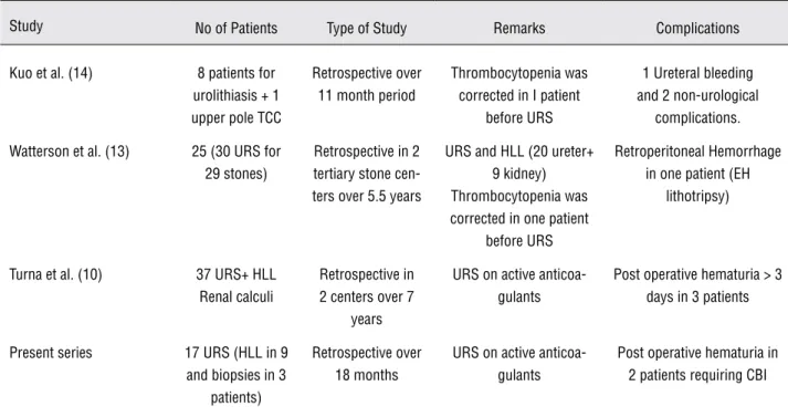
The first series describing URS in patients with coagulopathies was by Kuo et al. (Table-3) (14). Eight patients with stone disease and 1 pa-tient with upper tract TCC were treated by URS with the holmium laser (14). Six out of 7 patients who underwent laser fragmentation for calculi were stone free at 1 month, and no tumor recur-rence was noted in the patient with TCC (follow-up of 4 months). One patient only had a post-operative bleeding complication related to the procedure, involving an episode of oliguria sec-ondary to a small ureteral clot that was resolved with diuretics. Watterson et al. reported on a se-ries of 25 patients who were anticoagulated ei-ther pharmacologically or by underlying systemic diseases (13). The overall stone-free rate after a single ureteroscopic procedure was 93%. There were no hemorrhagic complications in patients undergoing laser lithotripsy. One patient who un-derwent electrohydraulic lithotripsy developed a retroperitoneal hematoma necessitating transfu-sion (Table-3). Therefore, electrohydraulic litho-Table 2 - Comparison of patients undergoing URS and laser lithotripsy.
Variable Group I
(n = 9)
Group II (n = 32)
P- Value
Median Age (yrs) 60.0 53.5 0.27
Male gender 7 (78%) 19 (59%) 0.49
Percent Renal Stones (# renal/ total) 8/14 (57%) 18/45 (40%) 0.35
Median stone size (mm) 14 9.2 0.01
Stone-free rate After 1st URS 69.2% 94.1 % 0.04
Stone-free rate After 2nd URS 92.3 % 100% 0.9
Median URS time (min.) 60 60 0.21
Median Fluoroscopy time (Sec) 63 114 0.24
tripsy must be avoided in this highly selected group of patients with coagulopathies (18). The Holmium: YAG laser has the ability to fragment calculi of all compositions including calcium oxalate monohydrate and it is an ideal intracor-poreal lithotripter for ureteral calculi with a high success rate and low morbidity (19). Moreover, the Holmium: YAG laser has haemostatic proper-ties that would be beneficial for treating patients with bleeding disorders (20). Therefore, it is ideal for fulguration of bleeders during biopsy of ure-teral lesions in patients with coagulopathies.
Turna et al. compared a group of 37 pa-tients on aspirin, clopidogrel, or warfarin with a cohort of matched controls without coagulopathy and found similar stone-free rates (81.1% versus 78.4%, p = 0.7) (10). However, the peri-operative hemoglobin change was significantly higher in the anticoagulated group (6 g/L vs. 2 g/L, p < 0.0001). In that study, there were no procedures terminated because of poor visibility. The au-thors reported 3 cases of hematuria of more than 3 days in patients with coagulopathies (Table-3).
In the present study, after the first URS, the stone-free rate was significantly lower in Group I when compared with Group II (69.2% vs. 94.1%, p = 0.04). This could be related to the fact that patients in Group I had significantly larger median stone size (14 vs. 9.2 mm. p = 0.01). Furthermore, two thirds (6 out of 9) of patients had significant stone burden that are ideally managed by PCNL. In the present study Group I, 2 out of 9 (22.2%) patients underwent a second URS and one patient required a third URS to achieve stone-free status. However, after a second URS, the stone-free rates were compa-rable in both groups (92.3% vs. 100%, p > 0.05). Similarly, in the study by Watterson et al., there was a second URS in 5 patients. Furthermore, in that study, a thrombocytopenic patient had correction of thrombocytopenia prior to URS. Thus, although URS and HLL are safe in these highly selected patients with coagulopathies, they may require more than one URS session for stone clearance. Larger sample size is required to verify these results.
Table 3 - Comparison between the present series and previous published studies.
Study No of Patients Type of Study Remarks Complications
Kuo et al. (14) 8 patients for urolithiasis + 1 upper pole TCC
Retrospective over 11 month period
Thrombocytopenia was corrected in I patient
before URS
1 Ureteral bleeding and 2 non-urological
complications.
Watterson et al. (13) 25 (30 URS for 29 stones)
Retrospective in 2 tertiary stone cen-ters over 5.5 years
URS and HLL (20 ureter+ 9 kidney) Thrombocytopenia was corrected in one patient
before URS
Retroperitoneal Hemorrhage in one patient (EH
lithotripsy)
Turna et al. (10) 37 URS+ HLL Renal calculi
Retrospective in 2 centers over 7
years
URS on active anticoa-gulants
Post operative hematuria > 3 days in 3 patients
Present series 17 URS (HLL in 9 and biopsies in 3
patients)
Retrospective over 18 months
URS on active anticoa-gulants
Post operative hematuria in 2 patients requiring CBI
Previous reports of URS and laser litho-tripsy in patients with coagulopathies did not re-port stone composition. In the present series, most of the stones in both groups (67% in Group I and 43% in Group II) were composed of calcium ox-alate monohydrate, which is one of the hardest stones to fragment (21). Therefore, this may have contributed to the lower stone-free rate in Group I.
Except for one patient reported by Kuo et al., there are no other reports in the litera-ture about safety of URS and ureteral biopsy in patients with coagulopathies (14). In the pres-ent study, 3 patipres-ents underwpres-ent ureteral biopsies safely and efficiently while they were on antico-agulants. One of them developed a late hematuria (after 47 days). This was an 86-year old man on warfarin for repeated bilateral DVT and atrial fi-brillation in addition to past medical history of hypertension, diabetes mellitus, and chronic renal failure. His preoperative INR was 2.89. After his diagnostic URS and biopsy, he underwent exter-nal beam radiotherapy for his 4 cm mid-ureteral invasive TCC. Therefore, his delayed hematuria could be related to other factors than the proce-dure itself such as the invasive TCC, indwelling ureteral stent, or radiation ureteritis. The other 2 patients underwent 3 ureteral biopsies on 3 occa-sions without complications indicating the safety of ureteroscopic biopsies in these patients with coagulopathies.
There are several limitations of the present study. Although the data were collected prospec-tively, this still remains a retrospective review of highly selected small cohort of patients with co-agulopathies undergoing URS. Furthermore, the cohort with coagulopathies was diverse with mul-tiple different therapies (antiplatelet and antico-agulation (Coumadin, LMW heparin)) undergoing two different procedures (biopsy and lithotripsy). Another limitation was that the INR on the day of the URS was not confirmed to be in the therapeu-tic level. It was only checked in the pre-operative evaluation.
CONCLUSIONS
Although URS in selected patients with coagulopathies is safe, it is associated with
sig-nificantly lower stone-free rates and higher re-admission for management of gross hematuria. Prospective randomized studies with and without correction of the coagulopathy is needed to weigh the risks and benefits of correcting anticoagula-tion during ureteroscopy and laser lithotripsy or biopsy of ureteral lesions.
ACKNOWLEDGEMENTS
This work was supported in part by the Northeastern AUA Young Investigator Award and Montreal General Hospital Foundation Award to Sero Andonian.
ABBREVIATIONS
ASA: acetyl salicylic acid
CBI: Continuous Bladder Irrigation
DVT: Deep Vein Thrombosis
ESRD: End Stage Renal Disease
HLL: Holmium Laser Lithotripsy
INR: International Normalized Ratio
LMW: low molecular weight
PCNL: Percutaneous Nephrolithotomy
PE: Pulmonary Embolism
PLT: platelets
POD: post operative day
SWL: Extracorporeal Shockwave Lithotripsy
TCC: Transitional Cell Carcinoma
UPJ: uretero-pelvic junction
URS: ureteroscopy
CONFLICT OF INTEREST
None declared.
REFERENCES
1. Ono S, Fujishiro M, Hirano K, Niimi K, Goto O, Kodashima S, et al.: Retrospective analysis on the management of an-ticoagulants and antiplatelet agents for scheduled endos-copy. J Gastroenterol. 2009; 44: 1185-9.
2. Kefer JC, Turna B, Stein RJ, Desai MM: Safety and efficacy of percutaneous nephrostolithotomy in patients on antico-agulant therapy. J Urol. 2009; 181: 144-8.
4. Chassot PG, Delabays A, Spahn DR: Perioperative anti-platelet therapy: the case for continuing therapy in patients at risk of myocardial infarction. Br J Anaesth. 2007; 99: 316-28.
5. Iakovou I, Schmidt T, Bonizzoni E, Ge L, Sangiorgi GM, Stankovic G, et al.: Incidence, predictors, and outcome of thrombosis after successful implantation of drug-eluting stents. JAMA. 2005; 293: 2126-30.
6. Ong AT, McFadden EP, Regar E, de Jaegere PP, van Dom-burg RT, Serruys PW: Late angiographic stent thrombosis (LAST) events with drug-eluting stents. J Am Coll Cardiol. 2005; 45: 2088-92.
7. Spertus JA, Kettelkamp R, Vance C, Decker C, Jones PG, Rumsfeld JS, et al.: Prevalence, predictors, and outcomes of premature discontinuation of thienopyridine therapy after drug-eluting stent placement: results from the PRE-MIER registry. Circulation. 2006; 113: 2803-9.
8. Pfisterer M, Brunner-La Rocca HP, Buser PT, Rickenbacher P, Hunziker P, Mueller C, et al.: Late clinical events after clopidogrel discontinuation may limit the benefit of drug-eluting stents: an observational study of drug-drug-eluting ver-sus bare-metal stents. J Am Coll Cardiol. 2006; 48: 2584-91.
9. Di Minno MN, Prisco D, Ruocco AL, Mastronardi P, Massa S, Di Minno G: Perioperative handling of patients on anti-platelet therapy with need for surgery. Intern Emerg Med. 2009; 4: 279-88.
10. Turna B, Stein RJ, Smaldone MC, Santos BR, Kefer JC, Jackman SV, et al.: Safety and efficacy of flexible ureterore-noscopy and holmium:YAG lithotripsy for intrarenal stones in anticoagulated cases. J Urol. 2008; 179: 1415-9. 11. Streem SB, Yost A: Extracorporeal shock wave lithotripsy
in patients with bleeding diatheses. J Urol. 1990; 144: 1347-8.
12. Klingler HC, Kramer G, Lodde M, Dorfinger K, Hofbauer J, Marberger M: Stone treatment and coagulopathy. Eur Urol. 2003; 43: 75-9.
13. Watterson JD, Girvan AR, Cook AJ, Beiko DT, Nott L, Auge BK, et al.: Safety and efficacy of holmium: YAG laser litho-tripsy in patients with bleeding diatheses. J Urol. 2002; 168: 442-5.
14. Kuo RL, Aslan P, Fitzgerald KB, Preminger GM: Use of ure-teroscopy and holmium:YAG laser in patients with bleeding diatheses. Urology. 1998; 52: 609-13.
15. Sayed MA: Semen changes after extracorporeal shockwave lithotripsy for distal-ureteral stones. J Endourol. 2006; 20: 483-5.
16. Eisner BH, Kurtz MP, Dretler SP: Ureteroscopy for the man-agement of stone disease. Nat Rev Urol. 2010; 7: 40-5. 17. Brejcha M, Gumulec J, Penka M, Klodová D, Wróbel M,
Bo-goczová E: Preparation of patients on anticoagulant treat-ment for invasive surgery. Vnitr Lek. 2009; 55: 272-5. 18. Türk C, Knoll T, Petrik A, Sarica K, Seitz C, Straub M, et al.:
Guidelines on Urolithiasis. EAU update series. 2010; 44-70. 19. Gupta PK: Is the holmium:YAG laser the best intracorporeal
lithotripter for the ureter? A 3-year retrospective study. J Endourol. 2007; 21: 305-9.
20. Wollin TA, Denstedt JD: The holmium laser in urology. J Clin Laser Med Surg. 1998; 16: 13-20.
21. Turgut M, Unal I, Berber A, Demir TA, Mutlu F, Aydar Y: The concentration of Zn, Mg and Mn in calcium oxalate mono-hydrate stones
______________________
Correspondence address: Dr. Sero Andonian Assistant Professor of Urology Royal Victoria Hospital McGill University Health Centre 687 Pine Ave West, Suite S6.92 Montreal, Quebec, Canada H3A 1A1 Fax: +514 843 1552 E-mail: [email protected]
EDITORIAL COMMENT
A clinical problem that troubles urologists is how they should treat patients on anticoagu-lants. Interruption of anticoagulation therapy for elective urologic procedures in these patients gen-erates a complex situation in which competing risks of thrombosis and bleeding must be weighed up; when anticoagulation is discontinued patients
co-morbid-ities. However, the safety and efficacy of different procedures have not been well documented.
In the present study, Elkoushy et al. com-pared the outcomes of ureteroscopy (URS) in pa-tients with coagulopathies with those with nor-mal bleeding profile. It was found that although URS in selected coagulopathic patients was safe, it was associated with significantly lower stone-free rates and higher re-admission for gross hematuria. The main limitations of the study included its ret-rospective nature, the diversity of the study
popu-lation (patients under different drugs with differ-ent properties) and the small number of patidiffer-ents enrolled (e.g. one could argue that the difference in patients who underwent 2nd URS due to poor vision did not reach statistical significance due to the small sample size). However, this study is use-ful because the authors add their experience to the limited existing literature and provide informa-tion which help urologists to better inform their patients about the potential risks and benefits of URS without stopping anticoagulation.
REFERENCES
1. Kearon C, Hirsh J: Management of anticoagulation before and after elective surgery. N Engl J Med. 1997; 336: 1506-11.
2. Eberli D, Chassot PG, Sulser T, Samama CM, Mantz J, Dela-bays A, et al.: Urological surgery and antiplatelet drugs after cardiac and cerebrovascular accidents. J Urol. 2010; 183: 2128-36.
Dr. Stavros Gravas