Key words:
Urethra; Surgical Procedures, Operative; Tissue Engineering; Microcirculation
Int Braz J Urol. 2013; 39: 414-23
__________________
Submitted for publication: February 27, 2012
__________________
Accepted after revision: April 29, 2013
Objective: To assess the integration of decellularized heterologous collagen matrices into the urethra, when implanted with no cells or when seeded with autologous smooth mus-cle cells.
Materials and Methods: Eighteen New Zealand rabbits were randomly assigned to two groups: Group I (n = 9) - animals undergoing urethral segment resection with interposi-tion of a patch of heterologous collagen matrix seeded with autologous smooth muscle cells; Group II (n = 9) - animals undergoing resection of a urethral segment with inter-position of a decellularized heterologous collagen matrix patch. Two animals from each group were sacrificed on postoperative days seven, fourteen and twenty-eight; three animals from each group were sacrificed at the end of three postoperative months. At the end of the third month one animal from each group underwent urethroscopy for urethral integrity assessment and one animal from each group had its microcirculation image captured by a SDF device (Side-stream Dark Field - Microscan Analysis Software). One animal from each group in each euthanasia period underwent cystourethrography so as the urethra could be viewed at flow time. The matrices integration was assessed through histological examination using hematoxylin and eosin (H&E), Masson trichrome (MT), Picrosirius red and Von Willebrand staining. In a blind study with two pathologists all the slides were studied.
Results: The matrices whether seeded or not with autologous muscle cells were able to restore the architecture of the urethra, but were eliminated from the first week on, before incorporation. Microcirculation of the neourethra, at the end of the third month, showed the same characteristics as a normal urethra in both groups of animals.
Conclusion: Natural heterologous matrices implanted in the urethra as onlay graft were not incorporated into its walls but were able to fully restore the cell architecture of the organ, regardless of being seeded or not with autologous muscle cells.
INTRODUCTION
Urethral reconstruction is frequently used in the treatment of both congenital and acquired anomalies (1-6). When the urethra needs to be re-paired or reconstructed, either partially or totally,
different surgical approaches can be used such as anastomosis of the urethral stumps, flaps or grafts. Although such techniques are still largely em-ployed, in several occasions adverse effects such as fistulas, stenosis, infections, stone formation, graft contracture or diverticula formation may
Integration of collagen matrices into the urethra when
implanted as onlay graft
_______________________________________________
Kleber Sayeg, Luiz G. Freitas-Filho, Ângela Flávia Logullo Waitzberg, Victor Eduardo Arrua Arias,
Marcus Laks, Fernanda Mattos Egydio, Andréia Silva Oliveira
Department of Surgery (KS, LGFFilho, ML, FME), Department of Nephrology (ASO) and Department of Pathology (AFLW, VEAA) – Federal University of São Paulo, Brazil
ABSTRACT
ARTICLE
INFO
appear. In addition to morbidity at the donor site, there is tissue scarcity when such patients undergo a number of surgical interventions (1,2,4-12).
Tissue engineering techniques are an al-ternative for such difficulties. A decellularized matrix, either natural or synthetic, may serve as a pattern for implantation of individual cells pre-viously obtained by harvesting a full fragment from the very organ. The different cell layers are separated by using different techniques, expanded “in vitro”, and then seeded in the matrix before reimplantation (5). The different researchers that use cell-seeded matrices describe the process of matrix incorporation into the new urethra, howe-ver, not observing the manner in which this incor-poration actually takes place in the early phases of tissue repair.
This paper aimed to study the evolution of decellularized heterologous matrices integration along time, whether or not seeded with autolo-gous smooth muscle cells and implanted into the urethra.
MATERIALS AND METHODS
All experimental procedures were appro-ved by the local Research Ethics Committee (CEP - 0939/07) and conducted in strict conformity with local institutional guidelines and with internatio-nal standards for manipulation and care of labo-ratory animals.
Eighteen New Zealand male rabbits, 6 to 8 months old, weighing approximately 3 kg, from the Center for Development of Experimental Models for Medicine and Biology of the Federal University of São Paulo, were used. The animals were assigned to 2 groups of nine animals each. The animals in both groups underwent resection of a 3.5 cm long by 0.5 cm wide fragment from the ventral portion of the penile urethra. The animals in Group I underwent interposition of a patch of decellularized heterolo-gous matrices, of same size as the urethral segment withdrawn, seeded with autologous muscle cells; the animals in Group II underwent interposition of decellularized heterologous matrix patches, of same size as the segment withdrawn, however with no cells seeded. The matrix was sutured to the urethra using 6-0 polypropylene in a continuous fashion.
The collagen matrices were taken from por-cine bladders submucosa obtained from animals un-dergoing different surgical procedures. They were kept in sterile tubes containing 50 ml PBS (Phospha-te-Buffered Saline - Sigma Chemical Co., Saint Louis, USA), to which 10.000 Ul/L penicillin and 50 mg/L streptomycin were added. They were continuously agitated for fourteen days in Triton X-100 1% de-tergent (Sigma Chemical CO, Saint Louis, USA) and 0.1% ammonia hydroxide, which solution was chan-ged every two days. On day fourteen the bladders were kept for twenty-four hours in distilled water in continuous agitation. Then, they underwent ultravio-let radiation for one hour and, after that, they were cut into 3 x 5 cm fragments under a laminar flow chamber. The fragments were then kept in tubes con-taining sterile PBS and stored at -20º C Celsius until they were used. A fragment of the material obtained was put in a 10% tamponade formaldehyde solution and forwarded for anatomopathological assessment.
For the surgical procedures the animals were anesthetized with intramuscular ketamine (50 mg/ kg) and xylazine (10 mg/kg). During the procedures a venous infusion of physiological solution was main-tained. The animals in Group I underwent median laparotomy. The bladder was isolated and a 1 cm2 segment was withdrawn from the anterior wall; the bladder was then sutured with 4-0 polyglactin and the abdominal wall was sutured with 3-0 polyglactin. The bladder fragment withdrawn was taken to the laminar flow chamber and cut into 2 mm2 fragments after full removal of the mucosa. The fragments were placed on culture plates by using the “explant” te-chnique, covered with Dulbecco’s modified Eagle’s medium (DMEM) and 10% fetal bovine serum, and incubated at 37º C in a 5% carbon dioxide atmos-phere. Around day fourteen, when the culture plates showed cell confluence of approximately 80%, they were subjected to trypsin enzymatic action. The so-lution was centrifuged, the cells were counted in a Neubauer chamber and seeded in the collagen ma-trix, where they remained for another seven days in the same culture solution before being implanted.
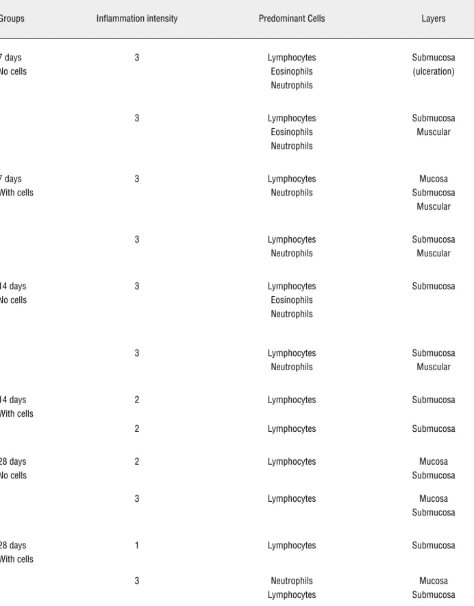
in block for a cystourethrography. The penises of all animals were removed in block and fixed in 10% buffered formaldehyde for 24 hours. The pieces were placed in paraffin blocks, cut into 5 micrometer sec-tions, stained with hematoxylin-eosin (H&E), Mas-son trichrome (MT), Picrosirius red and assessed with polarized light and immunohistochemistry using anti-Von Willebrand factor antibody and analyzed in optical microscopy with 150x augmentation. We observed the epithelial integrity, the inflammatory process and the amount of collagen, and scores 0 to 3 were created for every item assessed (Table-1).
At the end of the third postoperative month one animal in every group underwent urethrosco-py with an Olympus Winter & Ibe GMBH, Hamburg, Germany, 17Fr sheath, 12 degree optics cystoscope and was assessed for the new urethra microcircu-lation through an SDF device, 1 mm2 images from three different sites were captured, a 2 mmhh deep surface was assessed in 320x image augmentation; the images were saved in 10 second videos (13).
RESULTS
The urethroscopies performed showed the urethra with normal caliber - the 17Fr cystoscope was passed with no technical difficulty and an ade-quate urinary flow was obtained after bladder com-pression.
The microcirculation assessed by the SDF showed low flow through venules, scarce capillaries, absence of arterioles, and low vascular density; these results were similar to the ones observed in normal rabbits urethras (Figure-1).
The cystourethrografies performed showed urethras with normal caliber and adequate urinary flow (Figure-2).
Histological analyses performed on postope-rative day seven showed a disrupted area with a de-posit of fibrin-leukocitary material and stroma rich in leukocytes, with predominating polymorphonuclears and presence of eosinophils in the animals of both groups, suggestive of an acute exudative inflamtory process and of a recent intervention. The ma-trix was already being eliminated into the lumen of the organ, and an epithelial layer of cells was being formed right below (Figure-3). On day fourteen, in both groups, the matrix was found to be in the
ure-thral lumen, the epithelium not fully formed yet, and the stroma with mixed inflammatory infiltrate with lymphocytes predominating, suggestive of an early chronification. The process was being resolved from bottom to top with good formation of the muscle layer (Figure-4). On day twenty-eight full reepithe-lization could already be observed in the animals in both groups with inflammatory process, with predo-minating lymphocytes, limited to the submucosa (Fi-gure-5 a). A squamous metaplasia in the animals in the group without cells was observed (Figure-5 b). On day ninety the animals in both groups had urethral architecture similar to that of a normal urethra, ho-wever still with a less intense chronic inflammatory process, with predominating lymphocytes, showing the scarring not to be fully resolved on day ninety (Figure-6) The result of the assessment made by two pathologists in a blind study can be seen in Table-1.
Picrosirius red staining showed in all periods and in both groups, predominance of early type III collagen, even on day ninety (Figure-7).
Immunochemistry using anti-Von Wille-brand antibody demonstrated intense neoangioge-nisis in both groups, with no differences between them (Figure-8).
DISCUSSION
The need to partially or fully reconstruct the urethra is relatively usual given the high number of patients with congenital or acquired diseases, and trauma victims. Thus, a large number of techniques have been used in the attempt to restore the anatomy of the organ, aiming to achieve adequate micturition and, in the cases of male patients, to enable forward ejaculation with an adequate amount of spermato-zoids (1-5,7). All techniques used to date have a high rate of complications and, when such patients under-go several procedures, which is quite usual, the use of flaps or grafts is almost always necessary (5-7,14,15).
muco-Figure 1 - A - Photomicrography of the urethra - group I (seeded matrix), SDF 320x. B - Photomicrography of the urethra - group II (non-seeded matrix). SDF 320x.
A B
Figure 2 - A - Cystourethrography - Rabbit Group I (seeded heterologous matrix). Yellow line delimiting penile urethra subjected to matrix interposition. B - Cystourethrography Rabbit Group II (non-seeded heterologous matrix). Yellow line delimitating penile urethra subjected to matrix interposition.
Figure 3 - A - Photomicrography of the urethra - Group I animal, postoperative day 7, Black arrow = Matrix. Green arrow = inflammatory process. Blue arrow = epithelium. H&E 25x. B - Photomicrography of the urethra - Group II animal, postoperative day 7, Black arrow = Matrix. Green arrow = inflammatory process. Blue arrow = epithelium H&E, 25x.
A
A
B
Table 1 - Anatomopathologic findings.
Groups Inflammation intensity Predominant Cells Layers
7 days No cells
3 Lymphocytes Eosinophils Neutrophils
Submucosa (ulceration)
3 Lymphocytes Eosinophils Neutrophils
Submucosa Muscular
7 days With cells
3 Lymphocytes Neutrophils
Mucosa Submucosa
Muscular
3 Lymphocytes Neutrophils
Submucosa Muscular
14 days No cells
3 Lymphocytes Eosinophils Neutrophils
Submucosa
3 Lymphocytes Neutrophils
Submucosa Muscular
14 days With cells
2 Lymphocytes Submucosa
2 Lymphocytes Submucosa
28 days No cells
2 Lymphocytes Mucosa Submucosa
3 Lymphocytes Mucosa Submucosa
28 days With cells
1 Lymphocytes Submucosa
3 Neutrophils Lymphocytes
3 months No cells
1 Lymphocytes Eosinophils Neutrophils
Submucosa
2 Lymphocytes Submucosa
2 Lymphocytes Submucosa
3 months With cells
2 Neuthrophils Lymphocytes
Mucosa Submucosa
2 Neuthrophils Lymphocytes
Submucosa
3 Neutrophils Submucosa
0 - No inflammatory process 1 - Mild amount of inflammatory cells
2 - Moderate amount of inflammatory cells (more than 25% of the sample) 3 - Extended or severe inflammatory process (more than 50% of the sample)
Neutrophils - Acute Inflammation Lymphocytes - Chronic Inflammation
Eosinophils - Exsudation continuation
Figure 4 - A - Photomicrography of the urethra - group I animal, postoperative day 14, Blue arrow = epithelium Masson’s tri-chrome, 25x. B - Photomicrography of the urethra - Group II animal, postoperative day 14, Blue arrow = epithelium. Masson’s trichrome, 25x.
Figure 5 - A - Photomicrography of the urethra - Group I animal, postoperative day 28, H&E, 25x. B - Photomicrography of the urethra - Group II animal, postoperative day 28. Blue arrow = squamous metaplasia. Masson’s trichrome, 25x.
Figure 6 - A - Photomicrography of the urethra - Group I animal, postoperative day 90, Masson’s trichrome, 25x. B - Photo-micrography of the urethra - Group II animal, postoperative day 90, Masson’s trichrome, 25x.
A
A
B
B
sa appears to have become the gold standard in the treatment of major reconstructions, presenting the smallest rate of stenosis and fistulas, in the medium and long term. The number of adverse events, however, does not allow one to say yet that this would be the ideal urethral substitute (12,23-26).
The recent advance of molecular biology, the large scale production of cell nutrients, and the possibility of cultivating cells “in vitro” and then implanting them in synthetic or natural
ex-tracellular matrices, thus reconstructing an organ, have led to the introduction of Tissue Enginee-ring techniques. Combined with the employment of new materials these techniques may become important in the reconstruction or replacement of lost or insufficient organs or tissues, and a viable option in cases of lack or partial absence of tissues (5,27-29).
their slow degradation by lysosomal enzymes, in the case of synthetic matrices that gradually lead to their absorption, with minimal toxicity and in-flammatory reaction (3,5,7,9,30,31). Tissue engi-neering techniques may allow for the creation of a reservoir of biocompatible tissues that can be used in extensive urethral reconstructions when there is a lack of autologous tissues for flaps or grafts on account of various prior surgeries.
In this study we sought to assess integra-tion of a collagen matrix into the urethral wall. The collagen matrix was obtained of a porcine
bladder by employing tissue engineering tech-niques. Considering that in the different recons-truction techniques, urothelial growth frequently takes place starting from the lateral borders in the transition between the patient’s urethra and the graft, we decided to test the hypothesis that implantation of the autologous smooth muscle cells alone with no need to use the urothelial layer could reconstruct the entire architecture of the organ. It was found, through endoscopy and ra-diographic exams, that the macroscopic aspect of the urethral caliber of the animals in both groups Figure 7 - A - Photomicrography of the urethra - Group I animal, postoperative day 90, Picrosirius Red staining, 25x. Green area collagen (type III). B - Photomicrography of the urethra - Group II animal, postoperative day 90, Picrosirius Red staining, 25x. Blue area collagen (type III)
Figure 8 - A - Photomicrography of the urethra - Group I animal, postoperative day 90, immunohistochemistry with anti-Von Willebrand factor antibodies, 25x. B - Photomicrography of the urethra - Group II animal, postoperative day 90, immunohis-tochemistry with anti-Von Willebrand factor antibodies, 25x.
A
A
B
studied resembled that of a non-operated urethra, fact that had previously been observed by Chen et al., Atala et al. and El-Kassaby et al. using similar techniques (1,2,5,7,8).
By using the SDF it was possible to assess the microcirculation that was established on the new urethra around postoperative day ninety; it also showed the vascular network to resemble that of a normal urethra, regardless of the matrix having or not been seeded with muscle cells.
In order to verify the process of incor-poration of the matrices into the urethral bed, histological assessment with hematoxylin-eosin, Masson’s trichrome, Picrosirius red and Von Wil-lebrand staining were used. On day seven after implantation the animals in both groups had the same behavior in regard to epithelial integrity, to the inflammatory process and to the amount and type of collagen. The implanted matrix, instead of being incorporated and forming a portion of the urethral wall, was being eliminated into the organ’s lumen while, at the same time, the ana-tomy of the urethra was being restored. On pos-toperative days twenty-eight and ninety both groups showed epithelial integrity; the only diffe-rence found was that on day twenty-eight most of the animals in which the cells were not seeded had squamous metaplasia, which means the existence of an intense inflammatory process that turns the original epithelium into a barrier that is more re-sistant to aggressions.
Decellularized collagen matrices of diffe-rent origins are being used in urethral reconstruc-tions. Chen et al observed cell infiltration and neo-angiogenesis after two weeks, with disorganized migration of muscle fibers after two months (2). De Filippo et al comparing decellularized matri-ces seeded or not with epithelial and muscle cells observed scarce vascular organization in the non--seeded matrices, contrasting with the good orga-nization of the ones seeded (4). Our study was the first to make an early assessment of the heterolo-gous matrices integration process. We demonstra-ted that the matrix was actually eliminademonstra-ted into the lumen instead of being reincorporated, and the reason for such may be a possible greater matrix permeability to the urine, which can be explained, perhaps, by an exaggerated inflammatory process.
Regardless of being incorporated, however, the entire layer was reconstructed, which may have occurred in other studies, considering that, appa-rently, what really matters seems to be the presen-ce of the matrix at the beginning of scarring.
It can be said that the heterologous colla-gen matrix, although not having been integrated into the native tissue, was capable of stimulating urethral remodeling when the dorsal bed of the original urethra was maintained. In those cases muscle cells seeding was not necessary for the urethral remodeling to be completed, and auto-logous smooth muscle cells seeding, although ha-ving lessened the inflammatory process, was not found to be indispensable for regeneration of both the original muscle and the epithelial layer.
CONCLUSIONS
Natural heterologous matrices implanted in the urethra as onlay graft were not incorpo-rated into the urethral walls but were capable of restoring its entire cell architecture whether or not seeded with autologous muscle cells.
ACKNOWLEDGMENTS
We would like to thank Drs. Daniel Eberli and James Yoo, from the Wake Forest Institute for Regenerative Medicine, NC, USA, for their techni-cal support and Maria Cristina Carnevale for edi-torial assistance with this manuscript.
CONFLICT OF INTEREST
None declared.
REFERENCES
1. Chen F, Yoo JJ, Atala A: Acellular collagen matrix as a pos-sible “off the shelf” biomaterial for urethral repair. Urology. 1999; 54: 407-10.
2. Chen F, Yoo JJ, Atala A: Experimental and clinical experience using tissue regeneration for urethral reconstruction. World J Urol. 2000; 18: 67-70.
4. De Filippo RE, Yoo JJ, Atala A: Urethral replacement using cell seeded tubularized collagen matrices. J Urol. 2002; 168: 1789-92; discussion 1792-3.
5. Atala A: Tissue engineering for the replacement of organ function in the genitourinary system. Am J Transplant. 2004; 4(Suppl 6): 58-73.
6. Kropp BP, Ludlow JK, Spicer D, Rippy MK, Badylak SF, Ad-ams MC, et al.: Rabbit urethral regeneration using small in-testinal submucosa onlay grafts. Urology. 1998; 52: 138-42. 7. Atala A, Guzman L, Retik AB: A novel inert collagen matrix
for hypospadias repair. J Urol. 1999; 162: 1148-51. 8. El-Kassaby AW, Retik AB, Yoo JJ, Atala A: Urethral stricture
repair with an off-the-shelf collagen matrix. J Urol. 2003; 169: 170-3; discussion 173.
9. Fossum M, Svensson J, Kratz G, Nordenskjöld A: Autolo-gous in vitro cultured urothelium in hypospadias repair. J Pediatr Urol. 2007; 3: 10-8.
10. Dorin RP, Pohl HG, De Filippo RE, Yoo JJ, Atala A: Tubu-larized urethral replacement with unseeded matrices: what is the maximum distance for normal tissue regeneration? World J Urol. 2008; 26: 323-6.
11. Feng C, Xu YM, Fu Q, Zhu WD, Cui L, Chen J: Evaluation of the biocompatibility and mechanical properties of naturally derived and synthetic scaffolds for urethral reconstruction. J Biomed Mater Res A. 2010; 94: 317-25.
12. Dessanti A, Rigamonti W, Merulla V, Falchetti D, Caccia G: Autologous buccal mucosa graft for hypospadias repair: an initial report. J Urol. 1992; 147: 1081-3; discussion 1083-4. 13. De Backer D, Hollenberg S, Boerma C, Goedhart P, Büchele
G, Ospina-Tascon G, et al.: How to evaluate the microcircula-tion: report of a round table conference. Crit Care. 2007; 11: R101.
14. Memmelaar J: Use of bladder mucosa in a one-stage repair of hypospadias. J Urol. 1947; 58: 68-73.
15. Caldamone AA, Edstrom LE, Koyle MA, Rabinowitz R, Hul-bert WC: Buccal mucosal grafts for urethral reconstruction. Urology. 1998; 51: 15-9.
16. Schmieden V: New method of operation for male hypospa-dias: free transplant of ureter to form urethra. Arch Klin Chir 1909; 90: 748.
17. El-Sakka AI, Lue TF: Venous grafting for the correction of penile curvature in Peyronie’s disease. Curr Opin Urol. 1998; 8: 541-6.
18. Lexer E: Free transplantation: Ann Surg. 1914; 60: 166-94. 19. Leveuf J, Godard H: La greffe temporaire de la verge sur le
scrotum dans le traitement de l’hypospadias. J Chir 1936; 48: 328.
20. McIndoe A: Deformities of the male urethra. Br J Plast Surg. 1948; 1: 29-47.
21. Humby G: A one stage operation for hypospadias. Brit. J. Surg. 1941; 29: 84-92.
22. Devine CJ Jr, Horton CE: A one stage hypospadias repair. J Urol. 1961; 85: 166-72.
23. Coleman JW, McGovern JH, Marshall VF: The bladder mu-cosal graft technique for hypospadias repair. Urol Clin North Am. 1981; 8: 457-62.
24. Hakky SI: Urethral replacement by “dacron” mesh. Lancet. 1976; 2: 1192.
25. Hakky SI: The use of fine double siliconised dacron in ure-thral replacement. Br J Urol. 1977; 49: 167-71.
26. Anwar H, Dave B, Seebode JJ: Replacement of partially re-sected canine urethra by polytetrafluoroethylene. Urology. 1984; 24: 583-6.
27. Hodde J: Naturally occurring scaffolds for soft tissue repair and regeneration. Tissue Eng. 2002; 8: 295-308.
28. Eberli D, Freitas Filho L, Atala A, Yoo JJ: Composite scaffolds for the engineering of hollow organs and tissues. Methods. 2009; 47: 109-15.
29. Raya-Rivera A, Esquiliano DR, Yoo JJ, Lopez-Bayghen E, Soker S, Atala A: Tissue-engineered autologous urethras for patients who need reconstruction: an observational study. Lancet. 2011; 377: 1175-82.
30. Wünsch L, Ehlers EM, Russlies M: Matrix testing for urothe-lial tissue engineering. Eur J Pediatr Surg. 2005; 15: 164-9. 31. Kim BS, Atala A, Yoo JJ: A collagen matrix derived from
blad-der can be used to engineer smooth muscle tissue. World J Urol. 2008; 26: 307-14.