Rev Bras Oftalmol. 2013; 72 (1): 8-11
O
RIGINALA
RTICLESecondary piggyback with PMMA IOL for the
correction of refractive surprise after
phacoemulsification.
Long-term results of 20 cases
Piggyback secundário com LIO de PMMA
para correção de surpresa refracional pós-facoemulsificação.
Resultados a longo prazo de 20 casos
Fernando Cançado Trindade
11Clínica de Oftalmologia – Belo Horizonte (MG), Brasil.
Study conducted at the author’s own clinic — Belo Horizonte (MG), Brazil.
The authors declare no conflicts of interest
Received for publication: 14/11/2011 - Accepted for publication: 26/06/2012
A
BSTRACTPurpose: To evaluate long-term results of the secondary piggyback technique used for the correction of undesired ametropia after
phacoemulsification. Methods: Retrospective study comprising of 20 eyes (19 patients). The IOL used was a single-piece PMMA with an overall length of 12.5mm, a 5x6mm oval thin-edged optic with a 10-degree haptic angulation. The same technique was used in all cases, consisting of a scleral-corneal tunnel with a 5mm opening, through which the secondary intraocular lens was implanted into the ciliary sulcus. Results: The undesired ametropia was corrected in all cases. No complications were observed during or after the secondary piggyback procedure. Conclusion: The use of a single-piece PMMA IOL proved to be safe and effective in secondary piggyback for the correction of refractive surprises after phacoemulsification.
Keywords: Refractive errors; Phacoemulsification/adverse effects; Intraocular lenses; Ophthalmologic surgical procedures/methods
R
ESUMOObjetivo: Avaliar os resultados a longo prazo da técnica do piggyback secundário utilizada para a correção de ametropia indesejá-vel pós-facoemulsificação. Métodos: Estudo retrospectivo que compreendeu 20 olhos (19 pacientes). A LIO utilizada foi de peça única de PMMA de 12,5 mm de comprimento total, com óptica oval de 5x6mm, com borda fina e arredondada e angulação de 10 graus com as hápticas. A mesma técnica cirúrgica foi utilizada em todos os casos, consistindo na confecção de túnel esclero-corneano com 5mm de largura, através do qual foi implantada a lente secundária no sulco ciliar. Resultados: A ametropia indesejável foi corrigida em todos os casos. Não foi observado qualquer tipo de complicação durante ou após a cirurgia do piggyback secundário.
Conclusão: A utilização de LIO de peça única de PMMA foi segura e eficaz no piggyback secundário para a correção das surpresas refracionais pós-facoemulsificação.
9
Rev Bras Oftalmol. 2013; 72 (1): 8-11
I
NTRODUCTIOND
espite the scientific and technological advances that enable us to perform biometry and keratometry with increasing precision, employ more accurate formulas to calculate IOLs, and manufacture IOLs in a more controlled man-ner, refractive surprises after phacoemulsification occasionally occur. Such undesirable postoperative refractive errors cause considerable frustration and should be corrected as soon as pos-sible. Surgical correction can be performed on the cornea or in the intraocular environment.The original IOL can be removed and exchanged for another lens with the right dioptric value to correct the refractive surprise. However, a more conservative intraocular approach can also be used, namely the implantation of a second IOL into the ciliary sulcus, while the first IOL is implanted within the capsular bag. This procedure, called secondary piggyback, is the object of this retrospective study.
M
ETHODSA total of 20 eyes (10 REs and 10 LEs) in 19 patients (15 women and 4 men) underwent secondary piggyback to correct refractive surprises. The age of patients ranged from 41 to 78 years, with a mean of 64.3 years.
In all cases, the lens used for secondary piggyback in the ciliary sulcus was the single-piece PMMA-Slim™ manufactured by Mediphacos, with the following specifications:
- Total length of 12.5 mm;
- 5x6 mm oval optic with a thin rounded edge; - Optic-haptic angulation: 10 degrees.
To calculate the dioptric power of the IOL needed to achieve emmetropia, the following formula was used:
Residual spherical equivalent x 1.0 in cases of myopic sur-prise and x 1.5 in cases of hyperopic sursur-prise.
The same surgical technique was used in all cases by the same surgeon, consisting of a curved scleral incision approximately 2 mm from the limbus at the central portion, construction of a 5-mm wide sclero-corneal tunnel, and entry into the anterior cham-ber with production of a corneal valve. This type of incision, called a “frown incision,” aims to reduce incision-induced astigmatism(1).
Secondary lens implantation was then performed in the ciliary sulcus at the meridian providing better IOL stability and centralisation (Figure 1).
Peribulbar anaesthetic block with 2% lidocaine was used in all cases.
R
ESULTSFor each case, the following data were collected (Table 1). Of the 20 cases of secondary piggyback included in the
Figure 1: Scleral “frown incision” with sclero-corneal tunnel and secondary piggyback with an oval PMMA IOL in the ciliary sulcus study, 10 were performed to correct myopic surprises (cases 1, 2, 5, 6, 7, 8, 13, 16, 17, 20), 8 were performed to correct hyperopic surprises (cases 4, 9, 10, 11, 12, 14, 15, 19) and 2 were performed to improve near vision (cases 3, 18) (Table 1).
Table 1 shows that the difference between desired and achieved refraction (equivalent spherical diopter) after second-ary piggyback was minimal.
The longest follow-up period after secondary piggyback was 6 years and 11 months (case 2) and the shortest was 3 months (case 19). The mean follow-up period was 31.6 months (Table 1).
D
ISCUSSIONUndesirable refractive errors post-cataract surgery can be treated surgically in various ways.
Explantation of the original IOL followed by insertion of a new IOL with the correct dioptric power is a difficult procedure that entails a higher risk than other alternatives. This procedure should be used only in the extremely rare cases of very large refractive errors, and surgical correction should be done as soon as possible, before the formation of capsular adhesions with the IOL.
Implantation of a second IOL in the ciliary sulcus in front of the original IOL, which must be fully implanted within the capsular bag, is a simpler, faster, more accurate and much safer procedure than the IOL exchange. Another important advan-tage of secondary piggyback versus IOL exchange is that it is not necessary to know the cause of the postoperative refractive surprise, i.e., whether the error occurred during keratometry or biometry, in manufacturing the IOL, in using an inadequate for-mula for calculating the IOL, etc. All these issues become irrel-evant to solving the problem, as the solution does not depend on knowing its cause.
Several types of IOL can be used in secondary piggyback: Foldable lenses of various materials and designs as well as tradi-tional hard polymethylmethacrylate (PMMA) lenses. The latter require a larger incision for implantation.
Importantly, an angulation should exist between the optic and haptic parts of the IOL. The edges of the optic part should preferably be rounded. All these recommendations aim to pre-vent complications that can occur after secondary piggyback in the ciliary sulcus, such as pupillary capture, pigment dispersion due to friction of the second lens with the posterior surface of the iris, pigmentary glaucoma, areas of transillumination of the iris, hyphema, and vitreous haemorrhage(2-7).
10
Rev Bras Oftalmol. 2013; 72 (1): 8-11 Trindade FC
T
a
b
le 1
Secondary piggyback with PMMA IOL to correct refractive surprises after phacoemulsification
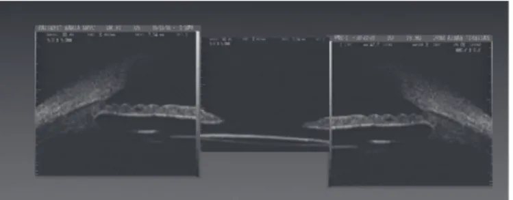
Figure 3. Ultrasound biomicroscopy showing the original lens in the capsular bag and the single-piece PMMA IOL, used for secondary piggyback, in the posterior chamber, with support in the ciliary sulcus Figure 2. Ultrasound biomicroscopy showing the intraocular lens in the capsular bag and a large posterior chamber
some reason, the haptic is outside the capsular bag, friction with the posterior surface of the iris or even with the ciliary body can cause complications such as uveitis, glaucoma and haemorrhage(5).
Therefore, their use is absolutely contraindicated for sec-ondary piggyback in the ciliary sulcus. Three-piece hydrophobic acrylic or silicone lenses with optic/haptic angulation are suit-able for this purpose. More recently, foldsuit-able lenses have been developed specifically for secondary piggyback in the ciliary sulcus: The single-piece hydrophilic acrylic IOL called Sulcoflex™, manufactured by Rayner, and the three-piece lens with a silicone optic and PMMA haptics called Add-On™, manu-factured by HumanOptics, with monofocal, both with toric and bifocal versions. However, the purpose of this retrospective study was to show that old single-piece PMMA lenses with an oval optic were useful in secondary piggyback for correcting refractive surprises after phacoemulsification.
Calculating the dioptric power of the second IOL is ex-tremely simple (dioptric power of the IOL = spherical lent of myopic surprise x 1, or dioptic power = spherical equiva-lent of hyperopic surprise x 1.5). This formula proved to be very accurate, as shown in Table 1.
postop-11 Secondary piggyback with PMMA IOL for the correction of refractive surprise after phacoemulsification. Long-term results of 20 cases
Rev Bras Oftalmol. 2013; 72 (1): 8-11 eratively, as it does not require the surgical wound to heal. Also,
the original incision can be used for implantation of the second IOL in the ciliary sulcus.
It is worth noting that secondary piggyback in the cili-ary sulcus is indicated only when the first IOL is implanted en-tirely within the capsular bag. In this situation, the posterior cham-ber is large, especially in high myopic eyes. This space between the anterior capsule and the posterior surface of the iris is well suited for implantation of the second IOL.
Ultrasound biomicroscopy is very useful in assessing the posterior chamber before (Figure 2) and after (Figure 3) sec-ondary piggyback in the ciliary sulcus.
Implantation of the second IOL within the capsular bag should be avoided. This technique is much more difficult and un-predictable than the secondary implantation in the ciliary sulcus. Moreover, interlenticular opacification could occur, especially when both IOLs are made of hydrophobic acrylic(6).
In secondary piggyback in the ciliary sulcus, an angula-tion should exist between the optic and haptic parts of the sec-ond IOL to avoid undesirable contact with the posterior surface of the iris and reduce the possibility of pupillary capture. Such contact causes pigment dispersion which can progress to sec-ondary glaucoma(7). Iritis and cystoid macular oedema are other
possible complications. Pupillary deviation can also occur when the haptics of the IOL are supported on the base of the iris and not in the ciliary sulcus. None of these complications were ob-served in this study.
It is also important to ensure that the piggyback IOL does not have an exaggerated size or angulation, as such a lens would cause posterior axial displacement of the primary IOL, thus caus-ing hyperopia as the final refractive outcome.
The PMMA IOLs used in all 20 cases of this study are much more easily available and less costly than foldable IOLs, especially when an unusual dioptric power is needed for the pig-gyback, such as -7.50 D (case 20).
Due to the characteristics of the PMMA IOLs used in this study, they should be implanted only within the capsular bag, otherwise there is a high probability of postoperative decentralisation. However, such IOLs provide stable fixation and excellent centralisation when implanted in the ciliary sulcus, as observed in all 20 cases of this study, even after several years of follow-up in some cases. Because these are hard IOLs, it advis-able to implant them using a scleral incision with a sclero-cor-neal tunnel in order to minimise the induction of astigmatism. In fact, this type of incision does not require suture as it does not induce clinically-significant astigmatism, as observed in this study. However, for safety reasons, in some cases two isolated mono-nylon 10-0 stitches were used on each side of the scleral incision. The astigmatism induced by these sutures was minimal and re-gressed over time.
Foldable lenses are obviously considered more appro-priate for secondary piggyback in the ciliary sulcus, as they can
Autor correspondente:
Fernando Cançado Trindade
Rua Manaus, nº 595
CEP 30150350 - Belo Horizonte (MG), Brasil
[email protected]
be implanted through the same incision of the original phacoemulsification. However, such lenses are not always easily available, especially when a dioptric power higher than -5.00 di-opters is needed (cases 2, 5 and 20).
The implantation of phakic posterior chamber lenses such as ICL™ is another alternative to secondary piggyback to correct refractive surprises. However, such lenses are not yet manufactured in low diopter and have a much higher cost.
In this study no complications were observed during the secondary piggyback procedure or in the immediate or late post-operative period. All patients were fully satisfied with the final visual outcome.
C
ONCLUSIONThe use of single-piece oval PMMA IOLs for secondary piggyback in the ciliary sulcus is a safe and effective procedure for correcting refractive surprises after phacoemulsification.
R
EFERENCES1. Singer JA. Frown incision for minimizing induced astigmatism after small incision cataract surgery with rigid optic intraocular lens im-plantation. J Cataract Refract Surg. 1991;17 Suppl:677-88.
2. Micheli T, Cheung LM, Sarma S, Assaad NN, Guzowski M, Francis IC, et al. Acute haptic-induced pigmentary glaucoma with an AcrySof intraocular lens. J Cataract Refract Surg. 2002;28(10):1869-72. 3 LeBoyer RM, Werner L, Snyder ME, Mamalis N, Riemann CD,
Augsberger JJ. Acute haptic-induced ciliary sulcus irritation associ-ated with single-piece AcrySof intraocular lenses. J Cataract Refract Surg. 2005;31(7):1421-7.
4 Rajak SN, Bahra A, Aburn NS, Warden NJ, Mossman SS. Recurrent anterior chamber hemorrhage from an intraocular lens simulating amaurosis fugax. J Cataract Refract Surg. 2007;33(8):1492-3. 5. Trindade FC. Haptic-induced recurrent vitreous hemorrhage and
in-creased intraocular pressure with a hydrophobic acrylic intraocular lens. J Cataract Refract Surg. 2009;35(2):399-402.
6. Gayton JL, Apple DJ, Peng Q, Visessook N, Sanders V, Werner L, et al. Interlenticular opacification: clinicopathological correlation of a com-plication of posterior chamber piggyback intraocular lenses. J Cata-ract RefCata-ract Surg. 2000;26(3):330-6.