https://doi.org/10.1590/0004-282X20170158
ARTICLE
Integrity of white matter structure is related
to episodic memory performance in the
low-educated elderly
Memória e integridade da substância branca em idosos de baixa escolaridade
Elisa de Paula França Resende1, Fernanda Freire Tovar-Moll2, Fernanda Meireles Ferreira2, Ivanei Bramati2,
Leonardo Cruz de Souza1, Karoline Carvalho Carmona1, Henrique Cerqueira Guimarães1, Viviane Amaral
Carvalho1, Maira Tonidandel Barbosa1, Paulo Caramelli 1
Memory decline occurs in normal aging and is also a core feature of the most common cause of dementia, Alzheimer’s disease1. As the prevalence of this disorder and other forms
of dementia is higher in developing countries2, it is
impor-tant to explore the neural correlates of episodic memory in the setting of low education, a situation extremely common among the elderly population in low- and middle-income
countries. Hence, understanding the anatomic substrate of memory performance, especially in vulnerable individuals, can help to develop strategies to prevent cognitive decline in this scenario.
Apart from the hippocampus and the medial tempo-ral lobe structures, well-established episodic memory corti-cal correlates3, recent studies have demonstrated that white
1Universidade Federal de Minas Gerais, Faculdade de Medicina da Belo Horizonte, Departamento de Clínica Médica,Grupo de Pesquisa em Neurologia
Cognitiva e do Comportamento, Belo Horizonte MG, Brasil;
2IDOR - Instituto D’Or de Pesquisa e Ensino, Rio de Janeiro RJ, Brasil.
Correspondence: Paulo Caramelli; Faculdade de Medicina da Universidade Federal de Minas Gerais; Av. Prof Alfredo Balena, 190 / sala 246; 30130-100 Belo Horizonte MG, Brasil. ; E-mail: [email protected]
Conflict of interest: There is no conflict of interest to declare.
Support: CNPq, Fundação de Apoio à Pesquisa de Minas Gerais and Instituto Hermes Pardini. Paulo Caramelli is funded by CNPq (bolsa de produtividade em pesquisa).
Received 26 April 2017; Received in final form 02 August 2017; Accepted 11 August 2017.
ABSTRACT
The low-educated elderly are a vulnerable population in whom studying the role of white matter integrity on memory may provide insights for understanding how memory declines with aging and disease. Methods: Thirty-one participants (22 women), 23 cognitively healthy and eight with cognitive impairment-no dementia, aged 80.4 ± 3.8 years, with 2.2 ± 1.9 years of education, underwent an MRI scan with diffusion tensor imaging (DTI) acquisition. We verified if there were correlations between the performance on the Brief Cognitive Screening Battery (BCSB) and the Rey Auditory Verbal Learning Test (RAVLT) with DTI parameters. Results: The BCSB delayed recall task correlated with frontotemporoparietal connection bundles, with the hippocampal part of the cingulum bilaterally and with the right superior longitudinal fasciculus. The RAVLT learning and delayed recall scores also correlated with the hippocampal part of the cingulum bilaterally. Conclusions: Although preliminary, our study suggests that the integrity of white matter frontotemporoparietal fasciculi seems to play a role in episodic memory performance in the low-educated elderly. This finding opens opportunities to study potential targets for memory decline prevention in vulnerable populations.
Keywords: episodic memory; aging; white matter; health vulnerability.
RESUMO
Idosos de baixo nível educacional representam uma população vulnerável em que o estudo do papel da integridade da substância branca na memória pode revelar como essa declina no envelhecimento. Métodos: Trinta e um indivíduos (22 mulheres), sendo 23 cognitivamente saudáveis, oito com comprometimento cognitivo não demência, 80,4 ± 3,8 anos de idade e 2,2 ± 1,9 anos de escolaridade, foram submetidos à RM com imagem de tensor de difusão, cujos parâmetros foram correlacionados com a Bateria Cognitiva Breve (BCSB) e o Teste Auditivo Verbal de Rey (RAVLT). Resultados: A evocação tardia da BCSB correlacionou-se com fascículos de conexão frontotemporoparietal, fascículo longitudinal superior direito e cíngulo parte hipocampal bilateral, sendo que esse último também correlacionou com o RAVLT (aprendizado e evocação tardia). Conclusão: Apesar de preliminar, nosso estudo sugere que a integridade da substância branca parece ser importante para a memória em idosos de baixa escolaridade, achado que revela alvo potencial na prevenção do seu declínio em populações vulneráveis.
matter also plays a crucial role. Previous studies have shown that memory function was associated with white matter integrity in the inferior and superior longitudinal fasciculi, the posterior and anterior cingulum4 the uncinate fasciculus,
the dorsal cingulum bundle5 and the hippocampal part of the
cingulum6. However, these studies have explored the
corre-lation between episodic memory and white matter integrity in highly-educated individuals, selected under strict crite-ria from research centers5,7. Hence, the role of white matter
integrity on episodic memory in community-dwelling elderly with a low educational level remains to be explored.
Advanced neuroimaging techniques, such as difusion
tensor imaging (DTI) acquired through magnetic reso-nance imaging (MRI), have allowed the study of the integ-rity of white matter tracts with fair precision8. his method
is based on the anisotropic property of water molecules, characterized by nonrandom movement when constricted by structures such as the neuron membrane and the myelin sheath. By acquiring a DTI image, it is possible to detect these movements and to establish the directionality using
diagonalization to calculate the vectors. hereafter, these
vectors can be used to calculate measures that correlate with white matter integrity, such as the fraction anisotropy
(FA) and the mean difusivity (MD). he former relects the
longitudinal directionality of the water molecules’ move-ment, and reduces with white matter microstructural integ-rity disruption9. Conversely, the latter measure relects the
radial directionality, which increases with white matter
lesions. hese two parameters can be correlated with cogni -tive performance, enabling us to make inferences about the role of the integrity of white matter on cognitive paradigms, such as episodic memory.
Although the relationship between white matter integrity and episodic memory has been explored in highly-educated individuals, in this study we used DTI to explore the role of white matter integrity on episodic memory performance
in a low-educated elderly sample. A high magnetic ield10
and an advanced post-processing approach – whole-brain tract-based spatial statistics – were used to generate a reli-able interpretation of multiple DTI analyses11. We
hypoth-esized that, in low-educated individuals, episodic memory performance would correlate with the white matter integ-rity of connection bundles between the medial temporal lobe and frontoparietal regions, similar to that which happens in highly-literate individuals.
METHODS
Participants
he participants were selected from the Pietà study,
a community-based investigation on brain aging conducted in Caeté (Minas Gerais state), Brazil12. Six hundred thirty-nine
individuals, corresponding to 51.1% of the city population
aged 75+ years were evaluated. he Ethics Committee of the
Federal University of Minas Gerais approved this study and all participants or their legally-authorized representatives
provided written informed consent. he clinical, functional,
neurological and psychiatric status of the participants were
established and they were classiied into three cognitive per -formance categories: cognitively healthy, cognitive impair-ment-no dementia, and dementia. For this study purpose, we included both cognitively healthy and cognitive impairment-no dementia individuals to improve the range of performance in the memory tests, allowing the detection of white matter micro structural changes. Because it was a population-based study, the cognitive impairment-no dementia concept was used instead of mild cognitive impairment. Individuals were considered cognitive impairment-no dementia when, regard-less of cognitive complaints by the patient or the informant, objective cognitive impairment was detected in the cognitive assessment13. Demented individuals were excluded from this study because our primary aim was to analyze the inluence
of white matter integrity on cognitive performance in indi-viduals with preserved functional status.
Cognitive assessment
Participants were evaluated using the Mini-Mental
State Evaluation (MMSE)14 and two episodic memory tests,
namely the memory test from the Brief Cognitive Screening Battery (BCSB)15 and the Rey Auditory Verbal Learning Test
(RAVLT)16. he BCSB memory test evaluates both visual and
verbal memory. It consists of the presentation of 10 simple
drawings that are irst identiied and named. Immediately
after, the participant is asked to recall the drawings
(inci-dental encoding). he sheet of paper is then shown again for 30 seconds, the patient is asked to memorize the igures, and recall is requested immediately after. he latter procedure is
performed twice, leading to a score for immediate memory and learning. After that, interference activities are performed,
namely, the category luency test (animals/minute) and the clock drawing test. hen, the participant is asked to recall as
many items as possible in a delayed recall task. Finally, the participant is confronted with the 10 previously-presented drawings along with 10 distractors, and is asked to recognize the initial drawings. On the other hand, the RAVLT essentially assesses verbal memory. It consists of 15 words read aloud for
ive consecutive trials (List A), followed by a free-recall test (A1 through A5). After the ifth trial, a new interference list
of 15 words is presented (List B) followed by a free-recall test of that list (B1). After that, a free-recall of List A is requested
(A6). After a 30-minute delay period illed with distractor
Neuroimaging data acquisition and processing
A random subsample of individuals from the Pietà study (n = 200) was asked to take part in the MRI examination. he efective sample used in this study (n = 49) included partici -pants with an acceptable quality of the DTI images acquired
in a 3 Tesla Philips Achieva scanner. he difusion-weighted
images were obtained in the axial plane with a single-shot, spin-echo, echo planar sequences in the axial plane
(TR/TE = 8000/65 ms, FOV = 240 mm, matrix = 96x96
(reconstructed 128x128), slice thickness = 2.0 mm with 1.99
mm gap between slices). Difusion sensitization gradients
were applied in 32 non-collinear directions, with a b factor
of 1000 sec/mm2. Cognitive testing was completed within
nine months of the MRI scans. An experienced radiologist, blind to the clinical diagnoses, calculated the Fazekas score17,
a semi-quantitative visual scale for white matter lesions, which ranges from 0-6, with 0 being no lesions and 6 severe
and difuse lesions, which was used to assess the partici -pants’ white matter lesions burden.
Diffusion tensor imaging post-processing
All difusion images were inspected for artifacts. Non-difusion and difusion images were co-registered to
correct for movement artifacts and eddy current distortion
efects on the Echo Planar Imaging readout18. he difusion tensor for each voxel was calculated using multivariate itting
and diagonalization (FSL 4.0 FMRIB software)19. Fractional
anisotropy and MD images were brain-extracted and regis-tered to the Montreal Neurological Institute space standard (MNI152) using constrained nonlinear registration (Image Registration Toolkit)20. he derived FA and MD data were fur
-ther analyzed using a priori regions of interest (ROI) analyses and voxelwise whole-brain tract-based spatial statistics11.
Whole-brain voxelwise analysis
Whole-brain voxelwise statistical analyses of FA and MD were conducted to assess global correlations between those values and memory (BCSB delayed recall task, RAVLT learn-ing and 30 minutes delayed recall uslearn-ing tract-based spa-tial statistics (FMRIB Software Library, FSL)21, controlling for age and education. he results were considered signii
-cant at p < 0.05, using cluster-based hreshold-Free Cluster Enhancement fully corrected for multiple comparisons Family Wise Error Rate (FWE). he threshold-skeletonized
resulting image was thickened for better visualization.
Regions of interest analysis
A Johns Hopkins University DTI-MRI atlas of human white matter22 was used to determinate iber tract orien
-tation, and the ROIs, and was considered the reference for anatomical labels, which were checked by an experienced
investigator (FMF) afterward. he ROIs for memory corre -lation were placed along the hippocampal part of the cingu-lum, which runs along the surface of the corpus callosum
and hippocampal parts, the superior longitudinal fascic-ulus, which connects the frontal, temporal, parietal and occipital lobes, and the uncinate fasciculus, which connects medial prefrontal and anterior temporal brain regions.
hese fasciculi were selected because they are either part
of the limbic system (hippocampal part of the cingulum and uncinate), which is classically associated with memory, or because they are important for attention and working visuospatial memory (superior longitudinal fasciculus)23.
Multiple regression analyses including age and education as covariates were carried out to investigate associations between the FA and MD values and memory performance tested by the RAVLT A1-A5 sum of scores, RAVLT A7 scores and BCSB delayed recall task score.
Statistical analyses of demographic and clinical variables
Statistical analyses were performed using the SPSS 16.0. Kolmogorov-Smirnov tests to assess parametric distribution.
Group diferences for demographic and clinical variables with
parametric distribution were undertaken with the Student’s t-test for age, RAVLT A1-A5 sum of scores, RAVLT A7 scores
and the BCSB memory test delayed recall task score. he
Mann-Whitney test was used for non-parametric
distribu-tion variables: years of educadistribu-tion, MMSE and Fazekas scores.
Associations between categorical variables were investigated
with Chi-square and exact Fisher’s test. he level of signii -cance (α) was set at 0.05, two-tailed, for all statistical tests.
RESULTS
Demographical and clinical results
Images were available for whole analyses for 31 par-ticipants (after excluding artifacts), of whom 22 (70%) were women, with a mean age of 80.4 ± 3.8 years and
2.2 ± 1.9 years of education. Eight (25%) participants were classiied as cognitive impairment-no dementia. he whole group’s mean score on the MMSE was 22.3 ± 4.3. he mean
performance on the delayed recall task from the BCSB
memory test was 7.1 ± 1.7 (maximum score = 10). he learn -ing composite from the RAVLT (A1-A5) mean score was 23.7 ± 8.8 (maximum score = 75) and the delayed recall task (A7) mean score was 4.3 ± 3.5 (maximum score = 15). Global white matter injury accessed through the Fazekas scale was moderate, with a mean score of 2.7 ± 1.6.
Voxelwise whole-brain DTI findings
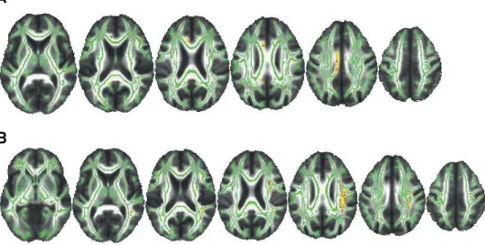
A signiicant negative correlation was found between the
BCSB memory test delayed recall task scores and the MD
values (p = 0.020) (Figure 1). he correlated fasciculi were difusely distributed and represented connections between frontal, temporal and parietal lobes. here was no signiicant
DTI regions of interest analyses
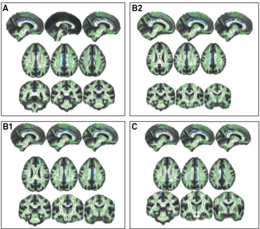
A signiicant positive correlation emerged between the
BCSB memory test delayed recall task and the FA values in the left hippocampal part of the cingulum (p = 0.033) (Figure 2A), and right superior longitudinal fasciculus (p = 0.022) (Figure 2B), and a negative correlation between the BCSB memory test and the MD values in the hippocam-pal part of the cingulum bilaterally (p = 0.025) (Figure 3A).
As well, a signiicant negative correlation was found between
the RAVLT A1-A5 sum of scores and the MD values in the hip-pocampal part of the cingulum bilaterally (p = 0.010 on the right side and p = 0.022 on the left side) (Figure 3B). Finally, the RAVLT A7 score had a positive correlation with the FA val-ues in the left hippocampal part of the cingulum (p = 0.048) and negative correlation with the MD values in the right hippocampal part of the cingulum (p = 0.036) (Figure 3C).
No signiicant correlation was observed between the RAVLT
A1-A5 sum of scores and RAVLT A7 scores and DTI metrics in the superior longitudinal fasciculus. Moreover, no signif-icant correlation emerged between both memory tests and the uncinate fasciculus in ROI analyses.
DISCUSSION
he study of white matter integrity opens a window to
understanding the neural basis of cognition, as connectivity is a major factor that supports cognitive functioning24. In the
present study, we performed DTI analyses to investigate the neural basis of episodic memory in elderly individuals with
low educational attainment. We found signiicant correla -tions between the BCSB, a visual-verbal episodic memory test, and the integrity of frontotemporoparietal connection bundles on whole-brain analyses and with the superior longi-tudinal fasciculus on ROI analyses. Moreover, the BCSB and
RAVLT signiicantly correlated with the hippocampal part of
the cingulum integrity bilaterally on ROI analyses.
Episodic memory consists of conscious storage and
retrieval of information about previous events, which enables a person to remember the past25. Traditionally, since
the seminal observations relating severe amnesia due to bilateral hippocampectomy26, its function has been
attrib-uted to the structure and function of the hippocampus and medial temporal lobe structures. However, research on the
A
B
Positive correlation between the Brief Cognitive Screening Battery delayed task score and fraction anisotropy values in the left cingulum (p = 0.033) (A), and right superior longitudinal fasciculus (p = 0.022) (B). All analyses were corrected for multiple comparisons across space (Family Wise Error Rate) using threshold-free cluster enhancement. Each panel shows the significant voxel clusters superimposed on the mean fractional anisotropy map in axial slices. Images are displayed in neurological convention (right cerebral hemisphere = right side).
Figure 2. Correlation between fractional anisotropy and memory in region of interest analyses.
Whole-brain track-based correlation between the mean diffusivity values and the Brief Cognitive Screening Battery memory test delayed recall task score (p = 0.020 corrected for multiple comparisons across space (Family Wise Error Rate) using threshold-free cluster enhancement). Each panel shows the significant voxel clusters superimposed on the mean diffusivity map in axial slices. Images are displayed in neurological convention (right cerebral hemisphere = right side).
neural basis of memory performance demonstrated that other structures besides the Papez circuit are implicated in memory performance such as the parietal regions, precu-neus, posterior cingulate and retrosplenial cortex27. Apart
from cortical involvement, white matter structures also seem to be critical for memory functioning, as lesions in frontoparietal and frontotemporal fasciculi, including the hippocampal part of the cingulum6 and parahippocampal
gyrus28, may damage the connections between the
prefron-tal cortex and posterior regions, leading to memory impair-ment5,29. he present study suggests a possible correlation
between a visual-verbal episodic memory test with the integrity of white matter bundles in the brain, especially in the temporal regions and their connections with fron-totemporal structures, according to the results from the voxelwise analysis. Goldstein et al. showed a similar ind -ing, correlating a visual memory test with white matter integrity in temporal regions in a highly-educated sample30.
Furthermore, both the visual-verbal and the verbal mem-ory correlated with the integrity of the hippocampal part of
the cingulum in the ROI analyses. his tract includes ibers
that link the hippocampal formation with the cingulate cor-tex, both parts of the default mode network. Connections
between those areas have been demonstrated to be impor-tant for mnemonic processing23 and episodic memory
per-formance4,6. he present study indings suggest the hip
-pocampal part of the cingulum might also play a role in memory performance in low-educated individuals. In con-trast, the right superior longitudinal fasciculus seems to be more relevant in the visual-verbal memory processing,
because, in ROI analysis, we only found a signiicant cor -relation between the BCSB and the right superior longitu-dinal fasciculus integrity. Possibly the fact that the supe-rior longitudinal fasciculus has been associated with visual processing23, and the strong visual component of the BCSB test, may make them responsible for this inding in only the
right hemisphere.
he population-based setting and the very low education
attainment of the participants allowed the exploration of the relationships between microstructural white matter integrity and episodic memory performance in this context, which is the reality in developing countries. However, the present study has important limitations. We did not control our anal-yses for gender and white matter lesions burden. Considering that our sample consisted mostly of women with moderate levels of white matter lesions assessed by the Fazekas scale,
A
B2
C
B1
Negative correlation between the Brief Cognitive Screening Battery delayed recall task score and the mean diffusivity values in the cingulum bilaterally (p= 0.025) (A). Negative correlations between the Rey Auditory Learning Verbal Test (RAVLT) A1-A5 sum of scores and the mean diffusivity values in the right (p = 0.010) (B1) and left cingulum (p =0.022) (B2). Negative correlation between the RAVLT A7 score and the mean diffusivity values in the right cingulum (p =0.036) (C). All analyses were corrected for multiple comparisons across space (Family Wise Error Rate) using threshold-free cluster enhancement. Each panel shows the significant voxel clusters superimposed on the mean fractional anisotropy map in axial slices. Images are displayed in neurological convention (right cerebral hemisphere = right side).
our indings may apply mainly to individuals with those char -acteristics. We were unable to perform DTI assessment on all
individuals submitted to MRI, which led to a small inal sam -ple size. Although relatively common in population-based
studies, the drop of in the sample size remained an impor -tant limitation for more assertive conclusions. Moreover,
because the performance on the RAVLT is inluenced by the educational level, there was a loor efect on its performance in the entire sample, with implication on the negative ind -ings regarding the whole-brain analysis. Although important for memory performance, we could not assess the fornix in our study because it is not part of the automated atlas we
used and it is diicult to delineate this structure when using
the tract-based spatial statistics approach. Furthermore, the
diferent sensitivities of the FA and MD parameters led to few overlaps between the FA and MD indings, which has been
described in previous neuroimaging studies7 and did not
impact on the interpretation of the results.
One of the main goals of aging research is to understand the causes and the pathophysiology of cognitive decline, especially within the memory domain. Our study suggests that the integrity of white matter bundles that connect fron-totemporoparietal regions is important for memory per-formance in the low-educated elderly, similar to that in the
highly-educated elderly. Moreover, we identiied possible
white matter neural correlates of the BCSB test. Because the correlates were consistent with bundles important for
mem-ory processing, we believe that our indings reinforce the role
of the BCSB test in the clinical setting as an important tool in the diagnostic workup for Alzheimer’s disease in individuals
with diferent educational attainments.
he vast majority of scientiic studies are conducted in
high-income countries, where the population’s characteristics frequently lack similarity with the low- and middle-income countries, especially considering the educational level and the socioeconomic status. Although the white matter neural correlates of memory have been studied before, most stud-ies were conducted in developed countrstud-ies4,5,6. Our results, although preliminary, contribute to the ield of under -standing memory processing in the low-educated elderly. We acknowledge that further studies in a broader sample are
necessary to conirm our indings and the next step will be to identify the inluence of progressive educational strata on the episodic memory white matter neural correlates. he pres
-ent indings may contribute to future studies in dem-entia
prevention, since keeping the white matter integrity in fron-totemporoparietal connection bundles may have a role in memory processing in the low-educated elderly. Controlling for cardiovascular risk factors to keep the integrity of brain connections might be an important intervention tool to be
tested in future studies, to analyze its speciic role in prevent -ing memory decline in the elderly.
Acknowledgments
he authors thank the Pietà Study Group and the D’Or Institute for Research and Education for data collection and
analyses. We are also very grateful for the elderly participants
of the Pietà study who dedicated their time and energy to our
research. We specially thank Luciana Costa Silva for the anal-ysis of the Fazekas score in all images.
References
1. Dubois B, Feldman HH, Jacova C, Cummings JL, Dekosky ST, Barberger-Gateau P et al. Revising the definition of Alzheimer’s disease: a new lexicon. Lancet Neurol. 2010;9(11):1118-27. https://doi.org/10.1016/S1474-4422(10)70223-4
2. Nitrini R, Bottino CM, Albala C, Custodio Capuñay NS, Ketzoian C, Llibre Rodriguez JJ et al. Prevalence of dementia in Latin America: a collaborative study of population-based cohorts. Int Psychogeriatr. 2009;21(4):622-30. https://doi.org/10.1017/S1041610209009430
3. Zola-Morgan S, Squire LR. Neuroanatomy of memory. Annu Rev Neurosci. 1993;16(1):547-63. https://doi.org/10.1146/annurev.ne.16.030193.002555
4. Kantarci K, Senjem ML, Avula R, Zhang B, Samikoglu AR, Weigand SD et al. Diffusion tensor imaging and cognitive function in older adults with no dementia. Neurology. 2011;77(1):26-34. https://doi.org/10.1212/WNL.0b013e31822313dc
5. Lockhart SN, Mayda AB, Roach AE, Fletcher E,
Carmichael O, Maillard P et al. Episodic memory function is associated with multiple measures of white matter integrity in cognitive aging. Front Hum Neurosci. 2012;16(6):56. https://doi.org/10.3389/fnhum.2012.00056
6. Irish M, Hornberger M, Wahsh SE, et al. Grey and White matter correlates of recent and remote autobiographical memory retrieval: insights from the dementias. PLoS ONE. 2014;9(11):e113081. https://doi.org/10.1371/journal.pone.0113081
7. Metzler-Baddeley C, Jones DK, Belaroussi B, Aggleton JP, O’Sullivan MJ. Frontotemporal connections in episodic memory and aging: a diffusion MRI tractography study. J Neurosci. 2011;31(37):13236-45.
https://doi.org/10.1523/JNEUROSCI.2317-11.2011
8. Ciccarelli O, Werring DJ, Wheeler-Kingshott CA, Barker GJ, Parker GJ, Thompson AJ et al. Investigation of MS normal-appearing brain using diffusion tensor MRI with clinical correlations. Neurology. 2001;56(7):926-33. https://doi.org/10.1212/WNL.56.7.926
9. Beaulieu C. The basis of anisotropic water diffusion in the nervous system: a technical review. NMR Biomed. 2002;15(7-8):435-55. https://doi.org/10.1002/nbm.782
10. Lavdas I, Miquel ME, McRobbie DW, Aboagye EO. Comparison between diffusion-weighted MRI (DW-MRI) at 1.5 and 3 tesla: a phantom study. J Magn Reson Imaging. 2014;40(3):682-90. https://doi.org/10.1002/jmri.24397
11. Smith SM, Jenkinson M, Johansen-Berg H, Rueckert D, Nichols TE, Mackay CE et al. Tract-based spatial statistics: voxelwise analysis of multi-subject diffusion data. Neuroimage. 2006;31(4):1487-505. https://doi.org/10.1016/j.neuroimage.2006.02.024
13. Caracciolo B, Palmer K, Monastero R, Winblad B, Bäckman L, Fratiglioni L. Occurrence of cognitive impairment and dementia in the community: a 9-year-long prospective study. Neurology. 2008;70(19 Pt 2):1778-85. https://doi.org/10.1212/01.wnl.0000288180.21984.cb
14. Brucki SM, Nitrini R, Caramelli P, Bertolucci PH, Okamoto IH. [Suggestions for utilization of the mini-mental state examination in Brazil]. Arq Neuropsiquiatr. 2003;61(3B):777-81. Portuguese. https://doi.org/10.1590/S0004-282X2003000500014
15. Nitrini R, Caramelli P, Herrera Júnior E, Porto CS,
Charchat-Fichman H, Carthery MT et al. Performance of illiterate and literate nondemented elderly subjects in two tests of long-term memory. J Int Neuropsychol Soc. 2004;10(4):634-8. https://doi.org/10.1017/S1355617704104062
16. Malloy-Diniz LF, Lasmar VA, Gazinelli LS, Fuentes D, Salgado JV. The Rey Auditory-Verbal Learning Test: applicability for the Brazilian elderly population. Rev Bras Psiquiatr. 2007;29(4):324-9. https://doi.org/10.1590/S1516-44462006005000053
17. Wahlund LO, Barkhof F, Fazekas F, Bronge L, Augustin M, Sjögren M et al. A new rating scale for age-related white matter changes applicable to MRI and CT. Stroke. 2001;32(6):1318-22. https://doi.org/10.1161/01.STR.32.6.1318
18. Woods RP, Grafton ST, Holmes CJ, Cherry SR, Mazziotta JC. Automated image registration: I. General methods and intrasubject, intramodality validation. J Comput Assist Tomogr. 1998;22(1):139-52. https://doi.org/10.1097/00004728-199801000-00027
19. Jiang H, Zijl PC, Kim J, Pearlson GD, Mori S. DtiStudio: resource program for diffusion tensor computation and fiber bundle tracking. Comput Methods Programs Biomed. 2006;81(2):106-16. https://doi.org/10.1016/j.cmpb.2005.08.004
20. Pajevic S, Pierpaoli C. Color schemes to represent the orientation of anisotropic tissues from diffusion tensor data: application to white matter fiber tract mapping in the human brain. Magn Reson Med. 1999;42(3):526-40. https://doi.org/10.1002/(SICI)1522-2594(199909)42:3<526::AID-MRM15>3.0.CO;2-J
21. Smith SM, Jenkinson M, Woolrich MW, Beckmann CF, Behrens TE, Johansen-Berg H et al. Advances in functional and structural MR image analysis and implementation as FSL. NeuroImage. 2004;23(Suppl 1):S208-19. https://doi.org/10.1016/j.neuroimage.2004.07.051
22. Hua K, Zhang J, Wakana S, Jiang H, Li X, Reich DS et al. Tract probability maps in stereotaxic spaces: analyses of white matter anatomy and tract-specific quantification. NeuroimageNeuroimage. 2008;39(1):336-47. https://doi.org/10.1016/j.neuroimage.2007.07.053
23. Catani M, Schotten MT. A diffusion tensor imaging tractography atlas for virtual in vivo dissections. Cortex. 2008;44(8):1105-32. https://doi.org/10.1016/j.cortex.2008.05.004
24. Mesulam M. Imaging connectivity in the human cerebral cortex: the next frontier? Annals of Neurology. 2005;57(1):5-7. https://doi.org/10.1002/ana.20368
25. Tulving E. Episodic memory: from mind to brain. Annu Rev Psychol. 2002;53(1):1-25. https://doi.org/10.1146/annurev.psych.53.100901.135114
26. Scoville WB, Milner B. Loss of recent memory after bilateral hippocampal lesions. J Neurol Neurosurg Psychiatry. 1957;20(1):11-21. https://doi.org/10.1136/jnnp.20.1.11
27. Cabeza R, Ciaramelli E, Olson IR, Moscovitch M. Parietal cortex and episodic memory: an attentional account. Nat Rev Neurosci. 2008;9(8):613-25. https://doi.org/10.1038/nrn2459
28. Zhuang L, Sachdev PS, Trollor JN, Kochan NA, Reppermund S, Brodaty H et al. Microstructural white matter changes in cognitively normal individuals at risk of amnestic MCI. Neurology.
2012;79(8):748-54. https://doi.org/10.1212/WNL.0b013e3182661f4d
29. Lee DY, Fletcher E, Martinez O, Ortega M, Zozulya N, Kim J et al. Regional pattern of white matter microstructural changes in normal aging, MCI, and AD. Neurology. 2009;73(21):1722-8. https://doi.org/10.1212/WNL.0b013e3181c33afb