354
DOI:
10.1590/0004-282X20160021
IMAGES IN NEUROLOGY
Diabetic hemichorea-hemiballismus with
nonketotic hyperglicemia: a rare cause of
hyperkinetic movement disorders
Hemibalismo-hemicoreia associada à hiperglicemia não-cetótica: uma rara causa de
distúrbios hipercinéticas do movimento
Lucas Giansante Abud
1,2, Thiago Giansante Abud
1,3, Rodolfo Mendes Queiroz
1, Giovanni Salton Pietroni
1,
Daniel Giansante Abud
4A 67-year-old woman was admitted with right
hemichorea-hemiballismus. Blood glucose: 831 mg/dl.
Magnetic resonance imaging (MRI) showed signal change in
the left striatum (Figures 1 and 2).
his entity is characterized by hyperintensity conined to
the striatum on T1-weighted MRI and contralateral hyperki
-netic movement disorders in diabetic patients (type 2) with
non-ketotic hyperglycemia
1. T2*-weighted gradient-echo MRI
can reveal low signal intensity related to petechial hemor
-rhage
2. Pathological studies demonstrated selective neuronal
loss, gliosis, reactive astrocytosis and hemorrhage
3.
he clinical symptoms usually improve markedly following
the correction of hyperglycemia. hus, the prompt recognition
of this potentially treatable disease is of paramount importance.
1Documenta, Hospital São Francisco, Divisão de Neurorradiologia, Ribeirão Preto SP, Brazil;
2Universidade de São Paulo, Hospital das Clínicas, Faculdade de Medicina de Ribeirão Preto, Divisão de Neurorradiologia Diagnóstica, Ribeirão Preto SP, Brazil; 3Universidade Federal de São Paulo, Departamento de Radiologia, São Paulo SP, Brazil;
4Universidade de São Paulo, Hospital das Clínicas, Faculdade de Medicina de Ribeirão Preto, Divisão de Neurorradiologia Intervencionista, Ribeirão Preto SP, Brazil.
Correspondence: Lucas Giansante Abud; Avenida Heráclito Fontoura Sobral Pinto, 751,, CEP: 14022-000, Ribeirão Preto SP, Brasil; E-mail: abud.lucas@gmail.com
Conflict of interest: There is no conlict of interest to declare.
Received 06 July 2015; Received in inal form 01 October 2015; Accepted 20 October 2015.
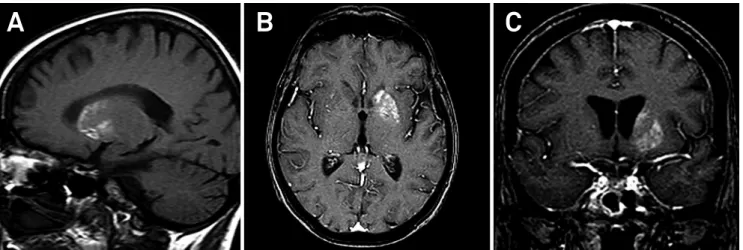
Figure 1.
A) Sagittal T1- weighted magnetic resonance imaging showing spontaneous diffuse high intensity of the left
striatum (caudate nucleus and putamen). B and C) There is no signiicant enhancement on axial and coronal T1-weighted MRI
after intravenous paramagnetic contrast administration.
355
Nome autores et al. Hemichorea-hemiballismus in diabetic hyperglycemia1. Lai PH, Tien RD, Chang MH, Teng MM, Yang CF, Pan HB et al. Chorea-Ballismus with nonketotic hyperglycemia in primary diabetes mellitus. AJNR Am J Neuroradiol. 1996;17(6):1057-64.
2. Suto Y, Mori M, Kagimoto H, Saito J. [A case of hemichorea with hyperglycemia presenting with low signal intensity in the striatum on T2*-weighted gradient-echo magnetic resonance imaging]. Rinsho Shinkeigaku. 2004;44(2):86-90. Japanese.
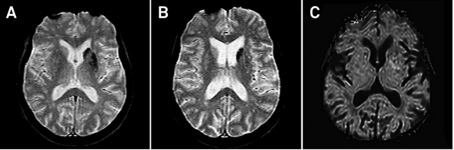
Figure 2.
A and B) Axial T2*-weighted gradient-echo MRI showing multiple conluent foci of hypointensities in left striatum
probably due to petechial hemorrhage. C) There is no signiicant signal change on axial diffusion-weighted image.
A
B
C
3. Abe Y, Yamamoto T, Soeda T, Kumagai T, Tanno Y, Kubo J et al. Diabetic striatal disease: clinical presentation, neuroimaging, and pathology. Intern Med. 2009;48(13):1135-41.