63
Sarcomatous degeneration of Paget’s disease in the calcaneus
Radiol Bras. 2009 Jan/Fev;42(1):63–65 Case Report • Relato de Caso
Sarcomatous degeneration of Paget’s disease
in the calcaneus: a case report*
Degeneração sarcomatosa de doença de Paget do calcâneo: relato de caso
Simone Berwig Matiotti1, Conrado Silva Tramunt1, Rogério Dias Duarte1, Rodrigo Dias Duarte1, Wolmir Lourenço Duarte1, Janine Bernardi Soder2
Neoplastic degeneration in Paget’s disease is a rare complication (approximately 1% of cases) and, despite the treatment, presents a poor prognosis. The authors report a case of a male, 82-year-old patient with long standing Paget’s disease who presented imaging findings of malignant degeneration in the calcaneus histopathologically diagnosed as sarcomatous degeneration.
Keywords: Paget’s disease; Sarcoma; Sarcomatous degeneration; Calcaneus; Osteitis deformans.
A degeneração maligna das lesões da doença de Paget é rara (cerca de 1% dos casos), sendo de mau prog-nóstico apesar do tratamento. Relatamos o caso de um paciente de 82 anos de idade, portador de doença de Paget há vários anos, em que se identificaram, nos exames de imagem, características de degeneração maligna no calcâneo, com anatomopatológico evidenciando degeneração sarcomatosa do osso.
Unitermos: Doença de Paget; Sarcoma; Degeneração sarcomatosa; Calcâneo; Osteitis deformans.
Abstract
Resumo
* Study developed at Fundação Saint Pastous, Clínica Serdil, Porto Alegre, RS, Brazil.
1. MDs, Radiologists, Clínica Serdil, Porto Alegre, RS, Brazil. 2. MD, Resident of Radiology at Fundação Saint Pastous, Clínica Serdil, Porto Alegre, RS, Brazil.
Mailing address: Dra. Simone Berwig Matiotti. Rua Felipe Camarão, 500/404, Bom Fim. Porto Alegre, RS, Brazil, 90035-140. E-mail: [email protected]
Received February 7, 2007. Accepted after revision August 14, 2007.
a new CT (Figure 3) were performed and demonstrated a significant increase in ra-diopharmaceutical uptake and subtle in-crease in bone volume, with cortical bone rupture on the lateroposterior surface of the calcaneus. Magnetic resonance imaging (MRI) demonstrated a volumetric increase, with heterogeneous signal intensity on all sequences, and some cystic and sclerotic areas. Cortical bone rupture and a mass in soft tissues of the lateral calcaneal region were observed, with peripheral contrast enhancement after gadolinium injection (Figure 4), and alterations suggesting
ma-Matiotti SB, Tramunt CS, Duarte RD, Duarte RD, Duarte WL, Soder JB. Sarcomatous degeneration of Paget’s disease in the calcaneus: a case report. Radiol Bras. 2009;42(1):63–65.
fected(1,3,4), calcaneal involvement, like in the present case, being extremely rare(6).
CASE REPORT
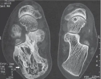
The present study reports the case of a male, 85-year-old patient with history of bone Paget’s disease for several years, and reporting pain and swelling in the right calcaneal region for three months. The first computed tomography (CT) study per-formed six years before (Figure 1) demon-strated characteristic findings of Paget’s disease. Bone scintigraphy (Figure 2) and
0100-3984 © Colégio Brasileiro de Radiologia e Diagnóstico por Imagem INTRODUCTION
Paget’s disease, firstly described by Sir James Paget in 1877 as osteitis deformans, is characterized by findings of a disturbed and extremely active bone remodeling caused by reactive osteoclastic and osteo-blastic activity, in a peculiar mosaic pat-tern(1,2).
The cause for this disease still remains unknown, the viral etiology theory being the most accepted among others such as genetic origin, parathormone-induced metabolic disorder, autoimmune disease, vascular disease, conjunctive disease tis-sue, or even neoplastic process(3–5).
Sarcomatous degeneration is rare, oc-curring in approximately 1% of cases of long-term disease activity(1). Osteosarcoma is the most frequently found histologic type of tumor (50–60% of cases). Most fre-quently, pelvis, hip or shoulder are
64
Matiotti SB et al.
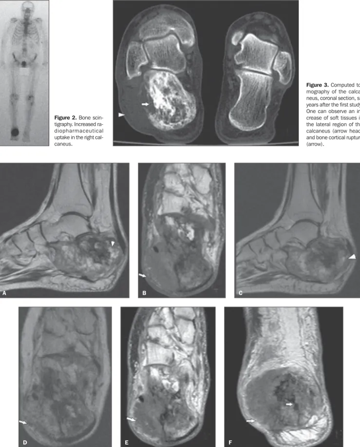
Radiol Bras. 2009 Jan/Fev;42(1):63–65 Figure 2. Bone
scin-tigraphy. Increased ra-diopharmaceutical uptake in the right cal-caneus.
Figure 3. Computed to-mography of the calca-neus, coronal section, six years after the first study. One can observe an in-crease of soft tissues in the lateral region of the calcaneus (arrow head) and bone cortical rupture (arrow).
Figure 4. Magnetic resonance imaging, T1-weighted, sagittal view (A) and axial view (B), and T2-weighted sequence, sagittal view (C) and axial view (D). Increase in calcaneal volume, with heterogeneous signal intensity and presence of some cystic and sclerotic areas (arrow heads). A soft tissue mass can be observed in the lateral region of the calcaneus (arrows). T1-weighted sequence after intravenous contrast injection, axial (E) and coronal (F) views demonstrate peripheral contrast-enhancement of the lesion (arrows).
A B C
65
Sarcomatous degeneration of Paget’s disease in the calcaneus
Radiol Bras. 2009 Jan/Fev;42(1):63–65 lignant degeneration. The patient was sub-mitted to biopsy of the lesion, whose result evidenced high-grade osteosarcoma.
DISCUSSION
Paget’s disease affects approximately 3–4% of the population aged above 40 years(1–3,7). Most frequently, the axial skel-eton (pelvis, spine and skull) is involved, but proximal long bones also can be fre-quently affected (25–35% of cases)(1,3,4,7). Involvement of other structures such as rips, fíbula, hand and feet bones, calcaneus and patella is not frequent(1,4,7). The poly-ostotic presentation represents 65-90% of cases. Involvement of an appendicular seg-ment is generally unilateral(1,3,7).
In most of cases, diagnosis can be reached by means of radiological examina-tion, presenting characteristic findings(2,8). These findings depend on the disease stage. At the osteolytic phase, radiolucent areas with well-defined margins are observed, with absence of areas of bone sclerosis. The mixed phase of the disease is characterized by the presence of gross and thickened tra-beculae, as well as cortical bone thicken-ing reflectthicken-ing osteoblastic activity. The blastic phase is characterized by the pres-ence of sclerotic areas and increased bone volume(2,3). Bone scintigraphy is a sensitive method, but poorly specific in the detection of hyperemia and osteoblastic activity, and can detect an increase in radionuclide up-take even before radiological findings be-come evident(3,7). Sometimes, the disease may be incidentally found at CT or MRI(9). Tomographic findings are similar to the radiographic ones, and trabecular thicken-ing can be better depicted by CT(2,3,8). Three patterns of bone marrow images can be identified at MRI. In most of cases the yel-low bone marrow presents normal signal intensity. In many cases the medullary space of the bone affected presents a larger amount of fat than a normal bone, which represents medullary atrophy. The volume of the medullary canal may be reduced by the thickening of the cortical layer(3). The second pattern identified presents hetero-geneous signal intensity on T1- and
T2-weighted sequences. On T1-T2-weighted se-quences, the bone marrow presents de-creased signal intensity, with intermingled foci of normal bone marrow which rules out the presence of malignant degeneration by the absence of masses. On T2-weighted sequences the bone marrow signal is het-erogeneously hyperintense(3,9). The third pattern is observed in the phase of de-creased blastic activity, with low bone marrow signal intensity on all the se-quences, corresponding to sclerosis. With the utilization of intravenous gadolinium as a contrast agent, a medullary enhancement can be observed, particularly at the most active phases of the disease(3).
Sarcomatous degeneration is rarely ob-served, occurring in approximately 1% of cases of long-term disease activity. The highest risk is observed in patients with polyostotic disease(1,3,4,7). Swelling and pain in the region affected are symptoms indicative of this condition(1,3). The most common histologic type of lesion is os-teosarcoma (50–60% of cases), besides cases of malignant fibrotic histiocytoma/ fibrosarcoma and chondrosarcoma(1,3,6,10). The prognosis for patients with sarcoma-tous degeneration is poor, with less than 10% for three-year disease-free survival after treatment(11). Metastases, particularly the pulmonary ones, are frequent(10). Typi-cal signs of sarcomatous degeneration are aggressive bone lysis, cortical destruction and presence of soft tissue masses(1,4,7). In most of cases, periosteal reaction is not observed(4). Neoplastic degeneration in bones affected by Paget´s disease may be hardly radiologically detected, requiring comparison with previous radiographies for identifying new osteolytic areas besides further studies by CT and MRI(3). MRI is superior to CT, allowing the visualization of replacement of bone marrow by tumor cells and cortical destruction in association with relatively large and infiltrating soft tissues mass. Sarcomatous degeneration is observed with intermediate signal intensity on T1-weighted images, with enhancement after gadolinium injection, and hyper-intense signal on T2-weighted images. Central necrosis is frequently found(1,3).
CONCLUSION
Malignant bone degeneration is rarely observed in Paget’s disease. However, in these cases, the main histologic types iden-tified are osteosarcoma (50-60%) and ma-lignant fibrotic histiocytoma/ fibrosarcoma (20–25%) and chondrosarcoma. Clinically, the findings that characterize malignant transformation are soft tissues swelling in the affected region, and radiologically, the presence of soft tissue mass and cortical bone destruction. In the suspicion of Paget’s disease, CT and MRI should be per-formed for findings characterization, even in bones were this disease is extremely rare, such as the calcaneus. Despite the CT su-periority for demonstrating cortical break-down and changes in the bone trabecular pattern, MRI also allows the identification of changes in the signal intensity produced by bone marrow and adjacent soft tissues.
REFERENCES
1. Resnick D, Niwayama G. Paget disease. In: Resnick D, editor. Diagnosis of bone and joint disorders. 4th ed. Philadelphia: Saunders; 2002. p. 1947–2000.
2. Greenspan A. Radiologia ortopédica. 3ª ed. Rio de Janeiro: Guanabara Koogan; 2001. 3. Smith SE, Murphey MD, Motamedi K, et al. From
the archives of the AFIP. Radiologic spectrum of Paget disease of bone and its complications with pathologic correlation. Radiographics. 2002;22: 1191–216.
4. Merkow RL, Lane JM. Paget’s disease of bone. Orthop Clin North Am. 1990;21:171–89.
5. Mirra JM, Brien EW, Tehranzadeh J. Paget’s dis-ease of bone: review with emphasis on radiologic features, part I. Skeletal Radiol. 1995;24:163–71. 6. de Waele S, Lonneux M, Vande Berg B, et al. Paget disease and osteosarcoma of the calcaneus. Clin Nucl Med. 2001;26:244–6.
7. Resnick D. Paget disease of bone: current status and a look back to 1943 and earlier. AJR Am J Roentgenol. 1988;150:249–56.
8. Pinho MC, Lima GAF, Rodrigues MB. Qual o seu diagnóstico? (Osteossarcoma periosteal). Radiol Bras. 2005;38(6):vii–ix.
9. Boutin RD, Spitz DJ, Newman JS, et al. Compli-cations in Paget’s disease at MR imaging. Radi-ology. 1998;209:641–51.
10. Smith J, Botet JF, Yeh SDJ. Bone sarcomas in Paget disease: a study of 85 patients. Radiology. 1984;152:583–90.