Prevalence and Clinical Significance of
Herpesvirus Infection in Populations of
Australian Marsupials
Kathryn Stalder1,2, Paola K. Vaz1, James R. Gilkerson1, Rupert Baker2, Pam Whiteley1, Nino Ficorilli1, Liliana Tatarczuch1, Timothy Portas3, Kim Skogvold4,5, Garry
A. Anderson1, Joanne M. Devlin1*
1The Faculty of Veterinary and Agricultural Sciences, The University of Melbourne, Melbourne, Victoria, Australia,2Australian Wildlife Health Centre, Healesville Sanctuary, Healesville, Victoria, Australia,
3Veterinary and Research Centre, Tidbinbilla Nature Reserve, Via Tharwa, Australian Capital Territory, Australia,4Conservation Medicine Program, School of Veterinary and Life Sciences, Murdoch University, Perth, Western Australia, Australia,5Perth Zoo Veterinary Department, Perth Zoo, South Perth, Western Australia, Australia
*devlinj@unimelb.edu.au
Abstract
Herpesviruses have been reported in several marsupial species, but molecular classifica-tion has been limited to four herpesviruses in macropodids, a gammaherpesvirus in two antechinus species (Antechinus flavipesandAntechinus agilis), a gammaherpesvirus in a potoroid, the eastern bettong (Bettongia gaimardi) and two gammaherpesviruses in koalas (Phascolarctos cinereus). In this study we examined a range of Australian marsupials for the presence of herpesviruses using molecular and serological techniques, and also assessed risk factors associated with herpesvirus infection. Our study population included 99 koalas (Phascolarctos cinereus), 96 eastern grey kangaroos (Macropus giganteus), 50 Tasmanian devils (Sarcophilus harrisii) and 33 common wombats (Vombatus ursinius). In total, six novel herpesviruses (one alphaherpesvirus and five gammaherpesviruses) were identified in various host species. The overall prevalence of detection of herpesvirus DNA in our study population was 27.2% (95% confidence interval (CI) of 22.6–32.2%), but this
var-ied between species and reached as high as 45.4% (95% CI 28.1–63.7%) in common
wom-bats. Serum antibodies to two closely related macropodid herpesviruses (macropodid herpesvirus 1 and 2) were detected in 44.3% (95% CI 33.1–55.9%) of animals tested. This
also varied between species and was as high as 92% (95% CI 74.0–99.0%) in eastern grey
kangaroos. A number of epidemiological variables were identified as positive predictors for the presence of herpesvirus DNA in the marsupial samples evaluated. The most striking association was observed in koalas, where the presence ofChlamydia pecorumDNA was strongly associated with the presence of herpesvirus DNA (Odds Ratio = 60, 95% CI 12.1–
297.8). Our results demonstrate the common presence of herpesviruses in Australian mar-supials and provide directions for future research.
OPEN ACCESS
Citation:Stalder K, Vaz PK, Gilkerson JR, Baker R, Whiteley P, Ficorilli N, et al. (2015) Prevalence and Clinical Significance of Herpesvirus Infection in Populations of Australian Marsupials. PLoS ONE 10 (7): e0133807. doi:10.1371/journal.pone.0133807
Editor:James P. Stewart, University of Liverpool, UNITED KINGDOM
Received:May 5, 2015
Accepted:July 1, 2015
Published:July 29, 2015
Copyright:© 2015 Stalder et al. This is an open access article distributed under the terms of the
Creative Commons Attribution License, which permits unrestricted use, distribution, and reproduction in any medium, provided the original author and source are credited.
Data Availability Statement:All relevant data are within the paper and its Supporting Information files.
Funding:This work was supported by the Ian Potter Foundation. PW works with Wildlife Health Surveillance Victoria and receives support from The Vizard Foundation. JMD is supported by a fellowship from the Australian Research Council. The funders had no role in study design, data collection and analysis, decision to publish or preparation of the manuscript.
Introduction
Herpesviruses are enveloped, double stranded DNA viruses that have been identified in species across the animal kingdom, including vertebrate and invertebrate species. Extensive coevolu-tion of herpesviruses with their host species is thought to be largely responsible for their excep-tional adaptation to their natural hosts, and plays an important role in their survival strategy [1,2]. Herpesviruses are well known for their capacity to induce lifelong infections. Herpesvirus infections are characterised by a primary infection event, with or without acute disease, fol-lowed by variable periods of subclinical latency, with subsequent episodes of virus reactivation and shedding during periods of stress or immune-compromise. It is this biological strategy that contributes significantly to the survival and dissemination success of herpesviruses in their host species [2].
Herpesviruses were first identified in Australian marsupials in 1975 when an outbreak of disease and sudden death in a group of captive parma wallabies (Macropus parma) led to the isolation of what is now known as Macropodid herpesvirus 1 (MaHV-1) from the renal tissue of an affected animal [3]. Affected wallabies exhibited various clinical and pathological abnor-malities; including rhinitis, conjunctivitis, pneumonia, cloacal ulceration, and variable splenic, pulmonic and hepatic necrosis. Intranuclear inclusion bodies were occasionally identified [3]. Since then, further outbreaks of disease in various macropod species have led to the discovery of three additional herpesvirus species. Macropodid herpesvirus 2 (MaHV-2), an alphaherpes-virus similar but distinct from MaHV-1 [4,5] was detected in samples from grey dorcopsis wal-labies (Dorcopsis luctuosa) and a quokka (Setonix brachyurus). Macropodid herpesvirus 3 (MaHV-3), a gammaherpesvirus, was identified in captive and free-living eastern grey kanga-roos (Macropus giganteus) [6,7]; and recently Macropodid herpesvirus 4 (MaHV-4), an alpha-herpesvirus associated with respiratory and possibly neurological disease was detected in a free-living eastern grey kangaroo [8]. A wide range of Australian marsupials (up to 23%) were found to have virus neutralising antibodies against MaHV-1 in an early seroprevalence study, with higher prevalence and antibody titres found to be associated with captivity and advancing age [9]. More recently a gammaherpesvirus, denoted Potoroid herpesvirus 1 (PotHV-1), was identified in four free-living eastern bettongs (Bettongia gaimardi) as part of a comprehensive health surveillance program during translocation [10]. Two gammaherpesviruses have also been recently identified in koalas (Phascolarctos cinereus) [11,12] and a novel gammaherpes-virus species was found in a yellow-footed antechinus (Antechinus flavipes) and an agile ante-chinus (Antechinus agilis) [13]. Whilst herpesvirus particles have been detected by electron microscopy in a common wombat (Vombatus ursinus) [14], molecular detection and classifica-tion of wombat herpesviruses have not previously been reported.
Many Australian marsupial species are now considered vulnerable and threats to the sur-vival of populations include habitat destruction through urbanisation or fire, introduced preda-tors and competipreda-tors, inbreeding and disease. These threats have been better characterised for some species than others, and the risk and consequences of herpesvirus infections in these pop-ulations remains to be quantified. Whilst most herpesvirus outbreaks reported in Australian marsupials have occurred within captive environments [3,15–17], the identification of MaHV-3 in a group of free-ranging eastern grey kangaroos exhibiting respiratory disease [7] and the isolation of MaHV-4 from a free-living eastern grey kangaroos with respiratory disease [8], indicates that some herpesviruses may have the potential to negatively impact free-living mar-supial populations.
infection in these animals. The marsupial populations targeted included koalas, eastern grey kangaroos(Macropus giganteus)and common wombats. Samples from other marsupial species were also included opportunistically.
Materials and Methods
Sample collection
Approval for this study (Animal Ethics ID 1112058.1) was granted from the Animal Ethics Committee for the Faculty of Veterinary Science, The University of Melbourne. In 2011, sam-ples were collected from free-living macropods, common wombats and koalas that presented to the Australian Wildlife Health Centre, Healesville Sanctuary, Healesville, Victoria (37.682° S, 145.532° E) and other local wildlife centres in Victoria as a result of trauma, disease or aban-donment. Sterile cotton swabs (Copan Italia) were used to collect swab samples from the con-junctivae, nasal cavity, oropharynx and cloaca from each animal and the prepuce of male animals. Blood samples were also collected from each animal where possible, and the serum stored at -20°C. Swab samples were stored in 500μl Dulbecco’s minimal essential medium
(DMEM, Sigma-Aldrich) supplemented with 1% v/v foetal bovine serum (Sigma-Aldrich), 10 mM HEPES, pH 7.6 and 50μg/ml gentamicin (Sigma-Aldrich) at–70°C. Demographic and
clinical data including species, sex, age, weight, location found, presence or absence of pouch young, body condition, clinical signs observed and concurrent diseases were recorded for each animal and entered into an electronic database (Microsoft Access, 2010). Swab samples and corresponding animal health data were also opportunistically gathered from other Australian marsupial species during their assessment for other purposes. This included captive and free-living Tasmanian devils from Healesville Sanctuary and Tasmania, respectively. Furthermore, swab samples and health data from an additional 68 free-ranging Victorian koalas collected in 2010 during a previous investigation intoChlamydiainfection [18] were included in our study. The study population is summarised inTable 1.
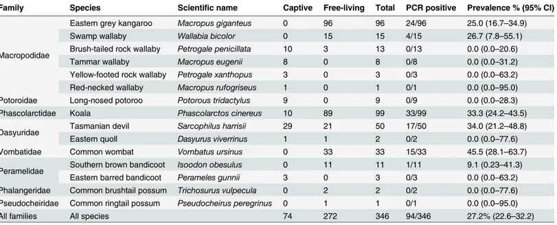
Table 1. Overview of the population of Australian marsupials sampled for this study during 2010 and 2011, and results from the PCR detection of herpesvirus DNA in the collected swab samples.
Family Species Scientific name Captive Free-living Total PCR positive Prevalence % (95% CI)
Macropodidae
Eastern grey kangaroo Macropus giganteus 0 96 96 24/96 25.0 (16.7–34.9)
Swamp wallaby Wallabia bicolor 0 15 15 4/15 26.7 (7.8–55.1)
Brush-tailed rock wallaby Petrogale penicillata 10 3 13 0/13 0.0 (0.0–20.6)
Tammar wallaby Macropus eugenii 8 0 8 0/8 0.0 (0.0–31.2)
Yellow-footed rock wallaby Petrogale xanthopus 3 0 3 0/3 0.0 (0.0–63.2)
Red-necked wallaby Macropus rufogriseus 1 0 1 0/1 0.0 (0.0–95.0)
Potoroidae Long-nosed potoroo Potorous tridactylus 9 0 9 0/9 0.0 (0.0–28.3)
Phascolarctidae Koala Phascolarctos cinereus 10 89 99 33/99 33.3 (24.2–43.5)
Dasyuridae Tasmanian devil Sarcophilus harrisii 29 21 50 17/50 34.0 (21.2–48.8)
Eastern quoll Dasyurus viverrinus 1 1 2 0/2 0.0 (0.0–77.6)
Vombatidae Common wombat Vombatus ursinus 0 33 33 15/33 45.5 (28.1–63.7)
Peramelidae Southern brown bandicoot Isoodon obesulus 0 11 11 1/11 9.1 (0.23–41.3)
Eastern barred bandicoot Perameles gunnii 3 0 3 0/3 0.0 (0.0–63.2)
Phalangeridae Common brushtail possum Trichosurus vulpecula 0 2 2 0/2 0.0 (0.0–77.6)
Pseudocheiridae Common ringtail possum Pseudocheirus peregrinus 0 1 1 0/1 0.0 (0.0–95.0)
All families All species 74 272 346 94/346 27.2% (22.6–32.2)
Molecular investigations
DNA was extracted from 200μl of each swab sample using VX Universal Liquid Sample DNA
Extraction Kits (Qiagen) and a Corbett X-tractor Gene Robot (Corbett Robotics). Negative extraction controls utilised sterile phosphate buffered saline (PBS) only. Positive extraction controls utilised supernatant from cell cultures infected with the avian alphaherpesvirus, infec-tious laryngotracheitis virus. Extracted DNA was then used as a template in a nested pan-her-pes PCR, using primers targeting a conserved region of the herpan-her-pesvirus DNA polymerase gene [19]. PCR negative controls containing no DNA template were also included. PCR products underwent DNA purification (QIAquick Gel Extraction Kit, Qiagen) and sequencing (Big Dye Terminator version 3.1, Applied Biosystems). Sequences were compared with published nucle-otide sequences in the GenBank database (NCBI, 2013) using the BLAST-N online algorithm. Further refinement and analysis of the sequence data was performed with Geneious Pro 5.1.6 (Biomatters Ltd) software. The predicted amino acid sequences of the detected herpesviruses were aligned with representative members from the threeHerpesviridaesubfamilies from a range of host species, and an unrooted maximum-likelihood phylogenetic tree was generated from this sequence alignment using the Jones–Taylor–Thornton model of amino acid replace-ment, as described previously [12].
Cell culture and virus isolation
Selected swab samples that were positive for the presence of novel herpesviruses, or herpesvi-ruses that had not been isolated previously, were used to inoculate wallaby fibroblast cells [20] or primary wombat kidney cell cultures in a closed culture system and incubated at 35–37°C. The primary wombat kidney cells were generated from kidney tissue harvested from a wombat joey using standard trypsin disaggregation techniques. Inoculated cells were evaluated daily for evidence of cytopathic effect (CPE) consistent with herpesvirus infection. Cell cultures were passaged as necessary. The presence of herpesvirus virions was confirmed using electron microscopy.
Serological analysis
Serum-virus neutralisation assays were used to detect antibodies to the closely related alpha-herpesviruses MaHV-1 and MaHV-2 in serum samples (n = 79) collected from animals from eight different marsupial species. Serum samples were thawed at room temperature before cen-trifugation to remove residual cellular debris. Samples were then heat inactivated at 56°C for 30 min before testing as previously described [8]. Briefly, serum samples were diluted 1:2 in sterile media and added to 100 times the median tissue culture infective dose (100 TCID50) of
MaHV-1 or MaHV-2. Serum samples that prevented viral CPE in wallaby fibroblast cells were recorded as positive for the presence of virus-neutralising antibodies. A serum sample that had previously tested positive in our laboratory using these methods was used as a positive control. Other control wells were cell-only controls, and no-serum (cell and virus only) controls. Virus titrations were performed to confirm the viruses were used at the correct dilution. Antibody levels were not determined by further dilution of positive serum samples.
Statistical analysis
individuals were present in the study population, and also for all marsupial species combined. The analysis of all marsupial species combined included data from the individual animals reported in this study (Table 1) in addition to data from 27 woylies (Bettongia penicillata) and 24 eastern bettongs which were originally tested as part of this project [10,21] but the results of which have been analysed at a species level separately as a component of studies focussed on those animals. Univariable analysis was initially performed for each variable under investiga-tion, and variables were assessed for collinearity using contingency tables. Reference levels were selected on the basis of greater sample size and biological sense. Inclusion in the prelimi-nary final model was based on a log likelihood p value0.25 and absence of collinearity with other included variables. A backward elimination method was used to determine the final mul-tivariable logistic regression model and eliminated variables were individually returned to the model to assess confounding (considered to be>25% change in the coefficient of retained
sig-nificant variables). A two-tailed p0.05 was considered to be statistically significant.
Results
Molecular investigations
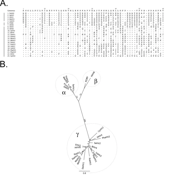
The prevalence of herpesvirus DNA detection by PCR in the marsupial species evaluated ranged from 0.0–45.5%, with an overall prevalence of 27.2% (Table 1). Nucleotide sequence analyses and comparison with sequences in the GenBank database identified six novel herpesviruses; three in wombats, one in swamp wallabies, one in Tasmanian devils and one in a southern brown bandi-coot. Using current herpesvirus nomenclature these novel herpesviruses were putatively desig-nated Vombatid herpesvirus 1–3 (VoHV1-3), Macropodid herpesvirus 5 (MaHV-5), Dasyurid herpesvirus 2 (DaHV-2) and Peramelid herpesvirus 1 (PeHV-1), respectively. Five of the six novel viruses were gammaherpesviruses, with one alphaherpesvirus (VoHV-3) detected in wombats. Sequence data and phylogenetic analysis for the novel herpesvirus species are presented inFig 1.
Each of the novel herpesviruses was restricted to a single host species. In wombats, VoHV-1, VoHV-2 and VoHV-3 were detected in 5/33 (15%), 7/33 (21%) and 3/33 (9%) of the sample population, respectively. In one wombat both VoHV-1 and VoHV-2 were detected. In swamp wallabies, Tasmanian devils and southern brown bandicoots, the novel herpesviruses (MaHV-5, DaHV-2 and PeHV-1, respectively) were detected in 4/15 (27%), 17/50 (34%) and 1/11 (9%) of animals, respectively.
In addition to the six novel herpesviruses identified in this study, nucleotide sequence analy-ses also revealed the presence of four previously described herpesviruanaly-ses in the samples col-lected from the animals listed inTable 1. Macropodid herpesviruses 3 and 4 were detected in 19/96 (20%) and 5/96 (5%) of eastern grey kangaroos, respectively. Phascolarctid herpesviruses 1 and 2 were detected in 10/99 (10%) and 23/99 (23%) of koalas, respectively. In one koala, both PhaHV-1 and PhaHV-2 were detected. Sequence data was unavailable for herpesvirus DNA detected in an additional koala.
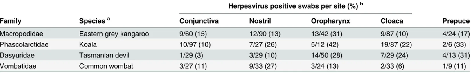
Herpesvirus DNA was detected in swabs collected from a number of different anatomical sites, with some variations between species (Table 2).
Cell culture, virus isolation and electron microscopy
Serological analysis
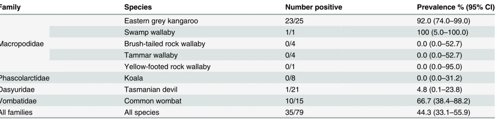
Serum samples from 79 animals from 8 different marsupial species were tested for antibodies against the two closely related macropodid alphaherpesviruses, MaHV-1 and MaHV-2. The Fig 1. Predicted amino acid alignment and phylogenetic tree of the novel marsupial herpesviruses.A: Alignment of the predicted amino acid sequence of a portion of the DNA polymerase gene of the novel marsupial herpesviruses, along with other herpesviruses from the three herpesvirus sub-families. B: Maximum likelihood tree generated from the alignment. Bootstrap values of 100 replicates are displayed on the tree branches. Novel herpesvirus species are underlined. Key: PaHV-2 = papiine herpesvirus 2 (AAN87165.1); SaHV-1 = saimiriine herpesvirus 1 (YP_003933809.1); HHV-1 = human herpesvirus 1 (NP_044632.1); MaHV-1 = macropodid herpesvirus 1 ([22]); VoHV-3 = vombatid herpesvirus 3 (novel sequence); PCMV = porcine cytomegalovirus (AF268042.1); HHV-6 = human herpesvirus 6A (NP_042931.1); HHV-4 = human herpesvirus 4 (YP_401712.1); HHV-8 = human herpesvirus 8 (ACY00400.1); SaHV-2 = saimiriine herpesvirus 2 (NP_040211.1); BHV-4 = bovine herpesvirus 4 (NP_076501.1); DaHV-1 = dasyurid herpesvirus 1 [13]; DaHV-2 = dasyurid herpesvirus 2 (novel sequence); MaHV-3 = macropodid herpesvirus 3 (ABO61861.1); MaHV-5 = macropodid herpesvirus 5 (novel sequence). PeHV-1 = peramelid herpesvirus 1 (novel sequence); PotHV-1 = potoroid herpesvirus 1 [10]; VoHV-1 = vombatid herpesvirus 1 (novel sequence); VoHV-2 = vombatid herpesvirus 2 (novel sequence); 1 = phascolarctid herpesvirus 1 (AEX15649.1); PhaHV-2 = phascolarctid herpesvirus PhaHV-2 (AFN665PhaHV-28.1); EHV-PhaHV-2 = equine herpesvirus PhaHV-2 (NP_04PhaHV-2605.1); CpHV-PhaHV-2 = caprine herpesvirus PhaHV-2 (ADV9PhaHV-2PhaHV-276.1).
seroprevalence of antibodies to MaHV-1 or -2 in the species evaluated ranged from 0.0–100%, with a pan-species prevalence of 44.3%. The results are summarised inTable 3.
Risk factors
The epidemiological variables of sex, age, body condition score, presence or absence of disease, season of sample collection, source (wild or captive) and the presence or absence of pouch young/lactation (in females only) were assessed as predictors for the presence of herpesvirus DNA in the collected swab samples in species in which 10 or more individual animals tested positive for the presence of herpesvirus DNA.Chlamydia pecorumstatus and geographical location were also assessed as variables for the sub-selection of koala samples (n = 68) for which this information was available [18]. Univariable analysis results for each of these species (eastern grey kangaroos, koalas, Tasmanian devils and common wombats) are shown in Tables 4–7. Statistically significant associations, as determined by multivariable analysis, are presented inTable 8. Similar analyses were performed for all marsupial species combined, including Table 2. Anatomical sites of herpesvirus DNA detection in swab samples collected from Australian marsupials in 2010 and 2011.
Herpesvirus positive swabs per site (%)b
Family Speciesa Conjunctiva Nostril Oropharynx Cloaca Prepuce
Macropodidae Eastern grey kangaroo 9/60 (15) 12/90 (13) 13/42 (31) 9/87 (10) 4/24 (17)
Phascolarctidae Koala 10/97 (10) 7/27 (26) 5/12 (42) 19/87 (22) 2/6 (33)
Dasyuridae Tasmanian devil 1/29 (3) 3/29 (10) 14/50 (28) 7/29 (24) 4/13 (31)
Vombatidae Common wombat 3/27 (11) 9/33 (27) 3/24 (13) 2/33 (6) 1/9 (11)
aOnly species that were sampled in relatively large numbers, from multiple anatomical sites, are included.
bHerpesvirus DNA was sometimes detected in more than one swab from the same animal, swabs were not collected from every anatomical site from
every animal.
doi:10.1371/journal.pone.0133807.t002
Fig 2. Electron micrographs of novel herpesviruses.Transmission electron microscopy was used to visualise herpesviruses in cultures of primary wombat kidney cells. Herpesvirus capsids (arrowheads) of VoHV-1 (A) and VoHV-2 (B) are shown. Bar = 100 nm.
Table 3. Seroprevalence of antibodies to MaHV-1 or MaHV-2 in serum samples collected from Australian marsupials in 2010 and 2011.
Family Species Number positive Prevalence % (95% CI)
Macropodidae
Eastern grey kangaroo 23/25 92.0 (74.0–99.0)
Swamp wallaby 1/1 100 (5.0–100.0)
Brush-tailed rock wallaby 0/4 0.0 (0.0–52.7)
Tammar wallaby 0/4 0.0 (0.0–52.7)
Yellow-footed rock wallaby 0/1 0.0 (0.0–95.0)
Phascolarctidae Koala 0/8 0.0 (0.0–31.2)
Dasyuridae Tasmanian devil 1/21 4.8 (0.1–23.8)
Vombatidae Common wombat 10/15 66.7 (38.4–88.2)
All families All species 35/79 44.3 (33.1–55.9)
doi:10.1371/journal.pone.0133807.t003
Table 4. Univariable analysis assessing select epidemiological variables as predictors for the presence of herpesvirus DNA in eastern grey kanga-roosa.
Variable Herpesvirus DNA positive Prevalence (%) Odds ratio 95% CI Wald p value Log likelihood p value
Sex 0.048
Female 10/57 17.5 1.0
Male 12/33 36.4 2.7 1.0–7.2 0.049
Unknown 2/6
Age 0.13
Pouch young/ sub-adult 12/35 34.3 2.1 0.8–5.5 0.13
Adult/aged 11/55 20.0 1.0
unknown 1/6
Wild/captive n/a
Wild 24/96 25.0 n/a n/a n/a
Captive 0/0
Pouch young/lactation (females) 0.005
No 8/23 34.8 8.5 1.6–45.2 0.012
Yes 2/34 5.9 1.0
Body condition score 0.19
2 12/36 33.3 1.9 0.7–5.3 0.20
3 9/44 20.5 1.0
Unknown 3/16
Disease present 0.048
No 12/62 19.4 1.0
Yes 12/31 38.7 2.6 1.0–6.9 0.048
Unknown 0/3
Season 0.19
Summer 0/2 0.0 0 n/a 0.99
Autumn 3/5 60.0 5.7 0.8–38.9 0.076
Winter 10/48 20.8 1.0
Spring 8/26 30.8 1.7 0.6–5.0 0.34
Unknown 3/15
aReference levels are indicated by odds ratio of 1.0. Results highlighted in bold (log likelihood p0.25) represent variables included in the initial
multivariable model, with the exception of presence of pouch young/lactation as it is correlated with sex and thus excluded. Backward elimination of non-significant variables yielded no significant variables. Multivariable analysis was repeated including presence of pouch young/lactation as a variable instead of sex. Age was excluded from the model due to collinearity. In thefinal model (n = 42) only the absence of pouch young/lactation was identified as a significant factor (Table 8). n/a = not applicable.
those species in which less than ten individual species tested positive for the presence of herpes-virus DNA. The results from this pan-species analysis are available as supplementary material (S1andS2Tables).
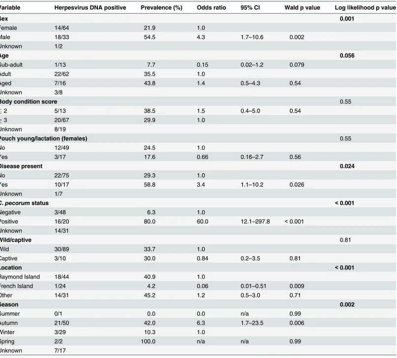
Table 5. Univariable analysis assessing select epidemiological variables as predictors for the presence of herpesvirus DNA in koalasa.
Variable Herpesvirus DNA positive Prevalence (%) Odds ratio 95% CI Wald p value Log likelihood p value
Sex 0.001
Female 14/64 21.9 1.0
Male 18/33 54.5 4.3 1.7–10.6 0.002
Unknown 1/2
Age 0.056
Sub-adult 1/13 7.7 0.15 0.02–1.2 0.079
Adult 22/62 35.5 1.0
Aged 7/16 43.8 1.4 0.5–4.3 0.54
Unknown 3/8
Body condition score 0.55
2 5/13 38.5 1.5 0.4–5.0 0.54
3 20/67 29.9 1.0
Unknown 8/19
Pouch young/lactation (females) 0.55
No 12/49 24.5 1.0
Yes 3/17 17.6 0.66 0.16–2.7 0.56
Disease present 0.024
No 22/75 29.3 1.0
Yes 10/17 58.8 3.4 1.1–10.2 0.026
Unknown 1/7
C.pecorumstatus <0.001
Negative 3/48 6.3 1.0
Positive 16/20 80.0 60.0 12.1–297.8 <0.001
Unknown 14/31
Wild/captive 0.81
Wild 30/89 33.7 1.0
Captive 3/10 30.0 0.84 0.2–3.5 0.81
Location <0.001
Raymond Island 18/44 40.9 1.0
French Island 1/24 4.2 0.06 0.01–0.51 0.009
Other 14/31 45.2 1.2 0.5–3.0 0.71
Season 0.002
Summer 0/1 0.0 0.0 n/a 0.99
Autumn 21/50 42.0 6.3 1.7–23.5 0.006
Winter 3/29 10.3 1.0
Spring 2/2 100.0 n/a n/a 0.99
Unknown 7/17
a
Reference levels are indicated by odds ratio of 1.0. Results highlighted in bold (log likelihood p0.25) represent variables included in the initial multivariable model, with the exception of season as the timing of sampling correlated with the location at which it occurred and was thus excluded. In the
final model (n = 68) only the presence ofChlamydia pecorumwas identified as a significant factor (Table 8). n/a = not applicable.
Discussion
This is the largest published study of herpesviruses in Australian marsupials to date, detecting DNA from 10 different herpesviruses in the samples tested. The findings demonstrate the com-mon presence of herpesvirus infection in Australian marsupials and describe a number of important epidemiological associations. The study identified six novel herpesviruses across four marsupial species. Three of these novel herpesviruses were successfully isolated on pri-mary wombat kidney cells or wallaby fibroblast cells. Herpesvirus DNA was detected from a number of different anatomical sites in the marsupial species tested, this information could be used to help inform sample collection strategies for future studies.
In this study three novel herpesviruses were detected in common wombats. This is the first time herpesvirus infections have been detected in common wombats by molecular techniques. It was found that adult and aged wombats had 17.4 times the odds of testing herpesvirus posi-tive than juvenile wombats (p = 0.012). These data are consistent with an earlier serological study of Australian marsupials which found seropositivity increased with age [9]. The observed association between poor body condition and the detection of herpesvirus DNA in the com-mon wombat (OR = 11.7, p = 0.041) may indicate the presence of disease caused by herpesvirus Table 6. Univariable analysis assessing select epidemiological variables as predictors for the presence of herpesvirus DNA in Tasmanian devilsa.
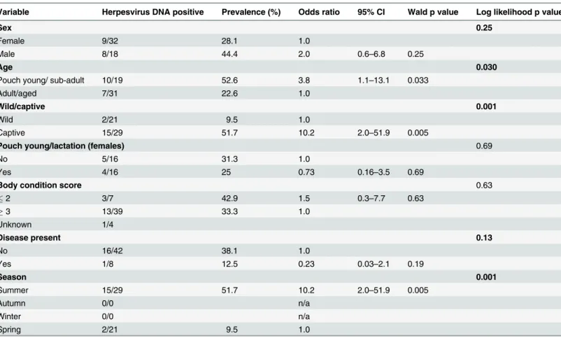
Variable Herpesvirus DNA positive Prevalence (%) Odds ratio 95% CI Wald p value Log likelihood p value
Sex 0.25
Female 9/32 28.1 1.0
Male 8/18 44.4 2.0 0.6–6.8 0.25
Age 0.030
Pouch young/ sub-adult 10/19 52.6 3.8 1.1–13.1 0.033
Adult/aged 7/31 22.6 1.0
Wild/captive 0.001
Wild 2/21 9.5 1.0
Captive 15/29 51.7 10.2 2.0–51.9 0.005
Pouch young/lactation (females) 0.69
No 5/16 31.3 1.0
Yes 4/16 25 0.73 0.16–3.5 0.69
Body condition score 0.63
2 3/7 42.9 1.5 0.3–7.7 0.63
3 13/39 33.3 1.0
Unknown 1/4
Disease present 0.13
No 16/42 38.1 1.0
Yes 1/8 12.5 0.23 0.03–2.1 0.19
Season 0.001
Summer 15/29 51.7 10.2 2.0–51.9 0.005
Autumn 0/0 n/a
Winter 0/0 n/a
Spring 2/21 9.5 1.0
aReference levels are indicated by odds ratio of 1.0. Results highlighted in bold (log likelihood p0.25) represent variables included in the initial
multivariable model, with the exception of season as it was directly influenced by timing of management procedures, and therefore correlated with captive status and was thus excluded. In thefinal model (n = 50) only captivity was identified as a significant factor (Table 8). n/a = not applicable.
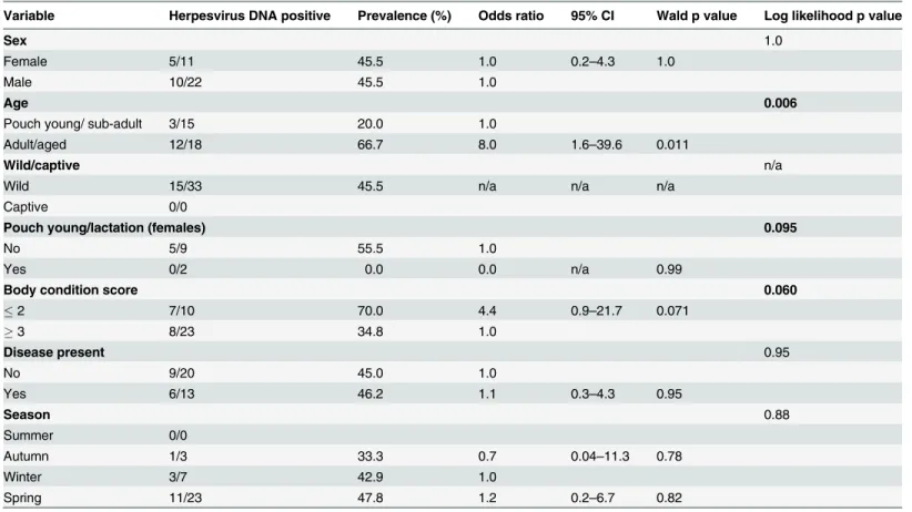
infection, or alternatively may be explained by a higher rate of new or reactivated herpesvirus infections as a consequence of immune-suppression associated with another disease process. The latter appears to be more likely in this study. With progressive urbanisation and habitat destruction in Australia, the generally solitary common wombat is increasingly being subjected Table 7. Univariable analysis assessing select epidemiological variables as predictors for the presence of active herpesvirus infection in common wombatsa.
Variable Herpesvirus DNA positive Prevalence (%) Odds ratio 95% CI Wald p value Log likelihood p value
Sex 1.0
Female 5/11 45.5 1.0 0.2–4.3 1.0
Male 10/22 45.5 1.0
Age 0.006
Pouch young/ sub-adult 3/15 20.0 1.0
Adult/aged 12/18 66.7 8.0 1.6–39.6 0.011
Wild/captive n/a
Wild 15/33 45.5 n/a n/a n/a
Captive 0/0
Pouch young/lactation (females) 0.095
No 5/9 55.5 1.0
Yes 0/2 0.0 0.0 n/a 0.99
Body condition score 0.060
2 7/10 70.0 4.4 0.9–21.7 0.071
3 8/23 34.8 1.0
Disease present 0.95
No 9/20 45.0 1.0
Yes 6/13 46.2 1.1 0.3–4.3 0.95
Season 0.88
Summer 0/0
Autumn 1/3 33.3 0.7 0.04–11.3 0.78
Winter 3/7 42.9 1.0
Spring 11/23 47.8 1.2 0.2–6.7 0.82
aReference levels are indicated by odds ratio of 1.0. Results highlighted in bold (log likelihood p0.25) represent variables included in the initial
multivariable model, with the exception of pouch young which was excluded due to zero prevalence of herpesvirus infection in females lactating/with pouch young. In thefinal model (n = 33) only age (adult/aged) and body condition score (2) were identified as significant factors (Table 8). n/a = not applicable.
doi:10.1371/journal.pone.0133807.t007
Table 8. Summary of epidemiological variables significantly associated with the presence of herpes-virus DNA in samples collected from different species of Australian marsupials in 2010 and 2011, as determined using multivariable analysis.
Population Variable Odds ratio (95% CI) p value
Eastern grey kangaroos (n = 42) Pouch young/lactation absenta 9.6 (1.3
–72.5) 0.028
Koalas (n = 68) Chlamydia pecorumpositive 60.0 (12.1–297.8) <0.001
Tasmanian devils (n = 50) Captivity 10.2 (2.0–51.9) 0.001
Common wombats (n = 33) Adult or aged 17.4 (1.9–162.4) 0.012 Poor body condition score (2) 11.7 (1.1–123.2) 0.041
aThe presence or absence of pouch young/lactation was assessed only in female animals.
to higher intraspecific competition for burrows and for food, with high rates of burrow sharing [23] and consequently higher stress levels and increased potential for disease transmission. Common wombats are particularly susceptible to infestation with the miteSarcoptes scabiei
var.wombatii. Sarcoptic mange is a major cause of debilitation, reduced reproduction and mortality in common wombats [24,25]. Such chronic debilitation could render infested ani-mals more susceptible to new or reactivated herpesvirus infection. The discovery of herpesvi-ruses (and especially alphaherpesvirus) infections in animals where sarcoptic mange is present is therefore of potential significance. The relatively high seroprevalence of antibodies to MaHV-1 and 2 in wombats in this study (66.7%) does not necessarily indicate infection with these viruses, but could indicate infection with another alphaherpesvirus (or multiple alphaher-pesviruses) that cross-neutralise with MaHV-1 or -2, potentially VoHV-3. Future research directed at understanding the significance of infection with the three novel herpesviruses iden-tified in this study is warranted. Expanding future studies to include the endangered northern hairy-nosed wombat (Lasiorhinus kreffiti) and vulnerable southern hairy-nosed wombat (Lasiorhinus latifrons) would also be informative.
In koalas, previous reports of herpesvirus infection (PhaHV-1 and PhaHV-2) have been lim-ited to the description of individual cases [11,12]. In the current study, herpesvirus DNA was detected in 33.3% of the koalas at the time of sampling and there was a significant association between herpesvirus infection and concurrent infection withC.pecorum(OR = 60, p<0.001).
Infections withChlamydiaspp. have been extensively studied in koalas.Chlamydiais a signifi-cant cause of infertility and morbidity in wild koala populations, although infections can also be subclinical [18,26]. Coinfection of herpesviruses with members of theChlamydiaceaefamily has also been observed in cats and humans [27–30], although co-infection appears to be less com-mon in these species than in the koalas examined in this study. The association betweenC.
pecorumand herpesvirus infection in koalas could represent concomitant transmission of both pathogens; reactivation of latent herpesvirus infection secondary to increased immunological burden associated with concurrentChlamydiainfection (or vice versa); or possibly a synergistic role in the pathogenesis of clinical disease in the koala. Further studies to investigate the relation-ship between herpesviruses andChlamydiain koalas are required to determine the reasons for this association and the impacts that coinfection may have on koalas health.
Our study demonstrated a high prevalence of herpesvirus infections in eastern grey kanga-roos. High levels of seropositivity to the alphaherpesviruses MaHV-1 and 2 have been reported in eastern grey kangaroos previously [4,5,9]. Interestingly, the only alphaherpesvirus detected in eastern grey kangaroos in this study was MaHV-4, which has only been recently described [8]. It is probable that the seropositivity to MaHV-1 and -2 detected in this study could reflect infection (including latent infection) with MaHV-4, given there is serological cross-reactivity between these macropodid alphaherpesviruses [8]. The gammaherpesvirus MaHV-3 was detected in 20% of eastern grey kangaroos in this study, but this virus was unlikely to contrib-ute to the detected seropositivity, as antibodies against MaHV-3 are unlikely to cross-neutralise the alphaherpesviruses MaHV-1 or -2. In multivariable analysis, non-lactation/absence of pouch young in female animals was the only factor associated significantly with the presence of herpesvirus DNA in this species (OR = 9.6, p = 0.028). This is contrary to observations in other species where lactation has been associated with reactivation of latent herpesvirus infection [31,32].
Whalley (1978) who identified a higher seroprevalence and higher titre level of anti-MaHV-1 antibodies in captive marsupials compared with free-living counterparts, although different species were represented in the two studies [9]. In general, disease outbreaks and mortality events as a result of marsupial herpesviruses have predominantly been reported in captive mals [3,6,15,16,34]. This trend is presumably due to the close proximity in which captive ani-mals are housed allowing for increased rates of transmission, including transmission to marsupial species that are not natural hosts of the transmitted herpesviruses, and the potential for higher rates of stress in the captive animals. High levels of stress have been reported in wild-caught Tasmanian devils introduced to captivity [35], and such stress could potentially be associated with the reactivation of latent herpesvirus infection. Importantly, no difference was observed in the rates of herpesvirus infection between healthy Tasmanian devils and those with disease (principally DFTD), indicating that DaHV-2 is not directly associated with disease. Serum from one captive Tasmanian devil tested positive in our serum-virus neutralisation assays, indicating infection with MaHV-1 or MaHV-2, or another related alphaherpesvirus. This could be an undetected alphaherpesvirus adapted to Tasmanian devils, or an alphaherpes-virus acquired from another host. Macropodoids can form part of the diet of both captive and wild Tasmanian devils and so this is a potential mechanism of herpesvirus transmission.
The identification and isolation of a novel gammaherpesvirus (MaHV-5) in healthy swamp wallabies, and the identification of a novel gammaherpesvirus (PeHV-1) in a southern brown bandicoot, are interesting findings. Further research involving larger numbers of animals is required to establish the prevalence of these viruses and to determine their significance to their hosts. At the pan-species level a number of parameters were identified as positive predictors for the presence of herpesvirus DNA (S1andS2), including male animals, animals sampled in summer months and animals in poor body conditions. Although the results obtained from analysis of data from multiple species should be interpreted with care due to the potential for confounding effects, this information could be useful for informing the design of future studies of herpesviruses in marsupial species where limited preliminary data is available.
This study is the first to demonstrate the widespread nature of herpesvirus infection in a range of Australian marsupial species. The relatively high prevalence of herpesviruses in a vari-ety of marsupial species and the large number of different herpesviruses identified have impli-cations for the management of captive animal collections and other programmes that relocate animals for the purpose of population management. Further systematic surveillance of wild and captive animal populations and the development of more marsupial specific reagents, such as cell lines for virus propagation, are integral to the further study of these viruses.
Supporting Information
S1 Table. Univariable analysis assessing select epidemiological variables as predictors for the presence of herpesvirus DNA in the study population of all marsupials.
(DOCX)
S2 Table. Summary of epidemiological variables significantly associated with the presence of herpesvirus DNA in samples collected in the study population of all marsupials, as deter-mined using multivariable analysis.
(DOCX)
Acknowledgments
others who collected or submitted samples. The authors also thank the veterinary support staff at the Australian Wildlife Health Centre, Healesville Sanctuary. This work was supported by the Ian Potter Foundation. PW works with Wildlife Health Surveillance Victoria and receives support from The Vizard Foundation. JMD is supported by a fellowship from the Australian Research Council.
Author Contributions
Conceived and designed the experiments: JMD JRG K. Stalder RB. Performed the experiments: K. Stalder PKV NF LT RB PW TP K. Skogvold. Analyzed the data: K. Stalder JMD JRG GAA. Contributed reagents/materials/analysis tools: K. Stalder RB PW NF TP K. Skogvold. Wrote the paper: K. Stalder PKV JRG RB PW NF TP GAA JMD.
References
1. McGeoch DJ, Cook S, Dolan A, Jamieson FE, Telford EA. Molecular phylogeny and evolutionary time-scale for the family of mammalian herpesviruses. J Mol Biol. 1995; 247: 443–458. PMID:7714900 2. Roizman B, Pellett PE. The Family Herpesviridae: A Brief Introduction. In: Knipe DM, Howley PM,
edi-tors. Fields Virology. 4 ed. Philadelphia: Lippincott Williams & Wilkins; 2001. pp. 2381–2397. 3. Finnie EP, Littlejohns IR, Acland HM. Letter: Mortalities in parma wallabies (Macropus parma)
associ-ated with probable herpesvirus. Aust Vet J. 1976; 52: 294. PMID:184775
4. Johnson MA, Whalley JM, Littlejohns IR, Dickson J, Smith VW, Wilks CR, et al. Macropodid herpesvi-ruses 1 and 2: two herpesviherpesvi-ruses from Australian marsupials differentiated by restriction endonucle-ases, DNA composition and hybridization. Brief report. Arch Virol. 1985; 85: 313–319. PMID:2992421 5. Kerr A, Whalley JM, Poole WE. Herpesvirus neutralising antibody in grey kangaroos. Aust Vet J. 1981;
57: 347–348. PMID:7340790
6. Smith JA, Wellehan JF Jr, Pogranichniy RM, Childress AL, Landolfi JA, Terio KA. Identification and iso-lation of a novel herpesvirus in a captive mob of eastern grey kangaroos (Macropus giganteus). Vet Microbiol. 2008; 129: 236–245. doi:10.1016/j.vetmic.2007.11.019PMID:18191922
7. Wilcox RS, Vaz P, Ficorilli NP, Whiteley PL, Wilks CR, Devlin JM. Gammaherpesvirus infection in a free-ranging eastern grey kangaroo (Macropus giganteus). Aust Vet J. 2011; 89: 55–57. doi:10.1111/j.
1751-0813.2010.00662.xPMID:21250958
8. Vaz PK, Motha J, McCowan C, Ficorilli N, Whiteley PL, Wilks CR, et al. Isolation and characterization of a novel herpesvirus from a free-ranging eastern grey kangaroo (Macropus giganteus). J Wildl Dis. 2013; 49: 143–151. doi:10.7589/2012-01-027PMID:23307380
9. Webber CE, Whalley JM. Widespread occurrence in Australian marsupials of neutralizing antibodies to a herpesvirus from a parma wallaby. Aust J Exp Biol Med Sci. 1978; 56: 351–357. PMID:213046 10. Portas T, Fletcher D, Spratt D, Reiss A, Holz P, Stalder K, et al. Health evaluation of free-ranging
east-ern bettongs (Bettongia gaimardi) during translocation for reintroduction in Australia. J Wildl Dis. 2014; 50: 210–223. doi:10.7589/2013-08-202PMID:24484484
11. Vaz P, Whiteley PL, Wilks CR, Browning GF, Gilkerson JR, Ficorilli N, et al. Detection of a second novel gammaherpesvirus in a free-ranging koala (Phascolarctos cinereus). J Wildl Dis. 2012; 48: 226–
229. PMID:22247398
12. Vaz P, Whiteley PL, Wilks CR, Duignan PJ, Ficorilli N, Gilkerson JR, et al. Detection of a novel gamma-herpesvirus in koalas (Phascolarctos cinereus). J Wildl Dis. 2011; 47: 787–791. PMID:21719855 13. Amery-Gale J, Vaz PK, Whiteley P, Tatarczuch L, Taggart DA, Charles JA, et al. Detection and
identifi-cation of a gammaherpesvirus in Antechinus spp. in Australia. J Wildl Dis. 2014; 50: 334–339. doi:10.
7589/2013-07-165PMID:24499331
14. Rothwell JT, Canfield PJ, Wilks CR. Death due to a probable herpesvirus infection in a common wom-bat (Vomwom-batus ursinus). Aust Vet J. 1988; 65: 360–361. PMID:2850789
15. Callinan RB, Kefford B. Mortalities associated with herpesvirus infection in captive macropods. J Wildl Dis. 1981; 17: 311–317. PMID:6264169
16. Dickson J, Hopkinson WI, Coackley W, Spence T, Fairfax R. Herpesvirus hepatitis in rat kangaroos. Aust Vet J. 1980; 56: 463–464.
18. Patterson J, Lynch M, Anderson GA, Noomohammadi AH, Legione A, Gilkerson JR, et al. The preva-lence and clinical significance of Chlamydia infection in island and mainland populations of Victorian koalas (Phascolarctos cinereus). J Wildl Dis. 2015; 51: 309–317 doi:10.7589/2014-07-176PMID:
25588005
19. Chmielewicz B, Goltz M, Lahrmann K-H, Ehlers B. Approach virus safety in xenotransplantation: a search for unrecognized herpesviruses in pigs. Xenotransplantation. 2003; 10: 349–356. PMID:
12795684
20. Uren J, Moore R, van den Brenk HA. Development of a pseudodiploid cell line, JU56, of wallaby fibro-blasts. Exp Cell Res. 1966; 43: 677–680. PMID:5957759
21. Stalder K (2013) Herpesviruses in Australian Marsupials. MVSc Thesis, The University of Melbourne. Available:https://minerva-access.unimelb.edu.au/handle/11343/38194
22. Guliani S, Smith GA, Young PL, Mattick JS, Mahony TJ. Reactivation of a macropodid herpesvirus from the eastern grey kangaroo (Macropus giganteus) following corticosteroid treatment. Vet Microbiol. 1999; 68: 59–69. PMID:10501162
23. Skerratt LF, Skerratt JHL, Banks S, Martin R, Handasyde KA. Aspects of the ecology of common wom-bats (Vombatus ursinus) at high density on pastoral land in Victoria. Aust J Zool. 2004; 52: 303–330. 24. Martin RW, Handasyde KA, Skerratt LF. Current distribution of sarcoptic mange in wombats. Aust Vet
J. 1998; 76: 411–414. PMID:9673766
25. Skerratt LF, Middleton D, Beveridge I. Distribution of life cycle stages of Sarcoptes scabiei var wombati and effects of severe mange on common wombats in Victoria. J Wildl Dis. 1999; 35: 633–646. PMID:
10574522
26. Polkinghorne A, Hanger J, Timms P. Recent advances in understanding the biology, epidemiology and control of chlamydial infections in koalas. Vet Microbiol. 2013; 165: 214–223. doi:10.1016/j.vetmic.
2013.02.026PMID:23523170
27. Cai Y, Fukushi H, Koyasu S, Kuroda E, Yamaguchi T, Hirai K. An etiological investigation of domestic cats with conjunctivitis and upper respiratory tract disease in Japan. J Vet Med Sci. 2002; 64: 215–219.
PMID:11999440
28. Kibur M, Koskela P, Dillner J, Leinikki P, Saikku P, Lehtinen M, et al. Seropositivity to multiple sexually transmitted infections is not common. Sex Transm Dis. 2000; 27: 425–430. PMID:10987446 29. Shizuma T. [Case of infectious mononucleosis with suspected primary coinfection with Chlamydophila
(Chlamydia) pneumoniae and Epstein-Barr virus]. Kansenshogaku Zasshi. 2008; 82: 451–454. PMID:
18975589
30. Sykes JE, Anderson GA, Studdert VP, Browning GF. Prevalence of feline Chlamydia psittaci and feline herpesvirus 1 in cats with upper respiratory tract disease. J Vet Intern Med. 1999; 13: 153–162. PMID:
10357102
31. Gaskell RM, Povey RC. Experimental induction of feline viral rhinotracheitis virus re-excretion in FVR-recovered cats. Vet Rec. 1977; 100: 128–133. PMID:191978
32. Meier J, Lienicke U, Tschirch E, Kruger DH, Wauer RR, Prosch S. Human cytomegalovirus reactivation during lactation and mother-to-child transmission in preterm infants. J Clin Microbiol. 2005; 43: 1318–
1324. PMID:15750102
33. Loh R, Bergfeld J, Hayes D, O'Hara A, Pyecroft S, Raidal S, et al. The pathology of devil facial tumor disease (DFTD) in Tasmanian Devils (Sarcophilus harrisii). Vet Pathol. 2006; 43: 890–895. PMID:
17099145
34. Acland HM. Parma wallaby herpesvirus infection. J Wildl Dis. 1981; 17: 471–477. PMID:6273601 35. Jones SM, Lockhart TJ, Rose RW. Adaptation of wild-caught Tasmanian devils (Sarcophilus harrisii) to